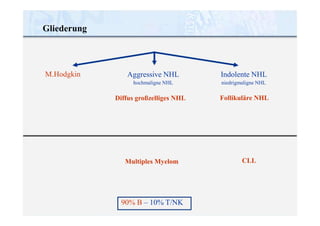
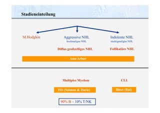
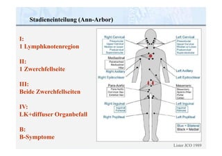
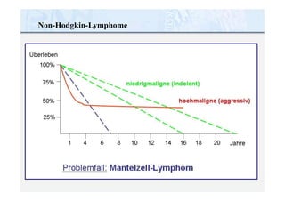
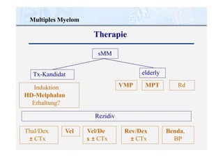
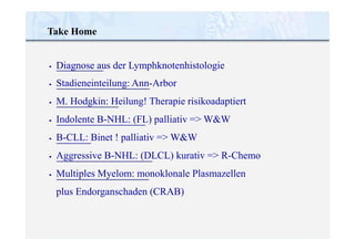
PD Dr. R. Schmidmaier
Medizinische Klinik Innenstadt · Ludwig - Maximilians- Universit ät Mü nchen
Maligne Lymphome und Multiples Myelom
•
•
•
Grundsätzliches zu Malignen Lymphomen
M. Hodgkin
indolente NHL: follikuläres B-NHL
• CLL
•
•
aggressive NHL: diffus großzelliges B-NHL
Multiples Myelom
Maligne Lymphome und Multiples Myelom
28-jährige Patientin mit ...
... Fieber und Nachtschweiß seit 3 Wochen
28-jährige Patientin mit ...
... Fieber und Nachtschweiß seit 3 Wochen
28-jährige Patientin mit ...
... Fieber und Nachtschweiß seit 3 Wochen
Die Diagnose eines Malignen
Lymphoms wird aus der
Lymphknotenhistologie gestellt.
Vorgehen bei Lymphadenopathie
Erfolgreiche Selektion
Malignität
1%
17 %
60 %
Jede Lymphadenpathie unklarer
Genese beim Primärarzt
Vom Primärarzt zum Hämatologen
überwiesen (alle Patienten)
Vom Hämatologen biopsiert
Vorgehen bei Lymphadenopathie
Zervikal: Häufig nicht maligne
Supraklavikulär: 50% Malignom
Axillär: >50% Malignom
Inguinal: Häufig nicht maligne
Lymphknotenhistologie
der NHL
Lymphknotenhistologie
M.Hodgkin
35% 45%
20%
Aggressive NHL
hochmaligne NHL
„blastär“
Indolente NHL
niedrigmaligne NHL
„zytisch“
• CLL
nodal
splenisch
• Immunozytom
M. Waldenström
• Haarzell-Leukämie
• Kutane T-NHL
• Diffus großzelliges NHL
centroblastisch
immunoblastisch
großzell.-anaplastisch
T-zellreiche B-NHL
• lymphozytenreich
• nodulär-sklerosierend
• gemischtzellig
• lymphozytenreich
• Mantelzell-Lymhome
• B-lymphoblastisches
• Burkitt-Lymphom
90% B – 10% T/NK
30-40%
der NHL
15-20%
der NHL
• Follikuläre NHL 25-30%
cbcc
• Marginalzonen-NHL
MALT
Gliederung
M.Hodgkin Aggressive NHL
hochmaligne NHL
Diffus großzelliges NHL
Multiples Myelom
90% B – 10% T/NK
Indolente NHL
niedrigmaligne NHL
Follikuläre NHL
CLL
Stadieneinteilung
M.Hodgkin Aggressive NHL
hochmaligne NHL
Diffus großzelliges NHL
Ann-Arbor
Multiples Myelom
ISS (Salmon & Durie)
90% B – 10% T/NK
Indolente NHL
niedrigmaligne NHL
Follikuläre NHL
CLL
Binet (Rai)
Stadieneinteilung (Ann-Arbor)
I:
1 Lymphknotenregion
II:
1 Zwerchfellseite
III:
Beide Zwerchfellseiten
IV:
LK+diffuser Organbefall
B:
B-Symptome
Lister JCO 1989
M. Hodgkin
CD30
modifiziert nach Diehl V, Internist 1998,39:917-26 und DeVita Annals Int Med 1970;73: 881-895.
nur alkylierende
Substanzen (1965)
keine Behandlung
(1940)
M. Hodgkin
Ziel: Heilung !
BEACOPP
(1993-97)
COPP/ABVD
(1988-93)
MOPP
(DeVita 1970)
Hodgkin’s Disease
Secondary Cancers
MDS
Cardiovascular
Pulmonary
Infection
Trauma/Suicide
Other/Unknown
Total
383
200
11
148
41
35
16
96
930
41.2 %
21.5 %
1.2 %
15.9 %
4.4 %
3.8 %
1.7 %
10.3 %
100 %
Stanford data, Hoppe et al.
M. Hodgkin
Todesursachen bei M. Hodgkin (N=2733)
M. Hodgkin
Therapierelevante Stadieneinteilung
limitierte Stadien:
intermediäre Stadien:
I (A,B), II (A,B)
I (A,B), II A
ohne RF
mit RF a-d
mit RF c,d
mit RF a,b
II B
fortgeschrittene Stadien: II B
III, IV
Risikofaktoren: a) großer Mediastinaltumor
b) Extranodalbefall
c) hohe BSG
d) 3 oder mehr befallene Areale
Chemo plus Involved-Field-Radiatio
2xABVD + 30 Gy IF-RT
Frühe Stadien:
6-JÜR > 90%
6-JÜR > 90%
Fortgeschr. Stadien:
6-JÜR > 90%
Intermediäre Stadien: Chemo plus Involved-Field-Radiatio
2xBEACOPP esc. + 2xABVD
+ 30 Gy IF-RT
Intensive Polychemotherapie
8xBEACOPPeskaliert + 30Gy@PET+
> 60 J.: 6-8x ABVD
M. Hodgkin
Therapie innerhalb von Studienprotokollen
Non-Hodgkin-Lymphome
Indolente NHL: Bsp. Follikuläres Lymphom
Therapieintention
Lokalisiertes Stadium (I/II): 15-20% d. Pat.: kurative Intention
- Bis 50% Langzeitremissionen bei Extended Field Bestrahlung
- Adjuvante Chemotherapie ohne Vorteil (Hiddemann 1997)
Fortgeschrittenes Stadium (III/IV): palliativer Therapieansatz
- Watch and wait (Horing, Ann Oncol 2000; Ardeshna, Lancet 2003)
- Therapie bei Symptomen
Indolente NHL: Bsp. Follikuläres Lymphom
Therapieindikation III-IV
Behandlungsindikation:
- Krankheitsassoziierte Symptome (B-Symptome)
- Hämatopoetische Insuffizienz
- Bulky disease
- Rasche Krankheitsprogredienz
- Vorhersehbare/drohende Organschäden
Indolente NHL: Bsp. Follikuläres Lymphom
Swenson JCO 2005
Human
CD20
Indolente NHL: Bsp. Follikuläres Lymphom
Rituximab
(Mabthera)
Mouse
ORR (%) 4y-PFS (%) 4y-OS (%) line
1. R-CHOP (6-8x)
2. R-CVP (8x)
3. R-FCM (4x)
4. R-MCP (8x)
96 vs. 90 *
81 vs. 57 *
94 vs. 70 *
92 vs. 75 *
62 vs. 28 *
47 vs. 17 *
(3) 78 vs 53 *
69 vs. 63 *
90 vs. 81 *
83 vs. 77 *
(2) 90 vs. 70
87 vs. 74 *
1st
1st
>1
1st
5. R-CHVP+I (6x) 94 vs. 85 * (3) 78 vs. 62 * (5) 84 vs. 79 1st
vs. CHVP+I (12x)
* signifikant
1. Hiddemann Blood 2005, Buske ASH 2006, 2. Marcus JCO 2008, 4.
Forstpointner Blood 2004, Blood 2006, 4. Herold JCO 2007, 5. Salles Blood 2008
Indolente NHL: Bsp. Follikuläres Lymphom
Immunochemotherapie
Follikuläres Lymphom
Immunochemotherapie
Hiddemann et al (GLSG), Blood 2005
Aggressive NHL: Bsp. diffus-großzelliges NHL
Fisher NEJM 1993, Shipp NEJM 1993
Aggressive NHL: Bsp. diffus-großzelliges NHL
Therapieintention
KURATIV ! => Diagnose ist Indikation zur Therapie !
Internationaler Prognoseindex (IPI):
• Alter > 60 Jahren
• Performance Status (ECOG ≥ 2)
• LDH-Erhöhung
• Extranodaler Befall > 1
• Stadium III/IV
Follikuläres Lymphom
MINT-Trial, Pfreundschuh Lancet Oncology 2006
Immunochemotherapie
Aggressive NHL: Bsp. diffus-großzelliges NHL
R-CHOP
CLL: chronische lymphatische Leukämie
 Anhaltende Lymphozytose > 5.000/µl
(morphologisch reife Lymphozyten)
 Charakteristischer Immunphänotyp
(CD5+, CD19+, CD23+, FMC7-
CD10-, CD20weak, surface-Igweak,
Leichtkettenrestriktion)
Cheson et al 1996 Blood 87: 4990-4997.
Diagnose
> 75 Jahre
23%
< 65 Jahre
65-75 Jahre
33%
 Altersverteilung:
44%
CLL: chronische lymphatische Leukämie
Epidemiologie
 Häufigste Leukämie der westlichen Welt (Inzidenz: 3/100.000)
Stadieneinteilung nach Binet:
A Keine Anämie, keine Thrombopenie
0-2 befallene Regionen
B 3-5 befallene Regionen
keine Anämie oder Thrombopenie
C Anämie und/oder Thrombopenie
Patienten
60%
30%
15%
Prognose
normal
7 Jahre
2 Jahre
Binet et al 1981 Cancer 48: 198-206.
(Hb < 10,0 g/dl, Thro <100.000/µl)
5 Regionen (klin. Untersuchung):




Axilläre Lymphknoten
Zervikale Lymphknoten
Inguinale Lymphknoten
Leber
 Milz
CLL: chronische lymphatische Leukämie
Stadieneinteilung
CLL: chronische lymphatische Leukämie
1. Krankheitsbedingte Symptome (Müdigkeit, B-Symptome)
2. Knochenmarkinsuffizienz (Hb<10g/dl; Thrombos<100.000/µl)
3. Immunzytopenie refraktär auf Kortikosteroide
4. Kurze Lymphozytenverdopplungszeit (< 6 Monate) und/oder
rasch zunehmende Lymphadenopathie und Splenomegalie
Montserrat Irsee 2002, Cheson Blood 1996, IWCLL Ann Intern Med 1991
Therapieindikation
Chronische lymphatische Leukämie
Knospe et al. Cancer 1974
Chlorambucil
CLL: chronische lymphatische Leukämie
CLL Trialists‘ Collaborative Group 1999 JNCI 91:861.
N=2022
Chlorambucil
CLL: chronische lymphatische Leukämie
CLL8-Studie: FCR versus FC
Binet A+B C
ORR 93% 88%
CR 57%/43% 29%
PFS P<0,000001 P=0,44
6 Zyklen:
ORR:
CR:
2yOS:
FCR FC
74% 67%
95% 88%*
52% 27%*
91% 88%
CTC°3/4: 62% 47%*
°3/4 ANC: 34% 21%*
°3/4 ID: 19% 15%
Hallek ASH 2008
N=817
009
SH 1
0,0
: P=
@3
CLL: chronische lymphatische Leukämie
CLL8-Studie: FCR versus FC
Binet A+B C
ORR 93% 88%
CR 57%/43% 29%
PFS P<0,000001 P=0,44
FCR FC
6 Zyklen:
ORR:
CR:
2yOS:
74% 67%
95% 88%*
52% 27%*
91% 88%
CTC°3/4: 62% 47%*
°3/4 ANC: 34% 21%*
°3/4 ID: 19% 15%
Hallek ASH 2008
N=817
:
A
OS
2
y
CLL: chronische lymphatische Leukämie
Dreger Leukemia 2007; 21:12.
„ ... cure seems to be possible in 1/3 – 2/3 of poor risk CLL.“
Junge, vorbehandelte Patienten mit Hochrisiko-CLL sollten
so früh als möglich bei einem Transplantationszentrum
vorgestellt werden.
Hochrisiko:
- Frührezidiv (<12 Monate) nach Fludarabin
- Rezidiv (<24 Monate) nach Fludarabin-Kombinationstherapie
- Rezidiv (<24 Monate) nach autologer Stammzelltransplantation
- behandlungsbedürftige CLL mit del17p13
Allogene Stammzelltransplantation
Multiples Myelom
Multiples Myelom
Spontanverlauf
Holland Blood 1966; 27: 328-342
Multiples Myelom
Prognoseverbesserung
Kumar Blood 2008; 111: 2516-2520
Multiples Myelom
Diagnose
Durie BG et al. Leukemia. 2006;20(9):1467–73.
International Myeloma Working Group. Br J Haematol. 2003;121(5):749–57.
asympt. MM:
Follow-Up alle 3-6 Monate
sympt. MM:
Bisphosphonate
systemische zytostatische Therapie
NCCN V.2.2009
MM-Risiko:
10%/Jahr x 5 Jahre
Multiples Myelom
Therapeutische Prinzipien
MGUS: MM-Risiko: 1%/Jahr
Follow-Up erst nach 6, dann alle 12 Monate
solitäres Plasmozytom (ossär/extraossär):
involved field Radiatio ≥ 45Gy
Follow-Up alle 6-12 Monate
Multiples Myelom
Hochdosis-Melphalan + autologe
Blutstammzelltransplantation
Ctx
G-CSF
MEL
PHA
LAN
PBSCs PBSCs
MEL
PHA
LAN
Attal NEJM 1996
14.8 30.9
Multiples Myelom
Melphalan und neue Substanzen
16 M
• Thalidomid
• Lenalidomid
• Bortezomib
Kumar Blood 2008; 111: 2516-2520
Multiples Myelom
Therapie
sMM
Induktion
HD-Melphalan
Erhaltung?
elderly
Rezidiv
Tx-Kandidat
MPT
VMP Rd
Vel/De
x ± CTx
Thal/Dex
± CTx
Rev/Dex
± CTx
Benda,
BP
Vel
Take Home
•
•
•
•
•
•
•
Diagnose aus der Lymphknotenhistologie
Stadieneinteilung: Ann-Arbor
M. Hodgkin: Heilung! Therapie risikoadaptiert
Indolente B-NHL: (FL) palliativ => W&W
B-CLL: Binet ! palliativ => W&W
Aggressive B-NHL: (DLCL) kurativ => R-Chemo
Multiples Myelom: monoklonale Plasmazellen
plus Endorganschaden (CRAB)

44a Leukämie. Lymphome. schmidmaier_ lymphome. Lymphome und multiples Myelom.

  • 1.
    PD Dr. R.Schmidmaier Medizinische Klinik Innenstadt · Ludwig - Maximilians- Universit ät Mü nchen Maligne Lymphome und Multiples Myelom
  • 2.
    • • • Grundsätzliches zu MalignenLymphomen M. Hodgkin indolente NHL: follikuläres B-NHL • CLL • • aggressive NHL: diffus großzelliges B-NHL Multiples Myelom Maligne Lymphome und Multiples Myelom
  • 3.
    28-jährige Patientin mit... ... Fieber und Nachtschweiß seit 3 Wochen
  • 4.
    28-jährige Patientin mit... ... Fieber und Nachtschweiß seit 3 Wochen
  • 5.
    28-jährige Patientin mit... ... Fieber und Nachtschweiß seit 3 Wochen Die Diagnose eines Malignen Lymphoms wird aus der Lymphknotenhistologie gestellt.
  • 6.
    Vorgehen bei Lymphadenopathie ErfolgreicheSelektion Malignität 1% 17 % 60 % Jede Lymphadenpathie unklarer Genese beim Primärarzt Vom Primärarzt zum Hämatologen überwiesen (alle Patienten) Vom Hämatologen biopsiert
  • 7.
    Vorgehen bei Lymphadenopathie Zervikal:Häufig nicht maligne Supraklavikulär: 50% Malignom Axillär: >50% Malignom Inguinal: Häufig nicht maligne
  • 8.
  • 9.
    der NHL Lymphknotenhistologie M.Hodgkin 35% 45% 20% AggressiveNHL hochmaligne NHL „blastär“ Indolente NHL niedrigmaligne NHL „zytisch“ • CLL nodal splenisch • Immunozytom M. Waldenström • Haarzell-Leukämie • Kutane T-NHL • Diffus großzelliges NHL centroblastisch immunoblastisch großzell.-anaplastisch T-zellreiche B-NHL • lymphozytenreich • nodulär-sklerosierend • gemischtzellig • lymphozytenreich • Mantelzell-Lymhome • B-lymphoblastisches • Burkitt-Lymphom 90% B – 10% T/NK 30-40% der NHL 15-20% der NHL • Follikuläre NHL 25-30% cbcc • Marginalzonen-NHL MALT
  • 10.
    Gliederung M.Hodgkin Aggressive NHL hochmaligneNHL Diffus großzelliges NHL Multiples Myelom 90% B – 10% T/NK Indolente NHL niedrigmaligne NHL Follikuläre NHL CLL
  • 11.
    Stadieneinteilung M.Hodgkin Aggressive NHL hochmaligneNHL Diffus großzelliges NHL Ann-Arbor Multiples Myelom ISS (Salmon & Durie) 90% B – 10% T/NK Indolente NHL niedrigmaligne NHL Follikuläre NHL CLL Binet (Rai)
  • 12.
    Stadieneinteilung (Ann-Arbor) I: 1 Lymphknotenregion II: 1Zwerchfellseite III: Beide Zwerchfellseiten IV: LK+diffuser Organbefall B: B-Symptome Lister JCO 1989
  • 13.
  • 14.
    modifiziert nach DiehlV, Internist 1998,39:917-26 und DeVita Annals Int Med 1970;73: 881-895. nur alkylierende Substanzen (1965) keine Behandlung (1940) M. Hodgkin Ziel: Heilung ! BEACOPP (1993-97) COPP/ABVD (1988-93) MOPP (DeVita 1970)
  • 15.
    Hodgkin’s Disease Secondary Cancers MDS Cardiovascular Pulmonary Infection Trauma/Suicide Other/Unknown Total 383 200 11 148 41 35 16 96 930 41.2% 21.5 % 1.2 % 15.9 % 4.4 % 3.8 % 1.7 % 10.3 % 100 % Stanford data, Hoppe et al. M. Hodgkin Todesursachen bei M. Hodgkin (N=2733)
  • 16.
    M. Hodgkin Therapierelevante Stadieneinteilung limitierteStadien: intermediäre Stadien: I (A,B), II (A,B) I (A,B), II A ohne RF mit RF a-d mit RF c,d mit RF a,b II B fortgeschrittene Stadien: II B III, IV Risikofaktoren: a) großer Mediastinaltumor b) Extranodalbefall c) hohe BSG d) 3 oder mehr befallene Areale
  • 17.
    Chemo plus Involved-Field-Radiatio 2xABVD+ 30 Gy IF-RT Frühe Stadien: 6-JÜR > 90% 6-JÜR > 90% Fortgeschr. Stadien: 6-JÜR > 90% Intermediäre Stadien: Chemo plus Involved-Field-Radiatio 2xBEACOPP esc. + 2xABVD + 30 Gy IF-RT Intensive Polychemotherapie 8xBEACOPPeskaliert + 30Gy@PET+ > 60 J.: 6-8x ABVD M. Hodgkin Therapie innerhalb von Studienprotokollen
  • 18.
  • 19.
    Indolente NHL: Bsp.Follikuläres Lymphom
  • 20.
    Indolente NHL: Bsp.Follikuläres Lymphom Therapieintention Lokalisiertes Stadium (I/II): 15-20% d. Pat.: kurative Intention - Bis 50% Langzeitremissionen bei Extended Field Bestrahlung - Adjuvante Chemotherapie ohne Vorteil (Hiddemann 1997) Fortgeschrittenes Stadium (III/IV): palliativer Therapieansatz - Watch and wait (Horing, Ann Oncol 2000; Ardeshna, Lancet 2003) - Therapie bei Symptomen
  • 21.
    Indolente NHL: Bsp.Follikuläres Lymphom Therapieindikation III-IV Behandlungsindikation: - Krankheitsassoziierte Symptome (B-Symptome) - Hämatopoetische Insuffizienz - Bulky disease - Rasche Krankheitsprogredienz - Vorhersehbare/drohende Organschäden
  • 22.
    Indolente NHL: Bsp.Follikuläres Lymphom Swenson JCO 2005
  • 23.
    Human CD20 Indolente NHL: Bsp.Follikuläres Lymphom Rituximab (Mabthera) Mouse
  • 24.
    ORR (%) 4y-PFS(%) 4y-OS (%) line 1. R-CHOP (6-8x) 2. R-CVP (8x) 3. R-FCM (4x) 4. R-MCP (8x) 96 vs. 90 * 81 vs. 57 * 94 vs. 70 * 92 vs. 75 * 62 vs. 28 * 47 vs. 17 * (3) 78 vs 53 * 69 vs. 63 * 90 vs. 81 * 83 vs. 77 * (2) 90 vs. 70 87 vs. 74 * 1st 1st >1 1st 5. R-CHVP+I (6x) 94 vs. 85 * (3) 78 vs. 62 * (5) 84 vs. 79 1st vs. CHVP+I (12x) * signifikant 1. Hiddemann Blood 2005, Buske ASH 2006, 2. Marcus JCO 2008, 4. Forstpointner Blood 2004, Blood 2006, 4. Herold JCO 2007, 5. Salles Blood 2008 Indolente NHL: Bsp. Follikuläres Lymphom Immunochemotherapie
  • 25.
  • 26.
    Aggressive NHL: Bsp.diffus-großzelliges NHL
  • 27.
    Fisher NEJM 1993,Shipp NEJM 1993 Aggressive NHL: Bsp. diffus-großzelliges NHL Therapieintention KURATIV ! => Diagnose ist Indikation zur Therapie ! Internationaler Prognoseindex (IPI): • Alter > 60 Jahren • Performance Status (ECOG ≥ 2) • LDH-Erhöhung • Extranodaler Befall > 1 • Stadium III/IV
  • 28.
    Follikuläres Lymphom MINT-Trial, PfreundschuhLancet Oncology 2006 Immunochemotherapie
  • 29.
    Aggressive NHL: Bsp.diffus-großzelliges NHL R-CHOP
  • 30.
    CLL: chronische lymphatischeLeukämie  Anhaltende Lymphozytose > 5.000/µl (morphologisch reife Lymphozyten)  Charakteristischer Immunphänotyp (CD5+, CD19+, CD23+, FMC7- CD10-, CD20weak, surface-Igweak, Leichtkettenrestriktion) Cheson et al 1996 Blood 87: 4990-4997. Diagnose
  • 31.
    > 75 Jahre 23% <65 Jahre 65-75 Jahre 33%  Altersverteilung: 44% CLL: chronische lymphatische Leukämie Epidemiologie  Häufigste Leukämie der westlichen Welt (Inzidenz: 3/100.000)
  • 32.
    Stadieneinteilung nach Binet: AKeine Anämie, keine Thrombopenie 0-2 befallene Regionen B 3-5 befallene Regionen keine Anämie oder Thrombopenie C Anämie und/oder Thrombopenie Patienten 60% 30% 15% Prognose normal 7 Jahre 2 Jahre Binet et al 1981 Cancer 48: 198-206. (Hb < 10,0 g/dl, Thro <100.000/µl) 5 Regionen (klin. Untersuchung):     Axilläre Lymphknoten Zervikale Lymphknoten Inguinale Lymphknoten Leber  Milz CLL: chronische lymphatische Leukämie Stadieneinteilung
  • 33.
    CLL: chronische lymphatischeLeukämie 1. Krankheitsbedingte Symptome (Müdigkeit, B-Symptome) 2. Knochenmarkinsuffizienz (Hb<10g/dl; Thrombos<100.000/µl) 3. Immunzytopenie refraktär auf Kortikosteroide 4. Kurze Lymphozytenverdopplungszeit (< 6 Monate) und/oder rasch zunehmende Lymphadenopathie und Splenomegalie Montserrat Irsee 2002, Cheson Blood 1996, IWCLL Ann Intern Med 1991 Therapieindikation
  • 34.
    Chronische lymphatische Leukämie Knospeet al. Cancer 1974 Chlorambucil
  • 35.
    CLL: chronische lymphatischeLeukämie CLL Trialists‘ Collaborative Group 1999 JNCI 91:861. N=2022 Chlorambucil
  • 36.
    CLL: chronische lymphatischeLeukämie CLL8-Studie: FCR versus FC Binet A+B C ORR 93% 88% CR 57%/43% 29% PFS P<0,000001 P=0,44 6 Zyklen: ORR: CR: 2yOS: FCR FC 74% 67% 95% 88%* 52% 27%* 91% 88% CTC°3/4: 62% 47%* °3/4 ANC: 34% 21%* °3/4 ID: 19% 15% Hallek ASH 2008 N=817
  • 37.
    009 SH 1 0,0 : P= @3 CLL:chronische lymphatische Leukämie CLL8-Studie: FCR versus FC Binet A+B C ORR 93% 88% CR 57%/43% 29% PFS P<0,000001 P=0,44 FCR FC 6 Zyklen: ORR: CR: 2yOS: 74% 67% 95% 88%* 52% 27%* 91% 88% CTC°3/4: 62% 47%* °3/4 ANC: 34% 21%* °3/4 ID: 19% 15% Hallek ASH 2008 N=817 : A OS 2 y
  • 38.
    CLL: chronische lymphatischeLeukämie Dreger Leukemia 2007; 21:12. „ ... cure seems to be possible in 1/3 – 2/3 of poor risk CLL.“ Junge, vorbehandelte Patienten mit Hochrisiko-CLL sollten so früh als möglich bei einem Transplantationszentrum vorgestellt werden. Hochrisiko: - Frührezidiv (<12 Monate) nach Fludarabin - Rezidiv (<24 Monate) nach Fludarabin-Kombinationstherapie - Rezidiv (<24 Monate) nach autologer Stammzelltransplantation - behandlungsbedürftige CLL mit del17p13 Allogene Stammzelltransplantation
  • 39.
  • 40.
  • 41.
  • 42.
    Multiples Myelom Diagnose Durie BGet al. Leukemia. 2006;20(9):1467–73. International Myeloma Working Group. Br J Haematol. 2003;121(5):749–57.
  • 43.
    asympt. MM: Follow-Up alle3-6 Monate sympt. MM: Bisphosphonate systemische zytostatische Therapie NCCN V.2.2009 MM-Risiko: 10%/Jahr x 5 Jahre Multiples Myelom Therapeutische Prinzipien MGUS: MM-Risiko: 1%/Jahr Follow-Up erst nach 6, dann alle 12 Monate solitäres Plasmozytom (ossär/extraossär): involved field Radiatio ≥ 45Gy Follow-Up alle 6-12 Monate
  • 44.
    Multiples Myelom Hochdosis-Melphalan +autologe Blutstammzelltransplantation Ctx G-CSF MEL PHA LAN PBSCs PBSCs MEL PHA LAN Attal NEJM 1996
  • 45.
    14.8 30.9 Multiples Myelom Melphalanund neue Substanzen 16 M • Thalidomid • Lenalidomid • Bortezomib Kumar Blood 2008; 111: 2516-2520
  • 46.
  • 47.
    Take Home • • • • • • • Diagnose ausder Lymphknotenhistologie Stadieneinteilung: Ann-Arbor M. Hodgkin: Heilung! Therapie risikoadaptiert Indolente B-NHL: (FL) palliativ => W&W B-CLL: Binet ! palliativ => W&W Aggressive B-NHL: (DLCL) kurativ => R-Chemo Multiples Myelom: monoklonale Plasmazellen plus Endorganschaden (CRAB)