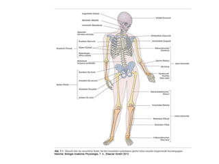
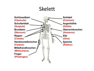
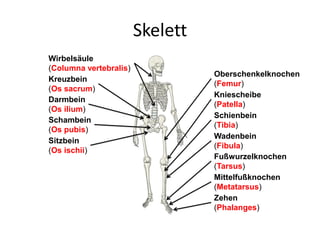
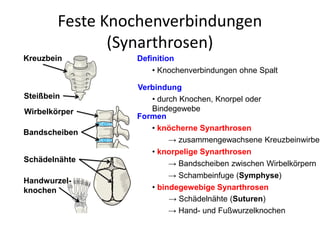
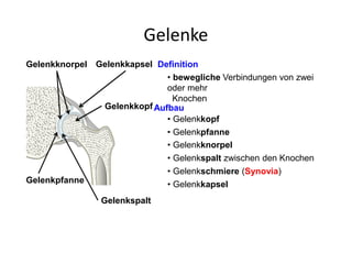
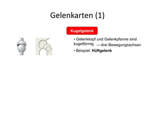
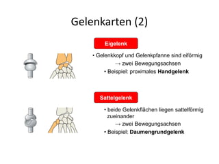
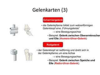
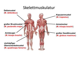
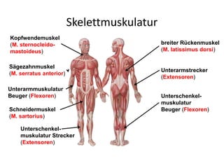
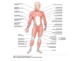
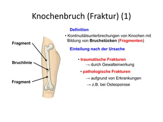
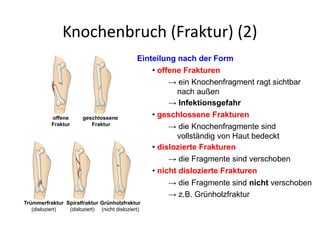
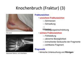
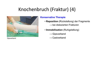
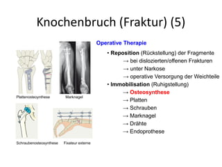
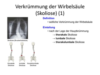
Anatomical description & illustration of:
- Ankle joint, it's relation with both leg and foot, movements.
- Foot bones, joints, ligaments, movements, arches and clinical significance in both ankle & foot.
*There are notes provided in some slides
Physiotherapists utilize sacroiliac joint special tests to diagnose pain or dysfunction within the joint connecting the sacrum and ilium in the pelvis. These tests focus on specific movements or palpation techniques that provoke discomfort or uncover irregularities in the joint. Common assessments like Gaenslen's, FABER, and the compression test aid in evaluating pain response, joint mobility, stability, and integrity. By identifying sacroiliac joint issues through these tests, physiotherapists can tailor treatment plans to alleviate pain and restore functionality for their patients.
Anatomy of skeleton system full/Human all BonesMdMehbubAlam
All about human skeleton system
Full Skeleton system
Appendicular Skeleton
Axial Skeleton
Rib cage
Skull bones
Facial bones
Ear bones
Humerus bone
Femur bone
Tarsals
Carpals
Falanges
The document summarizes the muscles of the foot in 4 layers. The intrinsic muscles are within the foot and responsible for fine motor control, while extrinsic muscles originate from the leg and control larger motions like plantarflexion and dorsiflexion. The first layer includes the abductor hallucis, abductor digiti minimi, flexor digitorum brevis. The second layer includes lumbricals and the tendons of flexor digitorum longus and flexor hallucis longus. The third layer includes the flexor hallucis brevis, adductor hallucis, and flexor digiti minimi brevis. The fourth layer includes the tendons of peroneus longus
This document provides information on basic vertebral structures and techniques for examining the spine and extremities. It describes the normal curves of the cervical, thoracic, and lumbar spine. It outlines the main anatomical structures of vertebrae. It then details techniques for examining range of motion, tenderness, and deformities of the spine, as well as tests for examining nerve root function in the upper and lower extremities.
Range of motion (ROM) refers to the measurement of the distance and direction a joint can move. It is affected by muscles, tendons, ligaments, bones, and other tissues. There are several types of ROM including active, passive, and active-assisted. Goniometers are used to measure ROM at joints in the upper body like the shoulder and elbow, and lower body like the hip and knee. Normal ROM is provided for many joints.
This document discusses proper wheelchair fitting for children with cerebral palsy. It covers positioning of the pelvis, lower body, and upper body to achieve ideal posture and accommodate various physical conditions. The pelvis is considered the keystone of positioning. Proper positioning can optimize independence, enhance function, and relieve pain while distributing pressure. Secondary supports like ankle huggers, adjustable footplates, and harnesses are discussed to stabilize and balance extremities.
The shoulder girdle consists of three bones - the clavicle, scapula, and humerus. It contains four joints - the sternoclavicular, acromioclavicular, scapulothoracic, and glenohumeral joints. The glenohumeral joint is a ball and socket joint that allows for great mobility but also instability. During shoulder movements, the humerus, scapula, and clavicle move in coordinated patterns to provide both mobility and stability.
The human foot has two basic functions: supporting body weight and propelling the body through locomotion. It is designed with elastic longitudinal and transverse arches that act as shock absorbers. The arches are maintained through bone shape, ligaments, tendons, and muscle slings that act as tie beams and suspension cables to prevent arch collapse under weight. Common foot deformities include talipes (clubfoot), pes planus (flat foot), pes cavus (high arch), hallux valgus (bunion), and hammertoe.
Pes Planus by Dr. Mohammad Azhar ud din Darokhanimazhardarokhan
This document discusses flat feet (pes planus) and its evaluation and treatment. It begins by defining flat feet and describing the anatomical changes that occur, including collapse of the longitudinal arch, hindfoot valgus, and forefoot abduction. It then covers the evaluation of flat feet using physical examination and radiographic measurements. Treatment options for flexible flat feet include arch supports, braces, heel inserts, and shoe modifications, with the goal of relieving symptoms without changing the underlying foot structure. Conservative treatment is recommended initially before considering surgical options.
This document summarizes the major muscles of the upper and lower extremities. It describes the muscles of the arm, forearm, thigh, and leg, organized by anatomical compartment. For each region, it lists the superficial and deep muscle groups from lateral to medial and their main actions. The upper extremity sections cover the muscles of the arm's anterior, posterior, and lateral compartments and the forearm's anterior and posterior groups. The lower extremity sections discuss the thigh muscles that act on the femur and the anterior, posterior, and lateral muscle compartments of the leg and their actions on the ankle.
The document summarizes anatomy and common injuries of the foot, ankle, and lower leg. It describes the 26 bones, 38 joints, ligaments, muscles, and four arches of the foot. The most commonly injured joint is the ankle, with sprains being most frequent. Other common injuries include blisters, calluses, athlete's foot, turf toe, arch strains, fractures, plantar fasciitis, shin splints, stress fractures, heel spurs, compartment syndrome, and Achilles tendon injuries. Special tests are used to evaluate injuries and determine if referral is needed.
This document provides an overview of hallux valgus, including its etiology, pathogenesis, clinical features, evaluation, classification systems, non-operative and operative treatment options. Key points include:
- Hallux valgus is a common foot deformity involving the big toe. Its prevalence increases with age and it is more common in females.
- Causes include biomechanical instability, arthritic/metabolic conditions, and trauma. The deformity progresses as the abductor hallucis muscle becomes unopposed.
- Evaluation involves assessing angles on weight-bearing radiographs and classifying severity.
- Treatment ranges from footwear modifications and orthoses to various osteotomy procedures depending on deform
Clinical examination of the spine/back covering the following sections:
INSPECTION
PALPATION
MOVEMENTS
MEASUREMENTS
SPECIAL TESTS
(Neurological examination covered separately in another slideshow : SPINE EXAMINATION - PART 2)
The document provides guidance on assessing the musculoskeletal system. It details how to inspect and palpate various joints and structures, including the temporomandibular joint, sternoclavicular joint, cervical, thoracic and lumbar spine, shoulders, arms, elbows, wrists, and tests range of motion and neurological function. Assessment findings considered normal include symmetry, smooth movement, and no pain. Abnormalities include tenderness, swelling, limited range of motion, muscle weakness or atrophy.
Rotational deformities of lower extremity in childrenAhmed Ashour dr.
Rotational deformities of the lower limbs in children can be caused by in-toeing or out-toeing. Common causes of in-toeing include metatarsus adductus, internal tibial torsion, and excessive femoral anteversion. These issues often resolve on their own with time and without intervention. Out-toeing may be due to external hip rotation contractures or excessive external tibial torsion. Diagnosis involves assessing the foot progression angle, hip rotation, and thigh-foot angle. Conservative management is typically recommended, with surgery reserved for severe cases that do not improve with growth.
The knee joint is the largest and most complicated joint in the body. It is a hinge joint made up of the femur, tibia, and patella bones. The knee joint contains three articulations - the medial and lateral condylar joints between the femur and tibia, and the patellofemoral joint between the patella and femur. Stability is provided by muscles, ligaments, a joint capsule, and synovial fluid within the capsule. The document describes the anatomy and structural components of the knee joint in detail.
1) The document provides an overview of the anatomy of the knee including osseous, extraarticular, and intraarticular structures.
2) It describes common knee examination procedures including inspection, palpation, and range of motion/stability tests performed with the patient in various positions.
3) A variety of clinical tests are outlined to evaluate the patellofemoral joint, ligament stability, and meniscal integrity including Apley's test, McMurray's test, and Lachman's test.
Anatomy: The study of the parts and structures of the human body
Physiology: The study of the functions of the human body
Gross anatomy: The study of the parts and structures of the human body that can be seen with the naked eye and without the use of a microscope
Microscopic anatomy: The study of the parts and structures of the human body that can NOT be seen with the naked eye and only seen with the use of a microscope
The frontal plane: Also referred to as the coronal plane, separates the front from the back of the body.
Ventral surface: The front of the body
Dorsal surface: The back of the body
The document provides an overview of the anatomy and structures of the knee joint. It describes the osseous structures including the femur, tibia, and patella. It also discusses the extra-articular tendinous structures, ligamentous structures including the capsule, collateral ligaments, and cruciate ligaments. The intra-articular structures of the menisci and synovial membrane are also outlined. Blood supply and innervation of the various structures is summarized.
Atlas der menschlichen Anatomie für PC/MacVisible Body
Visible Bodys Atlas der menschlichen Anatomie ist der meistverkaufte, anatomisch akkurateste 3D-Atlas des männlichen und weiblichen menschlichen Körpers. Er wurde von Experten im Bereich der Anatomiedarstellung entwickelt und von Anatomen und Fachkräften im Gesundheitswesen geprüft. Mit dem Atlas der menschlichen Anatomie wird an mehr Hochschulen und Universitäten unterrichtet als mit jedem anderen 3D-Atlas.
Hier erhalten Sie weitere Informationen zu Visible Body: http://www.visiblebody.com/de/
El documento clasifica las empresas en diferentes categorías. Menciona que las empresas se pueden clasificar según su sector económico en empresas del sector primario, secundario y terciario. También las clasifica según el origen de su capital en empresas públicas, privadas y mixtas. Además, proporciona ejemplos de diferentes tipos de empresas como industriales, comerciales, de servicios, financieras y fabricantes.
Este documento presenta 40 preguntas realizadas a un experto en inteligencia artificial sobre temas diversos como la fabricación de vehículos y estructuras con energías alternativas, investigación científica sin dañar especímenes, solución de problemas sociales y desastres naturales de forma ética. Para cada pregunta, el experto asigna puntajes entre 0 y 20 en cinco categorías como factibilidad, impacto positivo, seguridad y ética, para obtener un puntaje total.
Este documento describe los principales tipos de lípidos: grasas o acilglicéridos como los triglicéridos; ceras; fosfolípidos; y esteroides. Los fosfolípidos forman bicapas lipídicas que son la base de las membranas celulares, mientras que los esteroides incluyen compuestos como el colesterol y algunas hormonas. Cada tipo de lípido cumple funciones importantes como reserva energética, protección, estructura de membranas o señalización hormonal.
El documento proporciona instrucciones sobre cómo usar varias herramientas en un aula virtual, incluyendo cómo ingresar al aula virtual y actualizar la información personal, participar en foros, ver mensajes, agregar contactos, agregar una firma a los correos electrónicos, crear grupos en el correo electrónico, y usar Google Docs para crear y editar documentos.
El documento describe la historia y evolución de las tecnologías de información y comunicación (TIC) desde la década de 1960 hasta la actualidad. Resume las características clave de las TIC, incluidas sus ventajas como la facilitación de la información y el aprendizaje individualizado, y sus desventajas como la dependencia tecnológica. También resume dos proyectos internacionales clave para implementar las TIC en las escuelas, el Proyecto Red de Enlaces en Chile y su objetivo de conectar escuelas e impulsar
El CVN-65 Enterprise es el primer y más grande portaaviones que usa energía nuclear en la marina de los EEUU. Con 341 metros de longitud, usa 8 reactores nucleares para impulsar 4 turbinas de vapor que alimentan a los 94 aviones que transporta. El Enterprise marcó el comienzo de una nueva generación de portaaviones más grandes necesarios para los nuevos aviones a reacción pesados.
El documento presenta una propuesta para un Centro Cultural en la ciudad de Antuco, Chile. Propone ubicarlo en la esquina de las calles O'Higgins y Colon, aprovechando un terreno no construido. El centro cultural tendría un patio central público que conectaría el programa cultural con la sociedad. Contaría con salas de exposiciones, artesanías, cafetería, sala de eventos y otros espacios. La estructura espacial consistiría en tres volúmenes divididos por una terraza que daría luz natural.
Este documento es la Ley 3455 sobre la organización municipal en la República Dominicana. Establece las reglas para la creación, modificación y consolidación de municipios. Describe la composición y funcionamiento de los ayuntamientos municipales, incluyendo los requisitos para ser miembro de un ayuntamiento, las incompatibilidades de cargos, la elección anual de un presidente y vicepresidente, la frecuencia de sesiones y más. También establece las responsabilidades de los ayuntamientos y las sanciones por violaciones a disposiciones municipales.
El documento resume la historia y evolución de Internet. Comenzó como una red de computadoras para compartir información entre usuarios de todo el mundo. Luego, los avances en telecomunicaciones llevaron a una explosión en el uso de redes locales y globales, aumentando el interés social en acceder a grandes cantidades de conocimiento. La Web ha evolucionado de una versión 1.0 estática de solo texto a las versiones 2.0 y 3.0 que facilitan el compartir información y la colaboración entre usuarios en un entorno más dinámico e inter
Este documento presenta un plan de intervención para desarrollar habilidades de pensamiento a través de estrategias y actividades. El plan incluye secciones para el tema, objetivo, tiempo, conceptos, materiales, actividad de apertura, introducción, desarrollo y cierre de la unidad. Además, propone un plan de seguimiento para evaluar el progreso.
2. Aufgaben des Halte- und
Bewegungsapparates
Haltung des Körpers
Fortbewegung
Formgebung und Größe des Körpers
Bewegung des Körpers
3. Anteile des Halte- und
Bewegungsapparates
Passiver Bewegungsapparat
Aktiver Bewegungsapparat
• Skelettmuskulatur
•Sehnen
•Knochen
•Knorpel
•Bänder
4.
5. Skelett
Definition
Gliederung
• Knochengerüst des Körpers
→ besteht aus mehr als 200 Knochen
Gewicht
• ca. 15% des Körpergewichtes
• Schädel
• Rumpfskelett
• Schultergürtel
• Beckengürtel
• obere Extremitäten
• untere Extremitäten
12. Gelenkarten (2)
Sattelgelenk
Eigelenk
• Gelenkkopf und Gelenkpfanne sind eiförmig
→ zwei Bewegungsachsen
• Beispiel: proximales Handgelenk
• beide Gelenkflächen liegen sattelförmig
zueinander
→ zwei Bewegungsachsen
• Beispiel: Daumengrundgelenk
13. Gelenkarten (3)
Radgelenk
• die Gelenkpfanne bildet zum walzenförmigen
Gelenkkopf eine „Führungsleiste“
→ eine Bewegungsachse
• Beispiel: Gelenk zwischen Oberarmknochen
und Elle (Humero-Ulnar-Gelenk)
• der Gelenkkopf ist radförmig und dreht sich in
der Gelenkpfanne um eine Achse
→ eine Bewegungsachse
• Beispiel: Gelenk zwischen Speiche und
Elle (Radio-Ulnar-Gelenk)
Scharniergelenk
14.
15.
16. Schädel
Einteilung
• Gehirnschädel
→ umgibt schützend das Gehirn
• Gesichtsschädel
→ knöcherne Grundlage des Gesichtes
Knochenverbindung
• feste Verbindung durch stark gezackte
Knochennähte (Suturen)
• bewegliche Verbindung zwischen
Unterkiefer und Schädel (→ Kiefergelenk)
Knochennähte
Kiefergelenk
18. Kindlicher Gehirnschädel
Stirnbein
(Os frontale)
Scheitelbein
(Os parietale)
Stirnfontanelle
Seitenfontanellen
Definition
Besonderheit
• Schädel des Neugeborenen und
heranwachsenden Menschen
• die Schädelknochen sind noch nicht vollständig
ausgebildet
• zwischen den Gehirnschädelknochen sind
mit Bindegewebe verschlossene Lücken
(Fontanellen)
→ bewegliche Knochenverbindung
→ wichtig für den Geburtsvorgang
Hinterhauptbein
(Os occipitale)
Hinterhaupt-
fontanelle
→ beim Erwachsenen verknöchert
26. Zwischenwirbelscheiben
(Bandscheiben)
Lage
• zwischen den Wirbelkörpern der HWS,
BWS
und LWS
Aufgaben
• Stoßdämpfung
• Beweglichkeit der HWS, BWS und LWS
Aufbau
• innen: weicher Gallertkern
(→ elastische Verformbarkeit bei Beugung)
• außen: Fasermantel
(→ ringförmig verlaufender Faserknorpel)
Wirbelkörper
Bandscheibe
Gallertkern
Fasermantel
Stoßdämpfung Beweglichkeit
27. Grundform eines Wirbels
Aufbau und Aufgaben
• Wirbelkörper
→ trägt die cranial liegenden Anteile
• Wirbelbogen
→ begrenzt das Wirbelloch
• Dornfortsatz
• Querfortsätze
→ liegt ventral
→ liegt dorsal
→ Ansatz der Rückenmuskulatur
• Gelenkfortsätze
→ bewegliche Verbindung zwischen
den einzelnen Wirbeln
Wirbelkörper
Wirbelbogen
Wirbelloch
Querfortsatz
Dornfortsatz
Wirbelkörper
Gelenkfortsatz Dornfortsatz
Querfortsatz
von oben
von der Seite
28. Halswirbel - Atlas und Axis
Aufbau und Funktion
• erster Halswirbel (Atlas)
→ ringförmig
→ trägt den Schädel
• zweiter Halswirbel (Axis)
→ bildet die Drehachse mit dem Atlas
→ vorne zahnförmig (Dens axis)
Atlas
Axis
C3
C4
C5
C6
C7
Dens axis
Querband
des Atlas
Axis
29. Halswirbel (2)
• seitlich der Wirbelkörper befindet sich jeweils
eine Öffnung für eine Arterie (A. vertebralis)
• die Wirbelkörper sind relativ klein
→ Versorgung des Gehirns
• das Wirbelloch ist relativ groß
Wirbelkörper
Dornfortsatz
Wirbelloch
von oben
Gelenkfläche
Wirbelkörper Dornfortsatz
Gelenkfortsatz
von der Seite
Besonderheiten
30. Kreuzbein und Steißbein
• das Kreuzbein besteht aus 5 knöchern
verwachsenen Wirbeln
• das Steißbein besteht aus 4 knöchern
verwachsenen Wirbeln
Kreuzbein
Steißbein
5. Lendenwirbel Besonderheiten
31.
32. Krümmungen der Wirbelsäule
Formen
• Lordose
= Krümmung der Wirbelsäule nach ventral
→ Halswirbelsäule
→ Lendenwirbelsäule
• Kyphose
= Krümmung der Wirbelsäule nach dorsal
→ Brustwirbelsäule
→ Kreuzbein
Halslordose
Brustkyphose
Lendenlordose
Sakralkyphose
33.
34.
35. Schultergürtel (1)
Anteile
• rechtes und linkes Schulterblatt (Scapula)
Aufgabe
• rechtes und linkes Schlüsselbein (Clavicula)
• Verbindung der Arme mit dem Rumpf
• Schultergelenk (Kugelgelenk)
→ Gelenkpfanne der Scapula
→ runder Gelenkkopf des Humerus
Gelenk
Scapula
Clavicula
Humerus Scapula
36.
37. Obere Extremität (1)
Anteile
• Oberarmknochen (Humerus)
• Elle (Ulna)
• Speiche (Radius)
Aufgaben
• Bewegung der Arme
• Greifen und Festhalten von Gegenständen
• Handwurzel (Carpus)
• Mittelhand (Metacarpus)
• Finger (Phalanges)
• Pronation und Supination der Hände
Humerus
Radius
Uln
a
Carpus
Metacarpus
Phalanges
38. Obere Extremität (2)
• Ellenbogengelenk (Scharniergelenk)
→ zwischen Humerus und Ulna
Gelenke
• Ellenbogengelenk (Radgelenk)
→ zwischen Humerus, Radius und
Ulna
→ Beugen und Strecken des Arms
→ Pronation und Supination
Supination Pronation
Humerus
Radius
Ulna
46. Beckengürtel (2)
Aufgaben
• Fixierung der Wirbelsäule
• Halt und Stütze der Bauchorgane
Gelenk
• Hüftgelenk (Kugelgelenk)
→ Gelenkpfanne des Hüftbeins
→ Gelenkkopf des Femurs
Muskulatur
• Gesäß-, Hüft- und Beinmuskeln
arbeiten
beim Gehen zusammen•
•verbindet Becken und LWS mit dem Femur
Femur
Os coxae
Oberschenkel-
hals
Gelenkkopf
Gelenkpfanne
59. Knochenbruch (Fraktur) (1)
Definition
• Kontinuitätsunterbrechungen von Knochen mit
Bildung von Bruchstücken (Fragmenten)
Einteilung nach der Ursache
• traumatische Frakturen
→ durch Gewalteinwirkung
• pathologische Frakturen
→ aufgrund von Erkrankungen
→ z.B. bei Osteoporose
Fragment
Fragment
Bruchlinie
60. Knochenbruch (Fraktur) (2)
Einteilung nach der Form
• offene Frakturen
→ ein Knochenfragment ragt sichtbar
nach außen
• geschlossene Frakturen
→ die Knochenfragmente sind
vollständig von Haut bedeckt
• dislozierte Frakturen
→ die Fragmente sind verschoben
• nicht dislozierte Frakturen
→ die Fragmente sind nicht verschoben
→ z.B. Grünholzfraktur
→ Infektionsgefahr
offene
Fraktur
Trümmerfraktur
(disloziert)
Spiralfraktur
(disloziert)
geschlossene
Fraktur
Grünholzfraktur
(nicht disloziert)
64. Verkrümmung der Wirbelsäule
(Skoliose) (1)
Definition
• seitliche Verkrümmung der Wirbelsäule
Einteilung
• nach der Lage der Hauptkrümmung
→ thorakale Skoliose
→ lumbale Skoliose
→ thorakolumbale Skoliose
thorakale
Skoliose
lumbale
Skoliose
thorakolumbale
Skoliose
Skoliose (Brustwirbelsäule)
65. Typische Pflegeprobleme von
Menschen mit
Erkrankungen des
Bewegungssystems
• Schmerzen haben
Die Patientin kann:
• Probleme mit der Beweglichkeit haben
• beim An- und Auskleiden Probleme haben
• Probleme haben bei der Zubereitung und
oder Aufnahme von Nahrung
• bei der Körperpflege eingeschränkt sein
• sturzgefährdet sein