A02010106
•
0 gefällt mir•263 views

![Image Segmentation Methods for Dermatitis Disease: A Survey
www.ijeijournal.com P a g e | 2
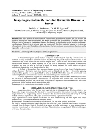
Fig.1. Image segmentation Process
III. IMAGE SEGMENTATION CLASSIFICATION
Image segmentation methods are broadly classified into three class viz. Region based, shape based and
gray–value based [2].
3.1 Region Based method
In this method, segmentation of images is to partition an image into regions. Region growing is a
procedure that group‟s pixels or sub-regions into larger regions based on predefined criteria.
The idea behind all region growing algorithms is that all pixels/voxels belonging to one object are connected
and similar according to some predicate. In this survey, we view region growing as a voxel-based procedure,
where an object is formed from a group of voxels [3].
3.1.1 Split and merge method
As the name split and merge, an image is initially split into smaller regions, e.g., down to individual
voxels. Neighboring regions are then merged if they fulfill a homogeneity criterion.
3.1.2 Seeded region growing method
Here, seed selection is crucial but can be seen as an external task, often done by hand in medical image
processing. Region growing algorithms start from an initial, incomplete segmentation and try to aggregate the
yet unlabelled pixels to one of the given regions. The initial regions are usually called seed regions or seeds. The
decision whether a pixel should join a region or not is based on some fitness function which reflects the
similarity between the region and the candidate pixel. The order in which the pixel is processed is determined by
a global priority queue which sorts all candidate pixels by their fitness values. This approach elegantly mixes
local (fitness) and global (pixel order) information.
3.1.3 The GMM Region-Based Method.
GMM approach groups the data points of an image into „k‟ segments, each assigned with a Gaussian
distribution. Probabilistic image modeling and segmentation is performed in the color and position (x, y) feature
space. The distribution of a d-dimensional random variable is a mixture of k Gaussians, if its density function is
as given below:
------------ (1)](data:image/gif;base64,R0lGODlhAQABAIAAAAAAAP///yH5BAEAAAAALAAAAAABAAEAAAIBRAA7)
Empfohlen
Weitere ähnliche Inhalte
Was ist angesagt?
Was ist angesagt? (20)
Andere mochten auch
Andere mochten auch (9)
Ähnlich wie A02010106
Ähnlich wie A02010106 (20)
Mehr von International Journal of Engineering Inventions www.ijeijournal.com
Kürzlich hochgeladen
Kürzlich hochgeladen (20)
A02010106
- 1. International Journal of Engineering Inventions ISSN: 2278-7461, ISBN: 2319-6491 Volume 2, Issue 1 (January 2013) PP: 01-06 www.ijeijournal.com P a g e | 1 Image Segmentation Methods for Dermatitis Disease: A Survey Prafulla N. Aerkewar1 , Dr. G. H. Agrawal2 , 1 Ph.D.Research scholar, B.D.College of Engg., Sevagram, Dist. Wardha, 2 Professor, Department of EE, K.D.K.College of Engineering, Nagpur. Abstract:-This paper presents a deep survey on various image segmentation methods that can be used for dermatitis disease that have been proposed and which are suitable for the processing of various images for different types of patches for various skin diseases. The focus is on thresholding, region growing, and shape– based methods. This review the original ideas and concepts of the above methods, because we believe this information to be important for judging when and under what circumstances a segmentation algorithm can be expected to work properly. Keywords:- Dermatology, Disease, Leprosy, Patches, Segmentation I. INTRODUCTION In the world twenty first century medical science and pharmacological, every day a new medicine and treatment is being invented for different diseases. But basically the job of diagnosis of the disease is still complicated one for the technician and even the surgeon also. It also becomes much more difficult when disease related dermatitis is under screening. So in the field of dermatology involvement of engineering technology not only can play a vital role to avoid the wrong diagnosis but could suggest the perfect methodology for detection of the disease related to skin. In the field of dermatology, there are many diseases such as Leprosy, Scar tissue, Contact dermatitis, Vitiligo, Psoriasis having the main issue that the skin is affected and may have the similar appearing symptoms like patches on some part of body or all over the body. Most of the time, it is complicated when such a thing happens complication like reaction of a treatment may cost a life of victim. So image segmentation suggests the methodology for perfect diagnosis of the dermatological disease and also helps in the analysis of such a disease which can analyze the prior situation also. There are different image segmentation procedure that can be apply to build up such a powerful electronics tool which may create revolution in field of medical as well as engineering field. This paper reviews various image segmentation methods. II. LITERATURE REVIEW IMAGE SEGMENTATION Image segmentation is the first and most important step for skin patch image detection so that we can diagnose the proper skin disease. For this task we require to extract the information from skin image using image extraction and image mining process. To achieve this it is necessary to study and survey all existing methods for image segmentation. This paper reviews various image segmentation methods. Image segmentation is often regarded as the first and most important step for other higher level image interpretation, e.g. information extraction and image mining. Segmentation is the task of recognizing objects in an image. The rest of the image is background. The result of image segmentation is a set of regions that collectively cover the entire image, or a set of contours extracted from the image. Each of the pixels in a region is similar with respect to some characteristic or computed property, such as color, intensity, or texture. Adjacent regions are significantly different with respect to the same characteristic(s).
- 2. Image Segmentation Methods for Dermatitis Disease: A Survey www.ijeijournal.com P a g e | 2 Fig.1. Image segmentation Process III. IMAGE SEGMENTATION CLASSIFICATION Image segmentation methods are broadly classified into three class viz. Region based, shape based and gray–value based [2]. 3.1 Region Based method In this method, segmentation of images is to partition an image into regions. Region growing is a procedure that group‟s pixels or sub-regions into larger regions based on predefined criteria. The idea behind all region growing algorithms is that all pixels/voxels belonging to one object are connected and similar according to some predicate. In this survey, we view region growing as a voxel-based procedure, where an object is formed from a group of voxels [3]. 3.1.1 Split and merge method As the name split and merge, an image is initially split into smaller regions, e.g., down to individual voxels. Neighboring regions are then merged if they fulfill a homogeneity criterion. 3.1.2 Seeded region growing method Here, seed selection is crucial but can be seen as an external task, often done by hand in medical image processing. Region growing algorithms start from an initial, incomplete segmentation and try to aggregate the yet unlabelled pixels to one of the given regions. The initial regions are usually called seed regions or seeds. The decision whether a pixel should join a region or not is based on some fitness function which reflects the similarity between the region and the candidate pixel. The order in which the pixel is processed is determined by a global priority queue which sorts all candidate pixels by their fitness values. This approach elegantly mixes local (fitness) and global (pixel order) information. 3.1.3 The GMM Region-Based Method. GMM approach groups the data points of an image into „k‟ segments, each assigned with a Gaussian distribution. Probabilistic image modeling and segmentation is performed in the color and position (x, y) feature space. The distribution of a d-dimensional random variable is a mixture of k Gaussians, if its density function is as given below: ------------ (1)
- 3. Image Segmentation Methods for Dermatitis Disease: A Survey www.ijeijournal.com P a g e | 3 Here αj are probabilities of occurrence of each Gaussian and μj , j are the mean and the covariance matrix of each Gaussian respectively. An immediate transition is possible between the GMM representation and probabilistic image segmentation. Each pixel of the original image is affiliated with the most probable Gaussian and a localized segmentation map, a map of convex superpixels, is formed [4]. The EM algorithm, which is utilized to estimate the model parameters, works by first estimating the probability of each pixel to belong to any of the k Gaussians (E-step), and then optimizing the GMM parameters to best fit the current segmentation (M-step). The E-step is composed of computing the posterior probability wtj of each feature vector xt to belong to the j-th component: ------------- (2) Where z is a normalization scalar. The end result of the region-based GMM is a mixture of k components, each consisting of similar points in feature space (e.g. color). However, in natural world images frequently points of similar features may actually belong to different objects in the image plane. Since the color- based model does not take into account edge-type information, the model cannot detect the existence of a boundary between spatially proximal objects. 3.1.4 Growing by Gray Value Method One of the most commonly used region growing criteria is based on the observation that an object‟s gray values are usually within some range around a mean value. Thus, while growing a region, its current mean and standard deviation are computed and a new voxel is added if its value lies within a range around the regions mean. 3.1.5 Adaptive Region Growing Method This is another region growing method based on criterion called adaptive region growing method for segmentation of the human cortex. Their idea was to adapt the decision function according to the region‟s size: Initially, for a region containing very few voxels, voxels are added as long as a homogeneity (gray value variance) threshold around the region is not exceeded. Then, when a certain number of voxels has been added to the region, one assumes that the gray level statistics of this region have approached the object‟s true distribution [5]. Thus, further voxels are added only if their gray values are close to the region‟s mean gray value. 3.1.6 Non-connected Region Growing Method In this algorithm the voxels may not only be added, but also removed from a region. To achieve this, the so-called “k-contraction” is used. K-contraction removes k voxels from the region, starting from the voxel with the lowest gray value, in increasing order. Note that this procedure assumes that object voxels have larger gray values than background voxels. The procedure is repeated until a homogeneous region is produced, where a region is called homogeneous if its gray value variance is below some threshold. Parameters of this method are the seeds for initialization of the region and the homogeneity parameter.The region R is grown until convergence by first applying a dilation, i.e., adding neighboring voxels, and then trimming the region using the k-contraction method described above. This algorithm differs significantly from the region growing methods described earlier. In this approach,growing and homogeneity testing are separate processes. 3.1.7 Parameter-Free Region Growing method Till now we have discussed various regions growing methods which needs to select seed voxels. Thresholding may be used for this initialization step. Here, one should apply thresholding conservatively. By this we mean that the purpose is not to segment the entire object. It is more important to choose a number of certain object voxels, from which to start region growing. In practice, this is often done manually. IV. SHAPE BASED METHOD: (MODEL-BASED APPROACHES) In this section, we discuss with some model-based approaches for image segmentation that have widely been applied in 2D and 3D medical image processing. 4.1.1 Deformable Surfaces In 2D image segmentation, active contours, also known as “snakes”, are parametric curves which one tries to fit to an image, usually to the edges within an image. 4.1.2 Level Set method
- 4. Image Segmentation Methods for Dermatitis Disease: A Survey www.ijeijournal.com P a g e | 4 This method deals with [Set99] lower dimensional structures such as surfaces via a function for some constant c, usually fixed to zero. In other words, a level set is nothing but an iso-surface of the function Level set methods are based on a partial differential equation and can be solved using finite difference methods [6]. The basic equation underlying level set methods is the level set equation, which describes the change of over time (or iteration) t, …………(3) is the temporal partial derivative of the level set function, denotes the spatial gradient operator and V describes an external force field guiding the evolution of the curve in the image. This equation is also known in physics, where it is used to represent the reaction surface of an evolving flame.In image processing, level set methods can for example be applied by defining the external force V as the gradient field of an image. We are then looking for a steady state solution of Eq. (2), where the surface S has locked onto object edges. This idea seems very similar to the deformable surface approach, and a level set method that implements deformable surfaces. 4.1.3 Implicit Deformable Surfaces The active contours of the level set method used by Caselles et al. [CKS97], called geodesic active contours. Their result has proven to be very useful because it combines the intuitive active contours concepts with efficient implementations of level set methods. Additionally, geodesic active contours have the advantage that they can change topology during evolution. The 3D analog to geodesic active contours, the evolution of an implicit surface S by modification of a level set function was described in [CKSS97]. The derivation therein is based on the concept of minimal surfaces: It is observed that finding S can be put as finding a surface of minimal weighted area, with the weight being given by a function h(f) of the image f. Minimization of this area via the calculus of variations leads to a gradient descent rule in image space, see [CKSS97]. 4.1.4 Edge detection methods This is a fundamental tool used in most image processing applications to obtain information from the frames as a precursor step to feature extraction and object segmentation. This technique detects outlines of an object and boundaries between objects and the background in the image. An edge-detection filter can also be used to improve the appearance of blurred or anti-aliased video streams [7]. The basic edge-detection operator is a matrix area gradient operation that determines the level of variance between different pixels. The edge-detection operator is calculated by forming a matrix centered on a pixel chosen as the center of the matrix area. If the value of this matrix area is above a given threshold, then the middle pixel is classified as an edge. Examples of gradient-based edge detectors are Roberts, Prewitt, and Sobel operators. All the gradient- based algorithms have kernel operators that calculate the strength of the slope in directions, which are orthogonal to each other, commonly vertical and horizontal. Later, the contributions of the different components of the slopes are combined to give the total value of the edge strength. V. GRAY-LEVEL BASED METHOD (THRESHOLDING) There are several methods based on gray level or thresholding for segmentation of images. Using this method, either global or local image information is used. In thresholding, one assumes that the foreground can be characterized by its brightness, which is often a valid assumption for 3D datasets [8]. 5.1.1 Global Thresholding The simplest and most widely used method for segmentation is global thresholding method. Where we selects a value -----------(4) and sets foreground voxels, according to following equation. …………..(5) Where equation(3) is a complete description of a binarization algorithm, it contains no indication on how to select the value The natural question that arises is whether there exists an optimal threshold value. There are in fact different solutions to this threshold selection problem, each being based on different model
- 5. Image Segmentation Methods for Dermatitis Disease: A Survey www.ijeijournal.com P a g e | 5 assumptions. However, even if was optimally selected using a model which is suitable for the present image data, global thresholding will give poor results whenever influences of noise are large compared to the image content (low signal to noise ratio) or when object and background gray value intensities are not constant throughout the volume. 5.1.2 Thresholding Using Prior Knowledge of image Sometimes it is require that, an imaged object will have known physical properties. For example, the manufacturer of a material may know the material‟s volume density from experiments. Then, the optimal threshold may be defined as the value for which the segmented image „g‟ reaches this priority known value. Such strategies can actually be applied for choosing parameters of any kind of image processing method. For low noise levels and suitable known parameters, i.e., parameters which can also be computed from binarized images, this pragmatic approach can often yield satisfactory results, especially if combined with proper filtering methods for noise reduction. 5.1.3 Otsu’s Method According to the value of one can analyze the distribution of gray values of an image (its histogram): Assume that the gray value histogram of the image contains two separate peaks, one each for fore- and background voxels (bimodal histogram). Then choosing the minimum value between these two peaks as threshold Otsu defines this choice for as the value minimizing the weighted sum of within-class variances [9]. This is equivalent to maximizing the between-class scatter. In case of 256 gray values, „T‟ can be simply determined by evaluating the term for each value and choosing the global minimum value. Note that, in the discrete case it can be entirely evaluated from the histogram by summing over the appropriate value ranges. Otsu‟s method is widely used because it proves to be a robust tool for threshold selection while segmentation. 5.1.4 Bayesian Thresholding In a statistical point of view, thresholding is an easily solvable task if we have a proper image model. In a Bayesian setting, the posterior probability of observing f(x) is simply is the unknown class label, i.e., background and foreground [10]. By Bayes‟ Theorem we know that …………...(6) This equation can be applied for determining the global image threshold This gives the optimal threshold for separating two regions which follow Gaussian gray value distributions with equal variance and different means. 5.1.5 Local Thresholding The another thresholding method is the Local thresholding method which will removes some shortcomings of the global thresholding approach. As mentioned above, intensity levels of an image can vary depending on the location within a data volume. One common approach for preprocessing in 2D image processing is to calculate the mean intensity value within a window around each pixel and subtract these sliding mean values from each pixel (“shading correction”). In the same spirit, but instead of modifying the image content prior to binarization, one can use a spatially varying threshold, to compensate for these inhomogeneous intensities. 5.1.6 Indicator Kriging Indicator kriging was described by Oh and Lindquist [OL99] for thresholding. Kriging is an interpolation method that is commonly used in geostatistics. It relies mainly on local covariance estimates for thresholding in that it estimates the value at voxel x using a linear combination of its neighbors. Kriging estimators build upon the linear combination; the central voxel is not included in the linear combination. Indicator kriging is a modification of this linear combination where the continuous voxel distribution F(x) is replaced by a binary variable i. 5.1.7 Hysteresis thresholding The common problem in image segmentation is that the segments of interest may well be defined by their intensities, but that there also exist other structures (e.g., noise) with high values. Global thresholding would either underestimate the size of the true segments (because was too large) or would include noise in
- 6. Image Segmentation Methods for Dermatitis Disease: A Survey www.ijeijournal.com P a g e | 6 the foreground (because was chosen too low).One way of dealing with such situations where the voxel value distributions of fore- and background voxels overlap is hysteresis thresholding, also known as “double thresholding”. It was proposed in [Can86] as a method to segment connected edges from an edge strength map. We can often do better than selecting one threshold by selecting two - an upper and a lower threshold.The rule for outputting a pixel is then that it is either greater than the upper threshold, or greater than the lower threshold and connected to a pixel who is greater than the upper threshold.This technique is called hysterisis thresholding and will generally give better connectivity without introducing many spurious edges. VI. HISTOGRAM BASED METHOD These are very efficient method when compared to other image segmentation methods because they typically require only one pass through the pixels. In this technique, a histogram is computed from all of the pixels in the image, and the peaks and valleys in the histogram are used to locate the clusters in the image. Color or intensity can be used as the measure. A refinement of this technique is to recursively apply the histogram- seeking method to clusters in the image in order to divide them into smaller clusters [11].This is repeated with smaller and smaller clusters until no more clusters are formed. The disadvantage of the histogram-seeking method is that it may be difficult to identify significant peaks and valleys in the image. In this technique of image classification distance metric and integrated region matching are familiar. VII. CONCLUSIONS From the above survey it is seen that there exist lot of segmentation methods. But no one can be suitable for all types of given images. It is clear that as per the requirement we choose any one that gives accurate segmentation. So there is a scope to develop a new algorithm for segmentation of dermatitis images of various skin disease patches. REFERENCES 1) P Rafael C. Gonzalez and Richard E. Woods, “Digital Image Processing”, 2nd Edition, Pearson Education Asia, 2 2) A. Chambolle. Inverse problems in image processing and image segmentation. In C.E. Chidume, editor, ICTP Lecture Notes, volume 2, pages 1–94. ICTP, 2001. 3) D. Verma, M. Meila, “A comparison of spectral clustering algorithms”, Univ of Washington technical report, 2001. 4) T. Zouagui, H. Benoit-Cattin and C. Odet, "Image segmentation functional model," Pattern Recognition, vol. 37, pp. 1785-1795, 2004. 5) E. Sharon, A Brandt, and R. Basri. Segmentation and boundary detection using multiscale intensity measurements. In Computer Vision and Pattern Recognition (CVPR), pages 469–476, 2001. 6) Johan Lie,Marius Lysaker and Xue-Cheng Tai “ABinary Level Set Model and Some Applications to Mumford Shah Image Segmentation. IEEE Trans. Image Processing, 15(5):May 2006 7) J. Canny. A computational approach to edge detection. IEEE Trans. Pattern Analysis Machine Intelligence, PAMI-8(6):679–698, Nov 1986. 8) M. Sezgin and B. Sankur, Survey over image thresholding techniques and quantitative performance evaluation”, Journal of Electronic Imaging, vol. 13(1), pp. 146-165, Jan. 2004. 9) N. Otsu. A threshold selection method from grey-level histograms. IEEE Trans. Systems, Man, and Cybernetics, 9(1):62–66, Jan 1979. 10) S. Geman and D. Geman. Stochastic relaxation, gibbs distributions, and the Bayesian restoration of images. IEEE Trans. Pattern Analysis Machine Intelligence, PAMI-6(6):721– 741, Nov 1984. 11) R.E.Broadhurst, Statistical estimation of histogram variation for texture classification, In Texture 2005: Proceedings of the 4th Internation Workshop on Texture Analysis and Synthesis, pp. 25-30, 2005.