SlideShare ist ein Scribd-Unternehmen logo
1 von 5
Downloaden Sie, um offline zu lesen
International Journal of Science and Research (IJSR), India Online ISSN: 2319-7064
Volume 2 Issue 8, August 2013
www.ijsr.net
Detection of Cysts in Ultrasonic Images of Ovary
Ashika Raj
M. Tech, Computer Science & Engineering, Calicut University, Calicut, India
Abstract: In this paper, cysts are detected in the ultrasonic images of ovary. PCOS is an endocrine disorder affecting women of
reproductive age. This syndrome is mainly seen in women whose age is in between 25 and 35. We are proposing methods for identifying
whether a person is suffering from Polycystic Ovary Syndrome (PCOS) or not. Ultrasound imaging of the follicles gives important
information about the size, number and mode of arrangement of follicles, position and response to hormonal stimulation. A
thresholding function is applied for denoising the image in the wavelet domain. Before the segmentation process the ultrasonic image is
preprocessed using contrast enhancement technique. Morphological approach is used for implementing contrast enhancement. This is
performed in order to improve the clarity and quality of the image. Fuzzy c-means clustering algorithm is applied to the resultant image.
Finally the cysts are detected with the help of clusters. The efficiency of the algorithm depends upon the value of Mean Square Error
(MSE) and Peak Signal to Noise Ratio (PSNR).
Keywords: Denoising, Contrast Enhancement, Morphological Operations, Segmentation, Fuzzy C-Means Clustering.
1. Introduction
Cysts are follicles which have abnormal size. Based on the
detection of follicles, the suspected patient can be treated
as normal or polycystic. Ovarian follicles are roughly
spherical, fluid-filled structures in which oocytes (eggs)
develop. Follicles imaged in two dimensions appear as
dark, roughly circular regions in ultrasound images since
fluid does not reflect ultrasonographic pulses. PCO
ovaries typically exhibit a larger number of smaller,
possibly irregularly shaped follicles, and a peripheral
distribution of follicles. The ultrasonographic morphology
of a polycystic ovary (PCO) is characterized by the
presence of 12 or more ovarian follicles which are 2-9 mm
in size. These follicles are termed as cysts. The symptoms
of PCOS are menstrual irregularity, obesity,
hyperandrogenism, diabetes, acne, increased risk of
cardiovascular disease, male-pattern facial and bodily hair
growth and balding, excessive production of male
hormones, infertility etc. An automatic detection of cysts
for a PCOS patient is implemented using fuzzy c-means
clustering. Before performing this algorithm we are
denoising and contrast enhancing the ultrasonic image
which is given as the input.
Diagnostic ultrasound uses frequency between 2 and 15
MHz. Ultrasonic waves are produced from the transducer
and penetrates into the body tissues and when the wave
reaches an object or a surface with different texture or
acoustic nature, some fraction of the energy is reflected
back. The echoes so produced are received by the
apparatus and changed into electric current. These signals
are then amplified and processed to get displayed on CRT
(Cathode Ray Tube) monitor. The image so obtained is
called ultrasound scan and the process is known as
ultrasonogram. This image is given as the input.
Ultrasound imaging technique is inexpensive and is very
effective for cyst recognition. The overall quality of the
ultrasound image is the end product of a combination of
many factors originating from the imaging system and the
performance of the operator. Ultrasonic image may
contain noises due to loss of proper contact or air gap
between transducer and body part. Noises can also be
formed during beam forming process or signal processing.
The noises may cause the image blurred and thereby lead
to poor segmentation. Hence we are performing denoising.
A soft thresholding function is proposed for the process of
removing noise.
Contrast enhancement increases the appearance of large-
scale light-dark transitions of an image. The edges of the
image become clearer. This process automatically
brightens images that appear dark or blurred and applies
appropriate correction to deliver optimal quality and
clarity. Morphological operations are performed for
contrast enhancement technique. Fuzzy c-means
clustering algorithm is applied for the segmentation of
follicles from the ultrasonic image. The purpose of
clustering is to identify natural groupings of data from a
large dataset to produce a concise representation of a
system’s behavior. The follicles are detected inside the
ovary and the obtained results are compared with the
manual results.
2. Proposed Methodology
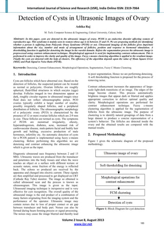
Figure 1 gives the schematic diagram of the proposed
methodology.
Figure 1: Block Diagram for cyst detection
185
International Journal of Science and Research (IJSR), India Online ISSN: 2319-7064
Volume 2 Issue 8, August 2013
www.ijsr.net
Figure 2: Original Ultrasonic Image (Input)
3. Denoising using Soft Thresholding
Ultrasonic images may be affected by noise in different
stages of image processing. Therefore image denoising is
an unavoidable step. Denoising is simply the process of
removing noise. Here we are adding noise to increase the
signal wavelength. The noisy image is shown in Figure3.
Figure 3: Noisy Image
We are proposing a soft thresholding function for image
denoising in the wavelet domain. Wavelet transform has
become an important tool to suppress the noise. Wavelet
transform of a noisy signal is a linear combination of the
wavelet transform of the original signal and the noise. The
power of the noise can be suppressed with a suitable
threshold while the main features can be preserved.
Wavelet coefficients of a noisy image are divided into
important and non-important coefficients and each of
these groups are modified by a certain rule called
thresholding rule.
At the initial stage, the noisy image is decomposed in the
wavelet domain. Based on a thresholding function,
detailed coefficients are modified by selecting a suitable
threshold value. The universal threshold is calculated as:
������������ � ��2ln ��� �1�
where σ is the standard deviation.
Thresholding function proposed by Ref [3] is defined by
Eq. (2):
���, ���, �� �
�
�
�
�
� � � ��� �
���
2� � 1
� � ����
1
�2� � 1������
����� |�| � ��� �2�
� � ��� �
���
2� � 1
� � ���
 
The proposed thresholding function has lower risk value
than others in fixed threshold value. Therefore it is a more
powerful function. In order to improve the flexibility and
capability of the proposed function, three shape tuning
factors have been added which may lead to a
comprehensive thresholding function that can be adjusted
to any desired thresholding function. The shape tuning
factors are ‘k’, ‘m’ and ‘n’. If the value of ‘k’ is 1, then
the function is a hard thresholding function and if the
value is 0 then it indicates soft thresholding function. The
value of ‘m’ and ‘n’ determines the shape of the function
for coefficients that are bigger and lesser than absolute
threshold value respectively. So here we are tuning the
parameter ‘k’ to 0 since we are performing soft
thresholding. The three shape tuning factors are added to
the proposed thresholding function as follows:
���, ���, �, �, ��
�
�
�
�
�
�� � 0.5�������
�
�
����
� �� � 1���� � � ����
0.5 � �
|�|�
������
������� |�| � ��� �3�
� � 0.5 ����
�
�
����
� �� � 1���� � � ���
 
where � � � � 2 � �/�.
Finally the denoised image can be obtained by
reconstructing the original image. The original image
which is free of noises can be reconstructed using Inverse
Discrete Wavelet Transform (IDWT).
The efficiency of the denoising method using thresholding
function can be calculated using the Mean Square Error
(MSE). Lesser the value of MSE, higher will be the
efficiency of the algorithm. The value of MSE can be
calculated using Eq. (4):
��� �
�
���
∑ ∑ �����, �� � ���, �����
���
�
��� �4�
Here, A (i,j) is the noisy image of size � � �, where ‘r’ is
the number of rows and ‘c’ is the number of columns. B
(i,j) is the denoised image.
186
International Journal of Science and Research (IJSR), India Online ISSN: 2319-7064
Volume 2 Issue 8, August 2013
www.ijsr.net
Figure 4: Denoised image
4. Contrast Enhancement
Contrast enhancement makes the denoised image more
clear and distinct. The edges of the follicles can be easily
identified by performing this technique, in other words the
edges are being sharpened so that the follicles can be
clearly seen. We are performing morphological operations
for contrast enhancement.
Morphology is a broad set of image processing operations
that process images based on certain shapes.
Morphological operations apply a structuring element to
an input image. The structuring element can be diamond,
disk, line or ball shaped. It can also take some other
shapes. In a morphological operation, the value of each
pixel in the output image is based on a comparison of the
corresponding pixel in the input image with its neighbors.
Here since we are detecting follicles and these follicles are
disk shaped, we are choosing a disk shaped structuring
element. Then we can construct morphological operation.
We are implementing two methods of morphological
operations: i) Morphological opening and closing. ii)
Tophat and Bottomhat filtering.
4.1Morphological opening and closing
The initial stage is to create a structuring element. A
structuring element is a matrix which consists of only
zeros and ones that can have a disk shape and a specific
size.
Dilation is the process of adding pixels to the boundaries
of objects in an image. Erosion is the process of removing
pixels on object boundaries. Dilation and erosion are used
in combination to perform image processing operations.
Erosion followed by dilation is termed as morphological
opening of an image where as dilation followed by erosion
is known as morphological closing of an image.
Morphological opening extracts bright features of an
image and morphological closing extracts dark features of
an image. By combining these two images, we get contrast
enhanced image.
4.2Tophat and Bottomhat filtering
Like morphological opening and closing, for this process
also create a structuring element. Morphological tophat
filtering is performed on the grayscale denoised input
image. This can be used to correct uneven illumination
when the background is dark. Therefore tophat filtering
extracts brighter features of an image. Bottomhat filtering
is implemented on the denoised image and as a result of
this the filtered image is obtained. This highlights darker
features of an image. Tophat filtering and bottomhat
filtering can be used together to enhance contrast in an
image.
The two methods mentioned above were performed for
the process of contrast enhancement. The second method
(tophat and bottomhat filtering) was found to be more
effective. This made the image clearer when compared to
the other method. Figure 5(a) gives the contrast enhanced
image using morphological opening and closing and
Figure 5(b) gives the contrast enhanced image using
tophat and bottomhat filtering.
Figure 5(a) Figure 5(b)
5. Fuzzy C-Means Clustering
Fuzzy C-Means (FCM) clustering is used for detecting the
follicles. FCM is a data clustering technique in which each
data point belongs to a cluster. These clusters have some
degree and those are specified by certain membership
functions. The intention behind clustering is that to
identify natural groupings of data from a given dataset.
Fuzzy C-means clustering algorithm is applied to the
contrast enhanced image. Clustering simply is a group of
data with similar characteristics. In a grayscale image, this
method allows one pixel to belong to two or more clusters.
A finite collection of pixels in an image are partitioned
into a collection of “C” fuzzy clusters with respect to a
given criterion. Based on the feature values, segmentation
can be performed.
Fuzzy C-means Clustering algorithm is based on the
objective function by eq. (5):
���, �1, �2, �3, … . . ��� � ∑ �� � ∑ ∑ ���
�
���
��
���
�
���
�
���
(5)
O : Objective function
M: Membership matrix ( M = [���] )
�� : Centroid of cluster �
��� ∶ Euclidian distance between ���
centroid ���� and ���
data point.
w: ∈ �1, ∞� is a weighting exponent
5.1Fuzzy C means Algorithm
Fuzzy c means algorithm is shown below :
Step1: Initialize the matrix, M = ����� which is the
membership matrix.
187
International Journal of Science and Research (IJSR), India Online ISSN: 2319-7064
Volume 2 Issue 8, August 2013
www.ijsr.net
Step2: At ���
number of iteration, calculate the vectors ��
:
�� � ∑ ���
� �� / ∑ ����
�
���
�
���
(6)
where y is the reduced dataset.
Step3: Update the membership matrix M for the ���
step
and �� � ����
step:
��� � 1/∑ ����
�
��� /��� ��/�����
(7)
where ��� � �� � ��
Step4: If ||� �� � 1� � ����|| � � then stop, otherwise
go to step 2.
The image obtained after performing the above algorithm
is the segmented image. The performance of segmentation
is calculated using Mean Square Error (MSE). The
formula for MSE is given in the eq.4. But here A (i,j) is
the manually segmented image and B(i,j) is the segmented
image formed using fuzzy c-means algorithm. If the value
of MSE is lesser, then we can conclude that the efficiency
of the algorithm is better. Figure5 gives the segmented
image.
Figure 6: Segmented image
6. Experiment and Results
The three proposed methods applied for different stages of
follicle detection ie thresholding function for denoising,
morphological operations for contrast enhancement and
fuzzy c-means algorithm for segmentation are tested on
about 40 ultrasonic images of ovary. The results obtained
after denoising, contrast enhancement and segmentation
are shown in Figure 4, 5 and 6 respectively. Figure 5(a)
and 5(b) gives the images which are contrast enhanced by
using morphological opening & closing and tophat &
bootomhat filtering respectively. Among these two
methods, tophat & bottomhat filtering are found to be
more effective. Figure 7(a), Figure 7(b) and Figure 7(c)
gives the original ultrasonic image of ovary1, manually
segmented image of ovary1 and fcm segmented image of
ovary1 respectively. Figure 8(a) Figure 8(b) and Figure
8(c) gives the original ultrasonic image of ovary2,
manually segmented image of ovary2 and fcm segmented
image of ovary2 respectively. Figure 9(a), Figure 9(b) and
Figure 9(c) gives the original ultrasonic image of ovary3,
manually segmented image of ovary3 and fcm segmented
image of ovary3 respectively.
The MSE of the segmented follicle of the image is lesser
when compared to the MSE of the manually segmented
image The PSNR values are also calculated. The value of
PSNR is comparatively higher. This proves the efficiency
of the proposed algorithm. The experimental results of 3
ultrasonic images are shown in the Table 1.
Figure 7(a) Figure 7(b) Figure 7(c)
Figure 8(a) Figure 8(b) Figure 8(c)
Figure 9(a) Figure 9(b) Figure 9(c)
Table 1: Experimental Results
In the Table 1, we can see that there are 3 ultrasonic
images of ovaries - Ovary1, ovary2 and ovary3 .We are
adding two types of noises- Gaussian and Speckle. The
standard values 0 for mean and 0.05 and 0.10 for variance
are given as input. Even though adding noise the PSNR
value calculated is high. This indicates the efficiency of
the proposed algorithm.
188
International Journal of Science and Research (IJSR), India Online ISSN: 2319-7064
Volume 2 Issue 8, August 2013
www.ijsr.net
7. Conclusion
In this paper we have used soft thresholding function for
the purpose of image denoising in the wavelet domain.
Morphological operations have been applied for contrast
enhancement and fuzzy c-means algorithm was
implemented for segmentation. Finally the cysts are
detected. This algorithm makes the detection of cysts
easier and less time consuming. The higher value of
PSNR indicates better performance of the algorithm. They
can reduce the burden of experts. Within a small period of
time, ultrasonic images of a number of suspected patients
can be screened.
8. Acknowledgement
The author would like to thank God.
References
[1] P.S.Hiremath and J.R.Tegnoor, “Automated detection
of follicle in ultrasound images of ovaries using edge
based method,” Recent trends in image processing
and pattern recognition (RTIPPR’10), pp. 120-125,
2010.
[2] M.Tamilarasi and V.Palanisamy, “Medical Image
Compression Using Fuzzy C-Means Based
Contourlet Transform”, Journal of Computer Science
7 (9): 1386-1392, 2011.
[3] M. J. Lawrence, R.A.Pierson, M.G.Eramian, E.
Neufeld, “Computer assisted detection of polycystic
ovary morphology in ultrasound images,” In Proc.
IEEE Fourth Canadian conference on computer and
robot vision (CRV’07), pp. 105-112, 2007.
[4] Jyothi R Tegnoor, “Automated Ovarian Classification
in Digital Ultrasound Images using SVM”,
International Journal of Engineering Research &
Technology (IJERT), ISSN: 2278-0181, Vol.1 Issue
6, 2012.
[5] X.-P. Zhang, M.D. Desai, Adaptive denoising based
on SURE risk, IEEE Signal Process. Lett. 5 (10)
(1998) 265–267.
[6] B.Potocnik, D.Zazula, (2002), The XUltra project-
Automated Analysis of Ovarian ultrasound images,
Proceedings of the 15th IEEE symposium on
computer based medical systems (CBMS’02). IEEE
Computer Society Washington, DC, USA, ISBN: 0-
7695-1614-9.
[7] P.S.Hiremath, Prema T.Akkasaligar, Sharan Badiger,
“Removal of Gaussian Noise in Despeckling Medical
Ultrasound Images”, International Journal of
Computer Science & Applications (TIJCA), Vol.1,
No.5, ISSN-2278-1080, 2012.
[8] Anthony Krivanek and Milan Sonka, “Ovarian
Ultrasound Image Analysis: Follicle Segmentation”,
IEEE Transactions on Medical imaging, Vol. 17, pp.
935-944, 1998.
[9] Mehdi Nasri, Hossein Nezamabadi-pour, “Image
denoising in the wavelet domain using a new adaptive
thresholding function”, Department of Electrical
Engineering, 2008.
[10] Kalpana Saini, M.L.Dewal, Manojkumar Rohit,
“Ultrasound Imaging and Image Segmentation in the
area of Ultrasound: A Review”, International Journal
of Advanced Science and Technology, Vol.24, 2010.
Author Profile
Ashika Raj has done M. Tech in Computer Science and
Engineering from Calicut University, B. Tech in
Computer Science and Engineering from Anna University.
189

Weitere ähnliche Inhalte

Was ist angesagt?

IRJET- Image Segmentation using Classification of Radial Basis Function of Ne...
IRJET- Image Segmentation using Classification of Radial Basis Function of Ne...IRJET- Image Segmentation using Classification of Radial Basis Function of Ne...
IRJET- Image Segmentation using Classification of Radial Basis Function of Ne...IRJET Journal
 
Ijiir journal of vib and control paper
Ijiir journal of vib and control paperIjiir journal of vib and control paper
Ijiir journal of vib and control paperkarhtikeyanvv
 
Detection of hard exudates using simulated annealing based thresholding mecha...
Detection of hard exudates using simulated annealing based thresholding mecha...Detection of hard exudates using simulated annealing based thresholding mecha...
Detection of hard exudates using simulated annealing based thresholding mecha...csandit
 
ENHANCING SEGMENTATION APPROACHES FROM FUZZY K-C-MEANS TO FUZZY-MPSO BASED LI...
ENHANCING SEGMENTATION APPROACHES FROM FUZZY K-C-MEANS TO FUZZY-MPSO BASED LI...ENHANCING SEGMENTATION APPROACHES FROM FUZZY K-C-MEANS TO FUZZY-MPSO BASED LI...
ENHANCING SEGMENTATION APPROACHES FROM FUZZY K-C-MEANS TO FUZZY-MPSO BASED LI...Christo Ananth
 
IRJET- Skin Cancer Detection using Local and Global Contrast Stretching
IRJET- Skin Cancer Detection using Local and Global Contrast StretchingIRJET- Skin Cancer Detection using Local and Global Contrast Stretching
IRJET- Skin Cancer Detection using Local and Global Contrast StretchingIRJET Journal
 
MRI Tissue Segmentation basics
MRI Tissue Segmentation basicsMRI Tissue Segmentation basics
MRI Tissue Segmentation basicsPooja G N
 
Lec8: Medical Image Segmentation (II) (Region Growing/Merging)
Lec8: Medical Image Segmentation (II) (Region Growing/Merging)Lec8: Medical Image Segmentation (II) (Region Growing/Merging)
Lec8: Medical Image Segmentation (II) (Region Growing/Merging)Ulaş Bağcı
 
IRJET- Robust Edge Detection using Moore’s Algorithm with Median Filter
IRJET- Robust Edge Detection using Moore’s Algorithm with Median FilterIRJET- Robust Edge Detection using Moore’s Algorithm with Median Filter
IRJET- Robust Edge Detection using Moore’s Algorithm with Median FilterIRJET Journal
 
MEDICAL IMAGE TEXTURE SEGMENTATION USINGRANGE FILTER
MEDICAL IMAGE TEXTURE SEGMENTATION USINGRANGE FILTERMEDICAL IMAGE TEXTURE SEGMENTATION USINGRANGE FILTER
MEDICAL IMAGE TEXTURE SEGMENTATION USINGRANGE FILTERcscpconf
 
Classification and Segmentation of Glaucomatous Image Using Probabilistic Neu...
Classification and Segmentation of Glaucomatous Image Using Probabilistic Neu...Classification and Segmentation of Glaucomatous Image Using Probabilistic Neu...
Classification and Segmentation of Glaucomatous Image Using Probabilistic Neu...ijsrd.com
 
Image Classification And Skin cancer detection
Image Classification And Skin cancer detectionImage Classification And Skin cancer detection
Image Classification And Skin cancer detectionEman Othman
 
Inflammatory Cell Extraction in Pap smear Images: A Combination of Distance C...
Inflammatory Cell Extraction in Pap smear Images: A Combination of Distance C...Inflammatory Cell Extraction in Pap smear Images: A Combination of Distance C...
Inflammatory Cell Extraction in Pap smear Images: A Combination of Distance C...TELKOMNIKA JOURNAL
 
HYBRID APPROACH FOR NOISE REMOVAL AND IMAGE ENHANCEMENT OF BRAIN TUMORS IN MA...
HYBRID APPROACH FOR NOISE REMOVAL AND IMAGE ENHANCEMENT OF BRAIN TUMORS IN MA...HYBRID APPROACH FOR NOISE REMOVAL AND IMAGE ENHANCEMENT OF BRAIN TUMORS IN MA...
HYBRID APPROACH FOR NOISE REMOVAL AND IMAGE ENHANCEMENT OF BRAIN TUMORS IN MA...acijjournal
 
Spot Edge Detection in cDNA Microarray Images using Window based Bi-Dimension...
Spot Edge Detection in cDNA Microarray Images using Window based Bi-Dimension...Spot Edge Detection in cDNA Microarray Images using Window based Bi-Dimension...
Spot Edge Detection in cDNA Microarray Images using Window based Bi-Dimension...idescitation
 

Was ist angesagt? (19)

IRJET- Image Segmentation using Classification of Radial Basis Function of Ne...
IRJET- Image Segmentation using Classification of Radial Basis Function of Ne...IRJET- Image Segmentation using Classification of Radial Basis Function of Ne...
IRJET- Image Segmentation using Classification of Radial Basis Function of Ne...
 
Ijiir journal of vib and control paper
Ijiir journal of vib and control paperIjiir journal of vib and control paper
Ijiir journal of vib and control paper
 
Detection of hard exudates using simulated annealing based thresholding mecha...
Detection of hard exudates using simulated annealing based thresholding mecha...Detection of hard exudates using simulated annealing based thresholding mecha...
Detection of hard exudates using simulated annealing based thresholding mecha...
 
50120140506006
5012014050600650120140506006
50120140506006
 
ENHANCING SEGMENTATION APPROACHES FROM FUZZY K-C-MEANS TO FUZZY-MPSO BASED LI...
ENHANCING SEGMENTATION APPROACHES FROM FUZZY K-C-MEANS TO FUZZY-MPSO BASED LI...ENHANCING SEGMENTATION APPROACHES FROM FUZZY K-C-MEANS TO FUZZY-MPSO BASED LI...
ENHANCING SEGMENTATION APPROACHES FROM FUZZY K-C-MEANS TO FUZZY-MPSO BASED LI...
 
IRJET- Skin Cancer Detection using Local and Global Contrast Stretching
IRJET- Skin Cancer Detection using Local and Global Contrast StretchingIRJET- Skin Cancer Detection using Local and Global Contrast Stretching
IRJET- Skin Cancer Detection using Local and Global Contrast Stretching
 
MRI Tissue Segmentation basics
MRI Tissue Segmentation basicsMRI Tissue Segmentation basics
MRI Tissue Segmentation basics
 
Lec8: Medical Image Segmentation (II) (Region Growing/Merging)
Lec8: Medical Image Segmentation (II) (Region Growing/Merging)Lec8: Medical Image Segmentation (II) (Region Growing/Merging)
Lec8: Medical Image Segmentation (II) (Region Growing/Merging)
 
C04461620
C04461620C04461620
C04461620
 
IRJET- Robust Edge Detection using Moore’s Algorithm with Median Filter
IRJET- Robust Edge Detection using Moore’s Algorithm with Median FilterIRJET- Robust Edge Detection using Moore’s Algorithm with Median Filter
IRJET- Robust Edge Detection using Moore’s Algorithm with Median Filter
 
Breast boundary detection in mammogram using entropy
Breast boundary detection in mammogram using entropyBreast boundary detection in mammogram using entropy
Breast boundary detection in mammogram using entropy
 
MEDICAL IMAGE TEXTURE SEGMENTATION USINGRANGE FILTER
MEDICAL IMAGE TEXTURE SEGMENTATION USINGRANGE FILTERMEDICAL IMAGE TEXTURE SEGMENTATION USINGRANGE FILTER
MEDICAL IMAGE TEXTURE SEGMENTATION USINGRANGE FILTER
 
Ijmet 09 11_013
Ijmet 09 11_013Ijmet 09 11_013
Ijmet 09 11_013
 
Jr3616801683
Jr3616801683Jr3616801683
Jr3616801683
 
Classification and Segmentation of Glaucomatous Image Using Probabilistic Neu...
Classification and Segmentation of Glaucomatous Image Using Probabilistic Neu...Classification and Segmentation of Glaucomatous Image Using Probabilistic Neu...
Classification and Segmentation of Glaucomatous Image Using Probabilistic Neu...
 
Image Classification And Skin cancer detection
Image Classification And Skin cancer detectionImage Classification And Skin cancer detection
Image Classification And Skin cancer detection
 
Inflammatory Cell Extraction in Pap smear Images: A Combination of Distance C...
Inflammatory Cell Extraction in Pap smear Images: A Combination of Distance C...Inflammatory Cell Extraction in Pap smear Images: A Combination of Distance C...
Inflammatory Cell Extraction in Pap smear Images: A Combination of Distance C...
 
HYBRID APPROACH FOR NOISE REMOVAL AND IMAGE ENHANCEMENT OF BRAIN TUMORS IN MA...
HYBRID APPROACH FOR NOISE REMOVAL AND IMAGE ENHANCEMENT OF BRAIN TUMORS IN MA...HYBRID APPROACH FOR NOISE REMOVAL AND IMAGE ENHANCEMENT OF BRAIN TUMORS IN MA...
HYBRID APPROACH FOR NOISE REMOVAL AND IMAGE ENHANCEMENT OF BRAIN TUMORS IN MA...
 
Spot Edge Detection in cDNA Microarray Images using Window based Bi-Dimension...
Spot Edge Detection in cDNA Microarray Images using Window based Bi-Dimension...Spot Edge Detection in cDNA Microarray Images using Window based Bi-Dimension...
Spot Edge Detection in cDNA Microarray Images using Window based Bi-Dimension...
 

Ähnlich wie Detection of Cysts in Ultrasonic Images of Ovary

AN ADAPTIVE THRESHOLD SEGMENTATION FOR DETECTION OF NUCLEI IN CERVICAL CELLS ...
AN ADAPTIVE THRESHOLD SEGMENTATION FOR DETECTION OF NUCLEI IN CERVICAL CELLS ...AN ADAPTIVE THRESHOLD SEGMENTATION FOR DETECTION OF NUCLEI IN CERVICAL CELLS ...
AN ADAPTIVE THRESHOLD SEGMENTATION FOR DETECTION OF NUCLEI IN CERVICAL CELLS ...cscpconf
 
An adaptive threshold segmentation for detection of nuclei in cervical cells ...
An adaptive threshold segmentation for detection of nuclei in cervical cells ...An adaptive threshold segmentation for detection of nuclei in cervical cells ...
An adaptive threshold segmentation for detection of nuclei in cervical cells ...csandit
 
iaetsd Image fusion of brain images using discrete wavelet transform
iaetsd Image fusion of brain images using discrete wavelet transformiaetsd Image fusion of brain images using discrete wavelet transform
iaetsd Image fusion of brain images using discrete wavelet transformIaetsd Iaetsd
 
A Novel Framework For Preprocessing Of Breast Ultra Sound Images By Combining...
A Novel Framework For Preprocessing Of Breast Ultra Sound Images By Combining...A Novel Framework For Preprocessing Of Breast Ultra Sound Images By Combining...
A Novel Framework For Preprocessing Of Breast Ultra Sound Images By Combining...IRJET Journal
 
Visual Quality for both Images and Display of Systems by Visual Enhancement u...
Visual Quality for both Images and Display of Systems by Visual Enhancement u...Visual Quality for both Images and Display of Systems by Visual Enhancement u...
Visual Quality for both Images and Display of Systems by Visual Enhancement u...IJMER
 
DETECTION OF HARD EXUDATES USING SIMULATED ANNEALING BASED THRESHOLDING MECHA...
DETECTION OF HARD EXUDATES USING SIMULATED ANNEALING BASED THRESHOLDING MECHA...DETECTION OF HARD EXUDATES USING SIMULATED ANNEALING BASED THRESHOLDING MECHA...
DETECTION OF HARD EXUDATES USING SIMULATED ANNEALING BASED THRESHOLDING MECHA...cscpconf
 
Design of Ultrafast Imaging System for Thyroid Nodule Detection
Design of Ultrafast Imaging System for Thyroid Nodule DetectionDesign of Ultrafast Imaging System for Thyroid Nodule Detection
Design of Ultrafast Imaging System for Thyroid Nodule DetectionIRJET Journal
 
brain tumor detection by thresholding approach
brain tumor detection by thresholding approachbrain tumor detection by thresholding approach
brain tumor detection by thresholding approachSahil Prajapati
 
IRJET- Texture Analysis and Fracture Identification of Bones X-Ray Images...
IRJET-  	  Texture Analysis and Fracture Identification of Bones X-Ray Images...IRJET-  	  Texture Analysis and Fracture Identification of Bones X-Ray Images...
IRJET- Texture Analysis and Fracture Identification of Bones X-Ray Images...IRJET Journal
 
Detection of Skin Cancer using SVM
Detection of Skin Cancer using SVMDetection of Skin Cancer using SVM
Detection of Skin Cancer using SVMIRJET Journal
 
Detection of exudates draft
Detection of exudates draftDetection of exudates draft
Detection of exudates draftnitheshksuvarna
 
Face Recognition System Using Local Ternary Pattern and Signed Number Multipl...
Face Recognition System Using Local Ternary Pattern and Signed Number Multipl...Face Recognition System Using Local Ternary Pattern and Signed Number Multipl...
Face Recognition System Using Local Ternary Pattern and Signed Number Multipl...inventionjournals
 
Detection of exudates draft
Detection of exudates draftDetection of exudates draft
Detection of exudates draftnitheshksuvarna
 
Brain Image Fusion using DWT and Laplacian Pyramid Approach and Tumor Detecti...
Brain Image Fusion using DWT and Laplacian Pyramid Approach and Tumor Detecti...Brain Image Fusion using DWT and Laplacian Pyramid Approach and Tumor Detecti...
Brain Image Fusion using DWT and Laplacian Pyramid Approach and Tumor Detecti...INFOGAIN PUBLICATION
 
Brain Tumor Detection and Classification Using MRI Brain Images
Brain Tumor Detection and Classification Using MRI Brain ImagesBrain Tumor Detection and Classification Using MRI Brain Images
Brain Tumor Detection and Classification Using MRI Brain ImagesIRJET Journal
 
Lung cancer detection.pdf
Lung cancer detection.pdfLung cancer detection.pdf
Lung cancer detection.pdfSOHAMMADHAVI
 
D232430
D232430D232430
D232430irjes
 

Ähnlich wie Detection of Cysts in Ultrasonic Images of Ovary (20)

AN ADAPTIVE THRESHOLD SEGMENTATION FOR DETECTION OF NUCLEI IN CERVICAL CELLS ...
AN ADAPTIVE THRESHOLD SEGMENTATION FOR DETECTION OF NUCLEI IN CERVICAL CELLS ...AN ADAPTIVE THRESHOLD SEGMENTATION FOR DETECTION OF NUCLEI IN CERVICAL CELLS ...
AN ADAPTIVE THRESHOLD SEGMENTATION FOR DETECTION OF NUCLEI IN CERVICAL CELLS ...
 
An adaptive threshold segmentation for detection of nuclei in cervical cells ...
An adaptive threshold segmentation for detection of nuclei in cervical cells ...An adaptive threshold segmentation for detection of nuclei in cervical cells ...
An adaptive threshold segmentation for detection of nuclei in cervical cells ...
 
iaetsd Image fusion of brain images using discrete wavelet transform
iaetsd Image fusion of brain images using discrete wavelet transformiaetsd Image fusion of brain images using discrete wavelet transform
iaetsd Image fusion of brain images using discrete wavelet transform
 
A Novel Framework For Preprocessing Of Breast Ultra Sound Images By Combining...
A Novel Framework For Preprocessing Of Breast Ultra Sound Images By Combining...A Novel Framework For Preprocessing Of Breast Ultra Sound Images By Combining...
A Novel Framework For Preprocessing Of Breast Ultra Sound Images By Combining...
 
Visual Quality for both Images and Display of Systems by Visual Enhancement u...
Visual Quality for both Images and Display of Systems by Visual Enhancement u...Visual Quality for both Images and Display of Systems by Visual Enhancement u...
Visual Quality for both Images and Display of Systems by Visual Enhancement u...
 
A04450104
A04450104A04450104
A04450104
 
DETECTION OF HARD EXUDATES USING SIMULATED ANNEALING BASED THRESHOLDING MECHA...
DETECTION OF HARD EXUDATES USING SIMULATED ANNEALING BASED THRESHOLDING MECHA...DETECTION OF HARD EXUDATES USING SIMULATED ANNEALING BASED THRESHOLDING MECHA...
DETECTION OF HARD EXUDATES USING SIMULATED ANNEALING BASED THRESHOLDING MECHA...
 
Design of Ultrafast Imaging System for Thyroid Nodule Detection
Design of Ultrafast Imaging System for Thyroid Nodule DetectionDesign of Ultrafast Imaging System for Thyroid Nodule Detection
Design of Ultrafast Imaging System for Thyroid Nodule Detection
 
brain tumor detection by thresholding approach
brain tumor detection by thresholding approachbrain tumor detection by thresholding approach
brain tumor detection by thresholding approach
 
IRJET- Texture Analysis and Fracture Identification of Bones X-Ray Images...
IRJET-  	  Texture Analysis and Fracture Identification of Bones X-Ray Images...IRJET-  	  Texture Analysis and Fracture Identification of Bones X-Ray Images...
IRJET- Texture Analysis and Fracture Identification of Bones X-Ray Images...
 
F0164348
F0164348F0164348
F0164348
 
Detection of Skin Cancer using SVM
Detection of Skin Cancer using SVMDetection of Skin Cancer using SVM
Detection of Skin Cancer using SVM
 
D0361034037
D0361034037D0361034037
D0361034037
 
Detection of exudates draft
Detection of exudates draftDetection of exudates draft
Detection of exudates draft
 
Face Recognition System Using Local Ternary Pattern and Signed Number Multipl...
Face Recognition System Using Local Ternary Pattern and Signed Number Multipl...Face Recognition System Using Local Ternary Pattern and Signed Number Multipl...
Face Recognition System Using Local Ternary Pattern and Signed Number Multipl...
 
Detection of exudates draft
Detection of exudates draftDetection of exudates draft
Detection of exudates draft
 
Brain Image Fusion using DWT and Laplacian Pyramid Approach and Tumor Detecti...
Brain Image Fusion using DWT and Laplacian Pyramid Approach and Tumor Detecti...Brain Image Fusion using DWT and Laplacian Pyramid Approach and Tumor Detecti...
Brain Image Fusion using DWT and Laplacian Pyramid Approach and Tumor Detecti...
 
Brain Tumor Detection and Classification Using MRI Brain Images
Brain Tumor Detection and Classification Using MRI Brain ImagesBrain Tumor Detection and Classification Using MRI Brain Images
Brain Tumor Detection and Classification Using MRI Brain Images
 
Lung cancer detection.pdf
Lung cancer detection.pdfLung cancer detection.pdf
Lung cancer detection.pdf
 
D232430
D232430D232430
D232430
 

Mehr von International Journal of Science and Research (IJSR)

Mehr von International Journal of Science and Research (IJSR) (20)

Innovations in the Diagnosis and Treatment of Chronic Heart Failure
Innovations in the Diagnosis and Treatment of Chronic Heart FailureInnovations in the Diagnosis and Treatment of Chronic Heart Failure
Innovations in the Diagnosis and Treatment of Chronic Heart Failure
 
Design and implementation of carrier based sinusoidal pwm (bipolar) inverter
Design and implementation of carrier based sinusoidal pwm (bipolar) inverterDesign and implementation of carrier based sinusoidal pwm (bipolar) inverter
Design and implementation of carrier based sinusoidal pwm (bipolar) inverter
 
Polarization effect of antireflection coating for soi material system
Polarization effect of antireflection coating for soi material systemPolarization effect of antireflection coating for soi material system
Polarization effect of antireflection coating for soi material system
 
Image resolution enhancement via multi surface fitting
Image resolution enhancement via multi surface fittingImage resolution enhancement via multi surface fitting
Image resolution enhancement via multi surface fitting
 
Ad hoc networks technical issues on radio links security & qo s
Ad hoc networks technical issues on radio links security & qo sAd hoc networks technical issues on radio links security & qo s
Ad hoc networks technical issues on radio links security & qo s
 
Microstructure analysis of the carbon nano tubes aluminum composite with diff...
Microstructure analysis of the carbon nano tubes aluminum composite with diff...Microstructure analysis of the carbon nano tubes aluminum composite with diff...
Microstructure analysis of the carbon nano tubes aluminum composite with diff...
 
Improving the life of lm13 using stainless spray ii coating for engine applic...
Improving the life of lm13 using stainless spray ii coating for engine applic...Improving the life of lm13 using stainless spray ii coating for engine applic...
Improving the life of lm13 using stainless spray ii coating for engine applic...
 
An overview on development of aluminium metal matrix composites with hybrid r...
An overview on development of aluminium metal matrix composites with hybrid r...An overview on development of aluminium metal matrix composites with hybrid r...
An overview on development of aluminium metal matrix composites with hybrid r...
 
Pesticide mineralization in water using silver nanoparticles incorporated on ...
Pesticide mineralization in water using silver nanoparticles incorporated on ...Pesticide mineralization in water using silver nanoparticles incorporated on ...
Pesticide mineralization in water using silver nanoparticles incorporated on ...
 
Comparative study on computers operated by eyes and brain
Comparative study on computers operated by eyes and brainComparative study on computers operated by eyes and brain
Comparative study on computers operated by eyes and brain
 
T s eliot and the concept of literary tradition and the importance of allusions
T s eliot and the concept of literary tradition and the importance of allusionsT s eliot and the concept of literary tradition and the importance of allusions
T s eliot and the concept of literary tradition and the importance of allusions
 
Effect of select yogasanas and pranayama practices on selected physiological ...
Effect of select yogasanas and pranayama practices on selected physiological ...Effect of select yogasanas and pranayama practices on selected physiological ...
Effect of select yogasanas and pranayama practices on selected physiological ...
 
Grid computing for load balancing strategies
Grid computing for load balancing strategiesGrid computing for load balancing strategies
Grid computing for load balancing strategies
 
A new algorithm to improve the sharing of bandwidth
A new algorithm to improve the sharing of bandwidthA new algorithm to improve the sharing of bandwidth
A new algorithm to improve the sharing of bandwidth
 
Main physical causes of climate change and global warming a general overview
Main physical causes of climate change and global warming   a general overviewMain physical causes of climate change and global warming   a general overview
Main physical causes of climate change and global warming a general overview
 
Performance assessment of control loops
Performance assessment of control loopsPerformance assessment of control loops
Performance assessment of control loops
 
Capital market in bangladesh an overview
Capital market in bangladesh an overviewCapital market in bangladesh an overview
Capital market in bangladesh an overview
 
Faster and resourceful multi core web crawling
Faster and resourceful multi core web crawlingFaster and resourceful multi core web crawling
Faster and resourceful multi core web crawling
 
Extended fuzzy c means clustering algorithm in segmentation of noisy images
Extended fuzzy c means clustering algorithm in segmentation of noisy imagesExtended fuzzy c means clustering algorithm in segmentation of noisy images
Extended fuzzy c means clustering algorithm in segmentation of noisy images
 
Parallel generators of pseudo random numbers with control of calculation errors
Parallel generators of pseudo random numbers with control of calculation errorsParallel generators of pseudo random numbers with control of calculation errors
Parallel generators of pseudo random numbers with control of calculation errors
 

Kürzlich hochgeladen

How to Make a Pirate ship Primary Education.pptx
How to Make a Pirate ship Primary Education.pptxHow to Make a Pirate ship Primary Education.pptx
How to Make a Pirate ship Primary Education.pptxmanuelaromero2013
 
Hybridoma Technology ( Production , Purification , and Application )
Hybridoma Technology  ( Production , Purification , and Application  ) Hybridoma Technology  ( Production , Purification , and Application  )
Hybridoma Technology ( Production , Purification , and Application ) Sakshi Ghasle
 
18-04-UA_REPORT_MEDIALITERAСY_INDEX-DM_23-1-final-eng.pdf
18-04-UA_REPORT_MEDIALITERAСY_INDEX-DM_23-1-final-eng.pdf18-04-UA_REPORT_MEDIALITERAСY_INDEX-DM_23-1-final-eng.pdf
18-04-UA_REPORT_MEDIALITERAСY_INDEX-DM_23-1-final-eng.pdfssuser54595a
 
Mastering the Unannounced Regulatory Inspection
Mastering the Unannounced Regulatory InspectionMastering the Unannounced Regulatory Inspection
Mastering the Unannounced Regulatory InspectionSafetyChain Software
 
Beyond the EU: DORA and NIS 2 Directive's Global Impact
Beyond the EU: DORA and NIS 2 Directive's Global ImpactBeyond the EU: DORA and NIS 2 Directive's Global Impact
Beyond the EU: DORA and NIS 2 Directive's Global ImpactPECB
 
Web & Social Media Analytics Previous Year Question Paper.pdf
Web & Social Media Analytics Previous Year Question Paper.pdfWeb & Social Media Analytics Previous Year Question Paper.pdf
Web & Social Media Analytics Previous Year Question Paper.pdfJayanti Pande
 
Grant Readiness 101 TechSoup and Remy Consulting
Grant Readiness 101 TechSoup and Remy ConsultingGrant Readiness 101 TechSoup and Remy Consulting
Grant Readiness 101 TechSoup and Remy ConsultingTechSoup
 
URLs and Routing in the Odoo 17 Website App
URLs and Routing in the Odoo 17 Website AppURLs and Routing in the Odoo 17 Website App
URLs and Routing in the Odoo 17 Website AppCeline George
 
CARE OF CHILD IN INCUBATOR..........pptx
CARE OF CHILD IN INCUBATOR..........pptxCARE OF CHILD IN INCUBATOR..........pptx
CARE OF CHILD IN INCUBATOR..........pptxGaneshChakor2
 
Software Engineering Methodologies (overview)
Software Engineering Methodologies (overview)Software Engineering Methodologies (overview)
Software Engineering Methodologies (overview)eniolaolutunde
 
1029 - Danh muc Sach Giao Khoa 10 . pdf
1029 -  Danh muc Sach Giao Khoa 10 . pdf1029 -  Danh muc Sach Giao Khoa 10 . pdf
1029 - Danh muc Sach Giao Khoa 10 . pdfQucHHunhnh
 
Employee wellbeing at the workplace.pptx
Employee wellbeing at the workplace.pptxEmployee wellbeing at the workplace.pptx
Employee wellbeing at the workplace.pptxNirmalaLoungPoorunde1
 
BASLIQ CURRENT LOOKBOOK LOOKBOOK(1) (1).pdf
BASLIQ CURRENT LOOKBOOK  LOOKBOOK(1) (1).pdfBASLIQ CURRENT LOOKBOOK  LOOKBOOK(1) (1).pdf
BASLIQ CURRENT LOOKBOOK LOOKBOOK(1) (1).pdfSoniaTolstoy
 
mini mental status format.docx
mini    mental       status     format.docxmini    mental       status     format.docx
mini mental status format.docxPoojaSen20
 
“Oh GOSH! Reflecting on Hackteria's Collaborative Practices in a Global Do-It...
“Oh GOSH! Reflecting on Hackteria's Collaborative Practices in a Global Do-It...“Oh GOSH! Reflecting on Hackteria's Collaborative Practices in a Global Do-It...
“Oh GOSH! Reflecting on Hackteria's Collaborative Practices in a Global Do-It...Marc Dusseiller Dusjagr
 
Organic Name Reactions for the students and aspirants of Chemistry12th.pptx
Organic Name Reactions  for the students and aspirants of Chemistry12th.pptxOrganic Name Reactions  for the students and aspirants of Chemistry12th.pptx
Organic Name Reactions for the students and aspirants of Chemistry12th.pptxVS Mahajan Coaching Centre
 
Introduction to AI in Higher Education_draft.pptx
Introduction to AI in Higher Education_draft.pptxIntroduction to AI in Higher Education_draft.pptx
Introduction to AI in Higher Education_draft.pptxpboyjonauth
 

Kürzlich hochgeladen (20)

INDIA QUIZ 2024 RLAC DELHI UNIVERSITY.pptx
INDIA QUIZ 2024 RLAC DELHI UNIVERSITY.pptxINDIA QUIZ 2024 RLAC DELHI UNIVERSITY.pptx
INDIA QUIZ 2024 RLAC DELHI UNIVERSITY.pptx
 
How to Make a Pirate ship Primary Education.pptx
How to Make a Pirate ship Primary Education.pptxHow to Make a Pirate ship Primary Education.pptx
How to Make a Pirate ship Primary Education.pptx
 
Hybridoma Technology ( Production , Purification , and Application )
Hybridoma Technology  ( Production , Purification , and Application  ) Hybridoma Technology  ( Production , Purification , and Application  )
Hybridoma Technology ( Production , Purification , and Application )
 
18-04-UA_REPORT_MEDIALITERAСY_INDEX-DM_23-1-final-eng.pdf
18-04-UA_REPORT_MEDIALITERAСY_INDEX-DM_23-1-final-eng.pdf18-04-UA_REPORT_MEDIALITERAСY_INDEX-DM_23-1-final-eng.pdf
18-04-UA_REPORT_MEDIALITERAСY_INDEX-DM_23-1-final-eng.pdf
 
Mattingly "AI & Prompt Design: Structured Data, Assistants, & RAG"
Mattingly "AI & Prompt Design: Structured Data, Assistants, & RAG"Mattingly "AI & Prompt Design: Structured Data, Assistants, & RAG"
Mattingly "AI & Prompt Design: Structured Data, Assistants, & RAG"
 
Mastering the Unannounced Regulatory Inspection
Mastering the Unannounced Regulatory InspectionMastering the Unannounced Regulatory Inspection
Mastering the Unannounced Regulatory Inspection
 
Beyond the EU: DORA and NIS 2 Directive's Global Impact
Beyond the EU: DORA and NIS 2 Directive's Global ImpactBeyond the EU: DORA and NIS 2 Directive's Global Impact
Beyond the EU: DORA and NIS 2 Directive's Global Impact
 
Web & Social Media Analytics Previous Year Question Paper.pdf
Web & Social Media Analytics Previous Year Question Paper.pdfWeb & Social Media Analytics Previous Year Question Paper.pdf
Web & Social Media Analytics Previous Year Question Paper.pdf
 
Grant Readiness 101 TechSoup and Remy Consulting
Grant Readiness 101 TechSoup and Remy ConsultingGrant Readiness 101 TechSoup and Remy Consulting
Grant Readiness 101 TechSoup and Remy Consulting
 
URLs and Routing in the Odoo 17 Website App
URLs and Routing in the Odoo 17 Website AppURLs and Routing in the Odoo 17 Website App
URLs and Routing in the Odoo 17 Website App
 
CARE OF CHILD IN INCUBATOR..........pptx
CARE OF CHILD IN INCUBATOR..........pptxCARE OF CHILD IN INCUBATOR..........pptx
CARE OF CHILD IN INCUBATOR..........pptx
 
Software Engineering Methodologies (overview)
Software Engineering Methodologies (overview)Software Engineering Methodologies (overview)
Software Engineering Methodologies (overview)
 
Código Creativo y Arte de Software | Unidad 1
Código Creativo y Arte de Software | Unidad 1Código Creativo y Arte de Software | Unidad 1
Código Creativo y Arte de Software | Unidad 1
 
1029 - Danh muc Sach Giao Khoa 10 . pdf
1029 -  Danh muc Sach Giao Khoa 10 . pdf1029 -  Danh muc Sach Giao Khoa 10 . pdf
1029 - Danh muc Sach Giao Khoa 10 . pdf
 
Employee wellbeing at the workplace.pptx
Employee wellbeing at the workplace.pptxEmployee wellbeing at the workplace.pptx
Employee wellbeing at the workplace.pptx
 
BASLIQ CURRENT LOOKBOOK LOOKBOOK(1) (1).pdf
BASLIQ CURRENT LOOKBOOK  LOOKBOOK(1) (1).pdfBASLIQ CURRENT LOOKBOOK  LOOKBOOK(1) (1).pdf
BASLIQ CURRENT LOOKBOOK LOOKBOOK(1) (1).pdf
 
mini mental status format.docx
mini    mental       status     format.docxmini    mental       status     format.docx
mini mental status format.docx
 
“Oh GOSH! Reflecting on Hackteria's Collaborative Practices in a Global Do-It...
“Oh GOSH! Reflecting on Hackteria's Collaborative Practices in a Global Do-It...“Oh GOSH! Reflecting on Hackteria's Collaborative Practices in a Global Do-It...
“Oh GOSH! Reflecting on Hackteria's Collaborative Practices in a Global Do-It...
 
Organic Name Reactions for the students and aspirants of Chemistry12th.pptx
Organic Name Reactions  for the students and aspirants of Chemistry12th.pptxOrganic Name Reactions  for the students and aspirants of Chemistry12th.pptx
Organic Name Reactions for the students and aspirants of Chemistry12th.pptx
 
Introduction to AI in Higher Education_draft.pptx
Introduction to AI in Higher Education_draft.pptxIntroduction to AI in Higher Education_draft.pptx
Introduction to AI in Higher Education_draft.pptx
 

Detection of Cysts in Ultrasonic Images of Ovary

  • 1. International Journal of Science and Research (IJSR), India Online ISSN: 2319-7064 Volume 2 Issue 8, August 2013 www.ijsr.net Detection of Cysts in Ultrasonic Images of Ovary Ashika Raj M. Tech, Computer Science & Engineering, Calicut University, Calicut, India Abstract: In this paper, cysts are detected in the ultrasonic images of ovary. PCOS is an endocrine disorder affecting women of reproductive age. This syndrome is mainly seen in women whose age is in between 25 and 35. We are proposing methods for identifying whether a person is suffering from Polycystic Ovary Syndrome (PCOS) or not. Ultrasound imaging of the follicles gives important information about the size, number and mode of arrangement of follicles, position and response to hormonal stimulation. A thresholding function is applied for denoising the image in the wavelet domain. Before the segmentation process the ultrasonic image is preprocessed using contrast enhancement technique. Morphological approach is used for implementing contrast enhancement. This is performed in order to improve the clarity and quality of the image. Fuzzy c-means clustering algorithm is applied to the resultant image. Finally the cysts are detected with the help of clusters. The efficiency of the algorithm depends upon the value of Mean Square Error (MSE) and Peak Signal to Noise Ratio (PSNR). Keywords: Denoising, Contrast Enhancement, Morphological Operations, Segmentation, Fuzzy C-Means Clustering. 1. Introduction Cysts are follicles which have abnormal size. Based on the detection of follicles, the suspected patient can be treated as normal or polycystic. Ovarian follicles are roughly spherical, fluid-filled structures in which oocytes (eggs) develop. Follicles imaged in two dimensions appear as dark, roughly circular regions in ultrasound images since fluid does not reflect ultrasonographic pulses. PCO ovaries typically exhibit a larger number of smaller, possibly irregularly shaped follicles, and a peripheral distribution of follicles. The ultrasonographic morphology of a polycystic ovary (PCO) is characterized by the presence of 12 or more ovarian follicles which are 2-9 mm in size. These follicles are termed as cysts. The symptoms of PCOS are menstrual irregularity, obesity, hyperandrogenism, diabetes, acne, increased risk of cardiovascular disease, male-pattern facial and bodily hair growth and balding, excessive production of male hormones, infertility etc. An automatic detection of cysts for a PCOS patient is implemented using fuzzy c-means clustering. Before performing this algorithm we are denoising and contrast enhancing the ultrasonic image which is given as the input. Diagnostic ultrasound uses frequency between 2 and 15 MHz. Ultrasonic waves are produced from the transducer and penetrates into the body tissues and when the wave reaches an object or a surface with different texture or acoustic nature, some fraction of the energy is reflected back. The echoes so produced are received by the apparatus and changed into electric current. These signals are then amplified and processed to get displayed on CRT (Cathode Ray Tube) monitor. The image so obtained is called ultrasound scan and the process is known as ultrasonogram. This image is given as the input. Ultrasound imaging technique is inexpensive and is very effective for cyst recognition. The overall quality of the ultrasound image is the end product of a combination of many factors originating from the imaging system and the performance of the operator. Ultrasonic image may contain noises due to loss of proper contact or air gap between transducer and body part. Noises can also be formed during beam forming process or signal processing. The noises may cause the image blurred and thereby lead to poor segmentation. Hence we are performing denoising. A soft thresholding function is proposed for the process of removing noise. Contrast enhancement increases the appearance of large- scale light-dark transitions of an image. The edges of the image become clearer. This process automatically brightens images that appear dark or blurred and applies appropriate correction to deliver optimal quality and clarity. Morphological operations are performed for contrast enhancement technique. Fuzzy c-means clustering algorithm is applied for the segmentation of follicles from the ultrasonic image. The purpose of clustering is to identify natural groupings of data from a large dataset to produce a concise representation of a system’s behavior. The follicles are detected inside the ovary and the obtained results are compared with the manual results. 2. Proposed Methodology Figure 1 gives the schematic diagram of the proposed methodology. Figure 1: Block Diagram for cyst detection 185
  • 2. International Journal of Science and Research (IJSR), India Online ISSN: 2319-7064 Volume 2 Issue 8, August 2013 www.ijsr.net Figure 2: Original Ultrasonic Image (Input) 3. Denoising using Soft Thresholding Ultrasonic images may be affected by noise in different stages of image processing. Therefore image denoising is an unavoidable step. Denoising is simply the process of removing noise. Here we are adding noise to increase the signal wavelength. The noisy image is shown in Figure3. Figure 3: Noisy Image We are proposing a soft thresholding function for image denoising in the wavelet domain. Wavelet transform has become an important tool to suppress the noise. Wavelet transform of a noisy signal is a linear combination of the wavelet transform of the original signal and the noise. The power of the noise can be suppressed with a suitable threshold while the main features can be preserved. Wavelet coefficients of a noisy image are divided into important and non-important coefficients and each of these groups are modified by a certain rule called thresholding rule. At the initial stage, the noisy image is decomposed in the wavelet domain. Based on a thresholding function, detailed coefficients are modified by selecting a suitable threshold value. The universal threshold is calculated as: ������������ � ��2ln ��� �1� where σ is the standard deviation. Thresholding function proposed by Ref [3] is defined by Eq. (2): ���, ���, �� � � � � � � � � ��� � ��� 2� � 1 � � ���� 1 �2� � 1������ ����� |�| � ��� �2� � � ��� � ��� 2� � 1 � � ���   The proposed thresholding function has lower risk value than others in fixed threshold value. Therefore it is a more powerful function. In order to improve the flexibility and capability of the proposed function, three shape tuning factors have been added which may lead to a comprehensive thresholding function that can be adjusted to any desired thresholding function. The shape tuning factors are ‘k’, ‘m’ and ‘n’. If the value of ‘k’ is 1, then the function is a hard thresholding function and if the value is 0 then it indicates soft thresholding function. The value of ‘m’ and ‘n’ determines the shape of the function for coefficients that are bigger and lesser than absolute threshold value respectively. So here we are tuning the parameter ‘k’ to 0 since we are performing soft thresholding. The three shape tuning factors are added to the proposed thresholding function as follows: ���, ���, �, �, �� � � � � � �� � 0.5������� � � ���� � �� � 1���� � � ���� 0.5 � � |�|� ������ ������� |�| � ��� �3� � � 0.5 ���� � � ���� � �� � 1���� � � ���   where � � � � 2 � �/�. Finally the denoised image can be obtained by reconstructing the original image. The original image which is free of noises can be reconstructed using Inverse Discrete Wavelet Transform (IDWT). The efficiency of the denoising method using thresholding function can be calculated using the Mean Square Error (MSE). Lesser the value of MSE, higher will be the efficiency of the algorithm. The value of MSE can be calculated using Eq. (4): ��� � � ��� ∑ ∑ �����, �� � ���, ����� ��� � ��� �4� Here, A (i,j) is the noisy image of size � � �, where ‘r’ is the number of rows and ‘c’ is the number of columns. B (i,j) is the denoised image. 186
  • 3. International Journal of Science and Research (IJSR), India Online ISSN: 2319-7064 Volume 2 Issue 8, August 2013 www.ijsr.net Figure 4: Denoised image 4. Contrast Enhancement Contrast enhancement makes the denoised image more clear and distinct. The edges of the follicles can be easily identified by performing this technique, in other words the edges are being sharpened so that the follicles can be clearly seen. We are performing morphological operations for contrast enhancement. Morphology is a broad set of image processing operations that process images based on certain shapes. Morphological operations apply a structuring element to an input image. The structuring element can be diamond, disk, line or ball shaped. It can also take some other shapes. In a morphological operation, the value of each pixel in the output image is based on a comparison of the corresponding pixel in the input image with its neighbors. Here since we are detecting follicles and these follicles are disk shaped, we are choosing a disk shaped structuring element. Then we can construct morphological operation. We are implementing two methods of morphological operations: i) Morphological opening and closing. ii) Tophat and Bottomhat filtering. 4.1Morphological opening and closing The initial stage is to create a structuring element. A structuring element is a matrix which consists of only zeros and ones that can have a disk shape and a specific size. Dilation is the process of adding pixels to the boundaries of objects in an image. Erosion is the process of removing pixels on object boundaries. Dilation and erosion are used in combination to perform image processing operations. Erosion followed by dilation is termed as morphological opening of an image where as dilation followed by erosion is known as morphological closing of an image. Morphological opening extracts bright features of an image and morphological closing extracts dark features of an image. By combining these two images, we get contrast enhanced image. 4.2Tophat and Bottomhat filtering Like morphological opening and closing, for this process also create a structuring element. Morphological tophat filtering is performed on the grayscale denoised input image. This can be used to correct uneven illumination when the background is dark. Therefore tophat filtering extracts brighter features of an image. Bottomhat filtering is implemented on the denoised image and as a result of this the filtered image is obtained. This highlights darker features of an image. Tophat filtering and bottomhat filtering can be used together to enhance contrast in an image. The two methods mentioned above were performed for the process of contrast enhancement. The second method (tophat and bottomhat filtering) was found to be more effective. This made the image clearer when compared to the other method. Figure 5(a) gives the contrast enhanced image using morphological opening and closing and Figure 5(b) gives the contrast enhanced image using tophat and bottomhat filtering. Figure 5(a) Figure 5(b) 5. Fuzzy C-Means Clustering Fuzzy C-Means (FCM) clustering is used for detecting the follicles. FCM is a data clustering technique in which each data point belongs to a cluster. These clusters have some degree and those are specified by certain membership functions. The intention behind clustering is that to identify natural groupings of data from a given dataset. Fuzzy C-means clustering algorithm is applied to the contrast enhanced image. Clustering simply is a group of data with similar characteristics. In a grayscale image, this method allows one pixel to belong to two or more clusters. A finite collection of pixels in an image are partitioned into a collection of “C” fuzzy clusters with respect to a given criterion. Based on the feature values, segmentation can be performed. Fuzzy C-means Clustering algorithm is based on the objective function by eq. (5): ���, �1, �2, �3, … . . ��� � ∑ �� � ∑ ∑ ��� � ��� �� ��� � ��� � ��� (5) O : Objective function M: Membership matrix ( M = [���] ) �� : Centroid of cluster � ��� ∶ Euclidian distance between ��� centroid ���� and ��� data point. w: ∈ �1, ∞� is a weighting exponent 5.1Fuzzy C means Algorithm Fuzzy c means algorithm is shown below : Step1: Initialize the matrix, M = ����� which is the membership matrix. 187
  • 4. International Journal of Science and Research (IJSR), India Online ISSN: 2319-7064 Volume 2 Issue 8, August 2013 www.ijsr.net Step2: At ��� number of iteration, calculate the vectors �� : �� � ∑ ��� � �� / ∑ ���� � ��� � ��� (6) where y is the reduced dataset. Step3: Update the membership matrix M for the ��� step and �� � ���� step: ��� � 1/∑ ���� � ��� /��� ��/����� (7) where ��� � �� � �� Step4: If ||� �� � 1� � ����|| � � then stop, otherwise go to step 2. The image obtained after performing the above algorithm is the segmented image. The performance of segmentation is calculated using Mean Square Error (MSE). The formula for MSE is given in the eq.4. But here A (i,j) is the manually segmented image and B(i,j) is the segmented image formed using fuzzy c-means algorithm. If the value of MSE is lesser, then we can conclude that the efficiency of the algorithm is better. Figure5 gives the segmented image. Figure 6: Segmented image 6. Experiment and Results The three proposed methods applied for different stages of follicle detection ie thresholding function for denoising, morphological operations for contrast enhancement and fuzzy c-means algorithm for segmentation are tested on about 40 ultrasonic images of ovary. The results obtained after denoising, contrast enhancement and segmentation are shown in Figure 4, 5 and 6 respectively. Figure 5(a) and 5(b) gives the images which are contrast enhanced by using morphological opening & closing and tophat & bootomhat filtering respectively. Among these two methods, tophat & bottomhat filtering are found to be more effective. Figure 7(a), Figure 7(b) and Figure 7(c) gives the original ultrasonic image of ovary1, manually segmented image of ovary1 and fcm segmented image of ovary1 respectively. Figure 8(a) Figure 8(b) and Figure 8(c) gives the original ultrasonic image of ovary2, manually segmented image of ovary2 and fcm segmented image of ovary2 respectively. Figure 9(a), Figure 9(b) and Figure 9(c) gives the original ultrasonic image of ovary3, manually segmented image of ovary3 and fcm segmented image of ovary3 respectively. The MSE of the segmented follicle of the image is lesser when compared to the MSE of the manually segmented image The PSNR values are also calculated. The value of PSNR is comparatively higher. This proves the efficiency of the proposed algorithm. The experimental results of 3 ultrasonic images are shown in the Table 1. Figure 7(a) Figure 7(b) Figure 7(c) Figure 8(a) Figure 8(b) Figure 8(c) Figure 9(a) Figure 9(b) Figure 9(c) Table 1: Experimental Results In the Table 1, we can see that there are 3 ultrasonic images of ovaries - Ovary1, ovary2 and ovary3 .We are adding two types of noises- Gaussian and Speckle. The standard values 0 for mean and 0.05 and 0.10 for variance are given as input. Even though adding noise the PSNR value calculated is high. This indicates the efficiency of the proposed algorithm. 188
  • 5. International Journal of Science and Research (IJSR), India Online ISSN: 2319-7064 Volume 2 Issue 8, August 2013 www.ijsr.net 7. Conclusion In this paper we have used soft thresholding function for the purpose of image denoising in the wavelet domain. Morphological operations have been applied for contrast enhancement and fuzzy c-means algorithm was implemented for segmentation. Finally the cysts are detected. This algorithm makes the detection of cysts easier and less time consuming. The higher value of PSNR indicates better performance of the algorithm. They can reduce the burden of experts. Within a small period of time, ultrasonic images of a number of suspected patients can be screened. 8. Acknowledgement The author would like to thank God. References [1] P.S.Hiremath and J.R.Tegnoor, “Automated detection of follicle in ultrasound images of ovaries using edge based method,” Recent trends in image processing and pattern recognition (RTIPPR’10), pp. 120-125, 2010. [2] M.Tamilarasi and V.Palanisamy, “Medical Image Compression Using Fuzzy C-Means Based Contourlet Transform”, Journal of Computer Science 7 (9): 1386-1392, 2011. [3] M. J. Lawrence, R.A.Pierson, M.G.Eramian, E. Neufeld, “Computer assisted detection of polycystic ovary morphology in ultrasound images,” In Proc. IEEE Fourth Canadian conference on computer and robot vision (CRV’07), pp. 105-112, 2007. [4] Jyothi R Tegnoor, “Automated Ovarian Classification in Digital Ultrasound Images using SVM”, International Journal of Engineering Research & Technology (IJERT), ISSN: 2278-0181, Vol.1 Issue 6, 2012. [5] X.-P. Zhang, M.D. Desai, Adaptive denoising based on SURE risk, IEEE Signal Process. Lett. 5 (10) (1998) 265–267. [6] B.Potocnik, D.Zazula, (2002), The XUltra project- Automated Analysis of Ovarian ultrasound images, Proceedings of the 15th IEEE symposium on computer based medical systems (CBMS’02). IEEE Computer Society Washington, DC, USA, ISBN: 0- 7695-1614-9. [7] P.S.Hiremath, Prema T.Akkasaligar, Sharan Badiger, “Removal of Gaussian Noise in Despeckling Medical Ultrasound Images”, International Journal of Computer Science & Applications (TIJCA), Vol.1, No.5, ISSN-2278-1080, 2012. [8] Anthony Krivanek and Milan Sonka, “Ovarian Ultrasound Image Analysis: Follicle Segmentation”, IEEE Transactions on Medical imaging, Vol. 17, pp. 935-944, 1998. [9] Mehdi Nasri, Hossein Nezamabadi-pour, “Image denoising in the wavelet domain using a new adaptive thresholding function”, Department of Electrical Engineering, 2008. [10] Kalpana Saini, M.L.Dewal, Manojkumar Rohit, “Ultrasound Imaging and Image Segmentation in the area of Ultrasound: A Review”, International Journal of Advanced Science and Technology, Vol.24, 2010. Author Profile Ashika Raj has done M. Tech in Computer Science and Engineering from Calicut University, B. Tech in Computer Science and Engineering from Anna University. 189