SlideShare ist ein Scribd-Unternehmen logo
1 von 28
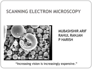
SCANNING ELECTRON MICROSCOPY

MUBASHSHIR ARIF
RAHUL RANJAN
P HARISH

“Increasing vision is increasingly expensive.”
Introduction
 Electron microscopes are scientific instruments that use

a beam of energetic electrons to examine objects on a very
fine scale.
 Electron microscopes were developed due to the
limitations of Light Microscopes which are limited by the
physics of light.
 In the early 1930's this theoretical limit had been reached
and there was a scientific desire to see the fine details of
the interior structures of organic cells (nucleus,
mitochondria...etc.).
 This required 10,000x plus magnification which was not
possible using optical microscopes.
The first scanning electron microscope (SEM)
debuted in 1938 ( Von Ardenne) with the first
commercial instruments around 1965. Its late
development was due to the electronics involved in
"scanning" the beam of electrons across the
sample.
An electron microscope is a type of microscope that uses a
beam of electrons to illuminate the specimen and produce a
magnified image. Electron microscopes (EM) have a
greater resolving power than a light-powered optical
microscope, because electrons have wavelengths about
100,000
times
shorter
than
visible
light
(photons),
and magnifications of up to about
10,000,000x, whereas ordinary, light microscopes are limited
by diffraction to about 200 nm resolution and useful
magnifications below 2000x.
TEM
 The original form of electron microscope, the transmission

electron microscope (TEM) uses a high voltage electron beam to
create an image. The electrons are emitted by an electron gun
and transmitted through the specimen that is in part transparent
to electrons and in part scatters them out of the beam.
 When it emerges from the specimen, the electron beam carries
information about the structure of the specimen that is magnified
by the objective lens system of the microscope.
 The spatial variation in this information (the "image") may be
viewed by projecting the magnified electron image onto a
fluorescent
viewing
screen
coated
with
a phosphor or scintillator material such as zinc sulfide.
 Image can be photographically recorded by exposing
a photographic film or plate directly to the electron beam, or a
high-resolution phosphor may be coupled by means of a lens
optical system or a fibre optic light-guide to the sensor of a CCD
(charge-coupled device) camera. The image detected by the CCD
may be displayed on a monitor or computer.
Characteristic Information:
SEM
 Topography:

The surface features of an object or "how it looks", its texture;
direct relation between these features and materials properties
 Morphology:
The shape and size of the particles making up the object; direct
relation between these structures and materials properties
 Composition:
The elements and compounds that the object is composed of
and the relative amounts of them; direct relationship between
composition and materials properties
 Crystallographic Information:
How the atoms are arranged in the object; direct relation between these
arrangements and material properties.
FIG: SCANNING ELECTRON MICROSCOPE
Diagram courtsey : Lowa State university
ELECTRON SOURCES

Anode [Hitachi S2300]
 A scanning electron microscope (SEM) is a

type of electron microscope that images a sample
by scanning it with a high-energy beam
of electrons in araster scan pattern. The electrons
interact with the atoms that make up the sample
producing signals that contain information about
the sample's
surfacetopography, composition, and other
properties such as electrical conductivity.
Specimen and Electron Detector Geometries:
-position of detectors is a function of relative
energies of the electrons
Final Lens

Backscatter
Detector

Secondary
Detector

SEM Sample Chamber [AMRAY 1830] showing positions
of SE and BSE Detectors and the Final Lens
Backscattered Electron
Generation
-SEM-BSE
-primary beam electrons
-high energy
-composition and topography
[specimen atomic number]
SEM Imaging Modes

Secondary Electron
Generation
-SEM-SE
-sample electrons ejected by the
primary beam [green line]
-low energy
-surface detail & topography
X ray is produced when outer shell electron
falls in to replace inner shell electron
WORKING OF SPUTTER COATER
 Switch power on with main switch.
 Flush working chamber several time with argon gas.

 Set sputter time with timer digit switch.
 Press start button to activate sputter process.
 Adjust appropriate gas pressure with argon valve.
 Set sputter current with current potentiometer.
 Process stops when selected sputter time elapses.
 To interrupt running sputter process press stop button.
 Switch power off/working chamber will be vented.
FIG: SPUTTER COATER
What happens when the Electron Beam hits the sample
When the electron is bombarded by the electron beam on the
specimen , electrons are ejected from the atoms of the specimen
surface.
Inelastic scattering, place the atom in the excited state. The atom
“wants ” to return to a ground or unexcited state. Hence the atoms
will relax giving off the excess energy.
X-rays, Cathodoluminescence and Auger electrons are the three
ways of relaxation.

 A resulting electron vacancy is filled by an electron from a higher
shell, and an X-ray is emitted to balance the energy difference
between the two electrons.
Limitations of Scanning Electron Microscopy (SEM)



Samples must be solid and they must fit into the microscope chamber.
Maximum size in horizontal dimensions is usually on the order of 10
cm, vertical dimensions should not exceed 40 mm.



For most instruments samples must be stable in vacuum . Samples likely
to outgas at low pressures (rocks saturated with hydrocarbons, "wet"
samples such as coal, organic materials or swelling clays, and samples
likely to depreciate at low pressure) are unsuitable for examination in
conventional SEM's.



SEM's cannot detect very light elements (H, He, and Li), and many
instruments cannot detect elements with atomic numbers less than 11.



An electrically conductive coating must be applied to electrically
insulating samples for study in conventional SEM's, unless the
instrument is capable of operation in a low vacuum mode.
Advantages of Using SEM


The SEM has a large depth of field, which allows a large amount of
the sample to be in focus at one time and produces an image that is a
good representation of the three-dimensional sample.



The combination of higher magnification, larger depth of
field, greater resolution, compositional and crystallographic
information makes the SEM one of the most heavily used instruments
in academic/national lab research areas and industry.
SEM at IIC, IIT ROORKEE
THANKYOU

Weitere ähnliche Inhalte

Was ist angesagt?

Transmission electron microscope
Transmission electron microscopeTransmission electron microscope
Transmission electron microscopeRaj Mohan
 
Scanning electron microscopy
Scanning electron microscopyScanning electron microscopy
Scanning electron microscopyJessa Ariño
 
Scanning Electron Microscopy (SEM) lecture
Scanning Electron Microscopy (SEM) lectureScanning Electron Microscopy (SEM) lecture
Scanning Electron Microscopy (SEM) lectureSaurabh Bhargava
 
Scanning Electron Microscope (SEM)
Scanning Electron Microscope (SEM)Scanning Electron Microscope (SEM)
Scanning Electron Microscope (SEM)Amna Jalil
 
2018 HM-Transmission electron microscope
2018 HM-Transmission electron microscope2018 HM-Transmission electron microscope
2018 HM-Transmission electron microscopeHarsh Mohan
 
Energy Dispersive Spectroscopy
Energy Dispersive SpectroscopyEnergy Dispersive Spectroscopy
Energy Dispersive SpectroscopySaad Shaukat
 
Scanning electron microscope (sem)
Scanning electron microscope (sem)Scanning electron microscope (sem)
Scanning electron microscope (sem)Mary Theresa
 
X ray photoelectron spectroscopy
X ray photoelectron spectroscopyX ray photoelectron spectroscopy
X ray photoelectron spectroscopyZubair Aslam
 
Scanning Electron microscopy
Scanning Electron microscopy Scanning Electron microscopy
Scanning Electron microscopy sagar sen sen
 
Scanning Electron Microscopy - SEM
Scanning Electron Microscopy - SEM Scanning Electron Microscopy - SEM
Scanning Electron Microscopy - SEM Stephen Raj D
 
EDS in TEM and SEM
EDS in TEM and SEMEDS in TEM and SEM
EDS in TEM and SEMHoang Tien
 
X-Ray Photo-electron Spectroscopy
X-Ray Photo-electron SpectroscopyX-Ray Photo-electron Spectroscopy
X-Ray Photo-electron SpectroscopyUzma Mhate
 
Transmission electron microscope (TEM)
Transmission electron microscope (TEM)Transmission electron microscope (TEM)
Transmission electron microscope (TEM)Nano Encryption
 
X-Ray Diffraction Technique
X-Ray Diffraction TechniqueX-Ray Diffraction Technique
X-Ray Diffraction Techniquetabirsir
 
Transmission Electron Microscopy
Transmission Electron MicroscopyTransmission Electron Microscopy
Transmission Electron MicroscopySejal9787
 

Was ist angesagt? (20)

Transmission electron microscope
Transmission electron microscopeTransmission electron microscope
Transmission electron microscope
 
Scanning electron microscopy
Scanning electron microscopyScanning electron microscopy
Scanning electron microscopy
 
Scanning Electron Microscopy (SEM) lecture
Scanning Electron Microscopy (SEM) lectureScanning Electron Microscopy (SEM) lecture
Scanning Electron Microscopy (SEM) lecture
 
Scanning Electron Microscope (SEM)
Scanning Electron Microscope (SEM)Scanning Electron Microscope (SEM)
Scanning Electron Microscope (SEM)
 
Scanning electron microscope
Scanning electron microscope  Scanning electron microscope
Scanning electron microscope
 
2018 HM-Transmission electron microscope
2018 HM-Transmission electron microscope2018 HM-Transmission electron microscope
2018 HM-Transmission electron microscope
 
Energy Dispersive Spectroscopy
Energy Dispersive SpectroscopyEnergy Dispersive Spectroscopy
Energy Dispersive Spectroscopy
 
SEM- scanning electron microscope
SEM- scanning electron microscope SEM- scanning electron microscope
SEM- scanning electron microscope
 
Scanning electron microscope (sem)
Scanning electron microscope (sem)Scanning electron microscope (sem)
Scanning electron microscope (sem)
 
X ray photoelectron spectroscopy
X ray photoelectron spectroscopyX ray photoelectron spectroscopy
X ray photoelectron spectroscopy
 
Scanning Electron microscopy
Scanning Electron microscopy Scanning Electron microscopy
Scanning Electron microscopy
 
Scanning electron microscopy (sem)
Scanning electron microscopy (sem)Scanning electron microscopy (sem)
Scanning electron microscopy (sem)
 
VSM
VSM VSM
VSM
 
Scanning Electron Microscopy - SEM
Scanning Electron Microscopy - SEM Scanning Electron Microscopy - SEM
Scanning Electron Microscopy - SEM
 
EDS in TEM and SEM
EDS in TEM and SEMEDS in TEM and SEM
EDS in TEM and SEM
 
X-Ray Photo-electron Spectroscopy
X-Ray Photo-electron SpectroscopyX-Ray Photo-electron Spectroscopy
X-Ray Photo-electron Spectroscopy
 
Transmission electron microscope (TEM)
Transmission electron microscope (TEM)Transmission electron microscope (TEM)
Transmission electron microscope (TEM)
 
X-Ray Diffraction Technique
X-Ray Diffraction TechniqueX-Ray Diffraction Technique
X-Ray Diffraction Technique
 
SEM & FESEM.pptx
SEM & FESEM.pptxSEM & FESEM.pptx
SEM & FESEM.pptx
 
Transmission Electron Microscopy
Transmission Electron MicroscopyTransmission Electron Microscopy
Transmission Electron Microscopy
 

Andere mochten auch

Scanning electron microscope
Scanning electron microscopeScanning electron microscope
Scanning electron microscopeAkhtar Kamal
 
Scanning electon microscope. Dr. GAURAV SALUNKHE
Scanning electon microscope. Dr. GAURAV SALUNKHEScanning electon microscope. Dr. GAURAV SALUNKHE
Scanning electon microscope. Dr. GAURAV SALUNKHEGaurav Salunkhe
 
Transmission Electron Microscope
Transmission Electron MicroscopeTransmission Electron Microscope
Transmission Electron MicroscopeManoranjan Ghosh
 
Electron Microscopy - Scanning electron microscope, Transmission Electron Mic...
Electron Microscopy - Scanning electron microscope, Transmission Electron Mic...Electron Microscopy - Scanning electron microscope, Transmission Electron Mic...
Electron Microscopy - Scanning electron microscope, Transmission Electron Mic...Sumer Pankaj
 
Scanning Electron Microscopy
Scanning Electron Microscopy Scanning Electron Microscopy
Scanning Electron Microscopy Waqas Javaid
 
transmission electron microscopy
transmission electron microscopytransmission electron microscopy
transmission electron microscopyJessa Ariño
 
Principle & Applications of Transmission Electron Microscopy (TEM) & High Res...
Principle & Applications of Transmission Electron Microscopy (TEM) & High Res...Principle & Applications of Transmission Electron Microscopy (TEM) & High Res...
Principle & Applications of Transmission Electron Microscopy (TEM) & High Res...Gulfam Raza
 
Electron microscopy
Electron microscopyElectron microscopy
Electron microscopysuniu
 
TRANSMISSION ELECTRON MICROSCOPY
TRANSMISSION ELECTRON MICROSCOPYTRANSMISSION ELECTRON MICROSCOPY
TRANSMISSION ELECTRON MICROSCOPYSutapa Saha
 
Scanning Electron Microscope
Scanning Electron MicroscopeScanning Electron Microscope
Scanning Electron MicroscopeAhmed AlAskalany
 
Microscopy
MicroscopyMicroscopy
Microscopykarade
 
Scanning electron microscope
Scanning electron microscopeScanning electron microscope
Scanning electron microscopeGajendra Kumar
 
Transmission electron microscopy new
Transmission electron microscopy newTransmission electron microscopy new
Transmission electron microscopy newDaiyanitha William
 
Electron Microscope Shanthakumar
Electron Microscope ShanthakumarElectron Microscope Shanthakumar
Electron Microscope Shanthakumarguestcd4662
 

Andere mochten auch (20)

Scanning electron microscope
Scanning electron microscopeScanning electron microscope
Scanning electron microscope
 
Scanning electon microscope. Dr. GAURAV SALUNKHE
Scanning electon microscope. Dr. GAURAV SALUNKHEScanning electon microscope. Dr. GAURAV SALUNKHE
Scanning electon microscope. Dr. GAURAV SALUNKHE
 
Sem and tem
Sem and temSem and tem
Sem and tem
 
Transmission Electron Microscope
Transmission Electron MicroscopeTransmission Electron Microscope
Transmission Electron Microscope
 
Electron Microscopy - Scanning electron microscope, Transmission Electron Mic...
Electron Microscopy - Scanning electron microscope, Transmission Electron Mic...Electron Microscopy - Scanning electron microscope, Transmission Electron Mic...
Electron Microscopy - Scanning electron microscope, Transmission Electron Mic...
 
Scanning Electron Microscopy
Scanning Electron Microscopy Scanning Electron Microscopy
Scanning Electron Microscopy
 
transmission electron microscopy
transmission electron microscopytransmission electron microscopy
transmission electron microscopy
 
Sem n tem
Sem n temSem n tem
Sem n tem
 
SEM,TEM & AFM
SEM,TEM & AFMSEM,TEM & AFM
SEM,TEM & AFM
 
Principle & Applications of Transmission Electron Microscopy (TEM) & High Res...
Principle & Applications of Transmission Electron Microscopy (TEM) & High Res...Principle & Applications of Transmission Electron Microscopy (TEM) & High Res...
Principle & Applications of Transmission Electron Microscopy (TEM) & High Res...
 
Electron microscopy
Electron microscopyElectron microscopy
Electron microscopy
 
TRANSMISSION ELECTRON MICROSCOPY
TRANSMISSION ELECTRON MICROSCOPYTRANSMISSION ELECTRON MICROSCOPY
TRANSMISSION ELECTRON MICROSCOPY
 
Light Microscopy
Light MicroscopyLight Microscopy
Light Microscopy
 
Scanning Electron Microscope
Scanning Electron MicroscopeScanning Electron Microscope
Scanning Electron Microscope
 
Microscopy
MicroscopyMicroscopy
Microscopy
 
Microscopy
MicroscopyMicroscopy
Microscopy
 
Microscopy
MicroscopyMicroscopy
Microscopy
 
Scanning electron microscope
Scanning electron microscopeScanning electron microscope
Scanning electron microscope
 
Transmission electron microscopy new
Transmission electron microscopy newTransmission electron microscopy new
Transmission electron microscopy new
 
Electron Microscope Shanthakumar
Electron Microscope ShanthakumarElectron Microscope Shanthakumar
Electron Microscope Shanthakumar
 

Ähnlich wie Scanning electron microscopy mubbu

Chapter-3. Electron Microscopy.pptx
Chapter-3. Electron Microscopy.pptxChapter-3. Electron Microscopy.pptx
Chapter-3. Electron Microscopy.pptxShivayogiPatil2
 
Scaning electron microscop
Scaning electron microscopScaning electron microscop
Scaning electron microscoptaameb
 
scanning electron microscope (SEM)
scanning electron microscope (SEM)scanning electron microscope (SEM)
scanning electron microscope (SEM)Himanshu Dixit
 
Scanning electron microscope
Scanning electron microscopeScanning electron microscope
Scanning electron microscopeSuganyaPaulraj
 
Electron microscope
Electron microscopeElectron microscope
Electron microscopeReena Rai
 
Advanced Characterization Technique - SEM
Advanced Characterization Technique - SEMAdvanced Characterization Technique - SEM
Advanced Characterization Technique - SEMIlyas Hussain
 
Scanning Electron Microscope
Scanning Electron Microscope Scanning Electron Microscope
Scanning Electron Microscope VrushankSalimath
 
ELECTRON MICROSCOPE AND ITS APPLICATION.pptx
ELECTRON MICROSCOPE AND ITS APPLICATION.pptxELECTRON MICROSCOPE AND ITS APPLICATION.pptx
ELECTRON MICROSCOPE AND ITS APPLICATION.pptxPallaviKumari112
 
Scanning Electron Microscope
Scanning Electron MicroscopeScanning Electron Microscope
Scanning Electron MicroscopeKetan Patil
 
Presentation forensic micoscopy SEM microscope.pptx
Presentation forensic micoscopy SEM microscope.pptxPresentation forensic micoscopy SEM microscope.pptx
Presentation forensic micoscopy SEM microscope.pptxHarishKumar330845
 
Materials Characterization
Materials CharacterizationMaterials Characterization
Materials CharacterizationVikas Barnwal
 
Typesofelectronmicroscope 130703030654-phpapp01
Typesofelectronmicroscope 130703030654-phpapp01Typesofelectronmicroscope 130703030654-phpapp01
Typesofelectronmicroscope 130703030654-phpapp01rupesh giri
 
Transmission Electron Microscopy (TEM)
Transmission Electron Microscopy (TEM) Transmission Electron Microscopy (TEM)
Transmission Electron Microscopy (TEM) Zohaib HUSSAIN
 

Ähnlich wie Scanning electron microscopy mubbu (20)

Chapter-3. Electron Microscopy.pptx
Chapter-3. Electron Microscopy.pptxChapter-3. Electron Microscopy.pptx
Chapter-3. Electron Microscopy.pptx
 
Sem Poster
Sem PosterSem Poster
Sem Poster
 
Scaning electron microscop
Scaning electron microscopScaning electron microscop
Scaning electron microscop
 
Electron microscope (SEM and TEM)
Electron microscope (SEM and TEM)Electron microscope (SEM and TEM)
Electron microscope (SEM and TEM)
 
scanning electron microscope (SEM)
scanning electron microscope (SEM)scanning electron microscope (SEM)
scanning electron microscope (SEM)
 
Scanning electron microscope
Scanning electron microscopeScanning electron microscope
Scanning electron microscope
 
Electron microscope
Electron microscopeElectron microscope
Electron microscope
 
electron microscopy
electron microscopyelectron microscopy
electron microscopy
 
Sem (2)
Sem (2)Sem (2)
Sem (2)
 
Advanced Characterization Technique - SEM
Advanced Characterization Technique - SEMAdvanced Characterization Technique - SEM
Advanced Characterization Technique - SEM
 
Scanning Electron Microscope
Scanning Electron Microscope Scanning Electron Microscope
Scanning Electron Microscope
 
ELECTRON MICROSCOPE AND ITS APPLICATION.pptx
ELECTRON MICROSCOPE AND ITS APPLICATION.pptxELECTRON MICROSCOPE AND ITS APPLICATION.pptx
ELECTRON MICROSCOPE AND ITS APPLICATION.pptx
 
Scanning and Transmission Electron Microscope
Scanning and Transmission Electron MicroscopeScanning and Transmission Electron Microscope
Scanning and Transmission Electron Microscope
 
Scanning Electron Microscope
Scanning Electron MicroscopeScanning Electron Microscope
Scanning Electron Microscope
 
Stem
StemStem
Stem
 
Presentation forensic micoscopy SEM microscope.pptx
Presentation forensic micoscopy SEM microscope.pptxPresentation forensic micoscopy SEM microscope.pptx
Presentation forensic micoscopy SEM microscope.pptx
 
Materials Characterization
Materials CharacterizationMaterials Characterization
Materials Characterization
 
Sem md ppt
Sem md pptSem md ppt
Sem md ppt
 
Typesofelectronmicroscope 130703030654-phpapp01
Typesofelectronmicroscope 130703030654-phpapp01Typesofelectronmicroscope 130703030654-phpapp01
Typesofelectronmicroscope 130703030654-phpapp01
 
Transmission Electron Microscopy (TEM)
Transmission Electron Microscopy (TEM) Transmission Electron Microscopy (TEM)
Transmission Electron Microscopy (TEM)
 

Kürzlich hochgeladen

Handwritten Text Recognition for manuscripts and early printed texts
Handwritten Text Recognition for manuscripts and early printed textsHandwritten Text Recognition for manuscripts and early printed texts
Handwritten Text Recognition for manuscripts and early printed textsMaria Levchenko
 
Top 5 Benefits OF Using Muvi Live Paywall For Live Streams
Top 5 Benefits OF Using Muvi Live Paywall For Live StreamsTop 5 Benefits OF Using Muvi Live Paywall For Live Streams
Top 5 Benefits OF Using Muvi Live Paywall For Live StreamsRoshan Dwivedi
 
Unblocking The Main Thread Solving ANRs and Frozen Frames
Unblocking The Main Thread Solving ANRs and Frozen FramesUnblocking The Main Thread Solving ANRs and Frozen Frames
Unblocking The Main Thread Solving ANRs and Frozen FramesSinan KOZAK
 
Mastering MySQL Database Architecture: Deep Dive into MySQL Shell and MySQL R...
Mastering MySQL Database Architecture: Deep Dive into MySQL Shell and MySQL R...Mastering MySQL Database Architecture: Deep Dive into MySQL Shell and MySQL R...
Mastering MySQL Database Architecture: Deep Dive into MySQL Shell and MySQL R...Miguel Araújo
 
The Codex of Business Writing Software for Real-World Solutions 2.pptx
The Codex of Business Writing Software for Real-World Solutions 2.pptxThe Codex of Business Writing Software for Real-World Solutions 2.pptx
The Codex of Business Writing Software for Real-World Solutions 2.pptxMalak Abu Hammad
 
The Role of Taxonomy and Ontology in Semantic Layers - Heather Hedden.pdf
The Role of Taxonomy and Ontology in Semantic Layers - Heather Hedden.pdfThe Role of Taxonomy and Ontology in Semantic Layers - Heather Hedden.pdf
The Role of Taxonomy and Ontology in Semantic Layers - Heather Hedden.pdfEnterprise Knowledge
 
2024: Domino Containers - The Next Step. News from the Domino Container commu...
2024: Domino Containers - The Next Step. News from the Domino Container commu...2024: Domino Containers - The Next Step. News from the Domino Container commu...
2024: Domino Containers - The Next Step. News from the Domino Container commu...Martijn de Jong
 
Strategies for Unlocking Knowledge Management in Microsoft 365 in the Copilot...
Strategies for Unlocking Knowledge Management in Microsoft 365 in the Copilot...Strategies for Unlocking Knowledge Management in Microsoft 365 in the Copilot...
Strategies for Unlocking Knowledge Management in Microsoft 365 in the Copilot...Drew Madelung
 
Workshop - Best of Both Worlds_ Combine KG and Vector search for enhanced R...
Workshop - Best of Both Worlds_ Combine  KG and Vector search for  enhanced R...Workshop - Best of Both Worlds_ Combine  KG and Vector search for  enhanced R...
Workshop - Best of Both Worlds_ Combine KG and Vector search for enhanced R...Neo4j
 
08448380779 Call Girls In Greater Kailash - I Women Seeking Men
08448380779 Call Girls In Greater Kailash - I Women Seeking Men08448380779 Call Girls In Greater Kailash - I Women Seeking Men
08448380779 Call Girls In Greater Kailash - I Women Seeking MenDelhi Call girls
 
🐬 The future of MySQL is Postgres 🐘
🐬  The future of MySQL is Postgres   🐘🐬  The future of MySQL is Postgres   🐘
🐬 The future of MySQL is Postgres 🐘RTylerCroy
 
How to Troubleshoot Apps for the Modern Connected Worker
How to Troubleshoot Apps for the Modern Connected WorkerHow to Troubleshoot Apps for the Modern Connected Worker
How to Troubleshoot Apps for the Modern Connected WorkerThousandEyes
 
Data Cloud, More than a CDP by Matt Robison
Data Cloud, More than a CDP by Matt RobisonData Cloud, More than a CDP by Matt Robison
Data Cloud, More than a CDP by Matt RobisonAnna Loughnan Colquhoun
 
08448380779 Call Girls In Civil Lines Women Seeking Men
08448380779 Call Girls In Civil Lines Women Seeking Men08448380779 Call Girls In Civil Lines Women Seeking Men
08448380779 Call Girls In Civil Lines Women Seeking MenDelhi Call girls
 
Driving Behavioral Change for Information Management through Data-Driven Gree...
Driving Behavioral Change for Information Management through Data-Driven Gree...Driving Behavioral Change for Information Management through Data-Driven Gree...
Driving Behavioral Change for Information Management through Data-Driven Gree...Enterprise Knowledge
 
Slack Application Development 101 Slides
Slack Application Development 101 SlidesSlack Application Development 101 Slides
Slack Application Development 101 Slidespraypatel2
 
WhatsApp 9892124323 ✓Call Girls In Kalyan ( Mumbai ) secure service
WhatsApp 9892124323 ✓Call Girls In Kalyan ( Mumbai ) secure serviceWhatsApp 9892124323 ✓Call Girls In Kalyan ( Mumbai ) secure service
WhatsApp 9892124323 ✓Call Girls In Kalyan ( Mumbai ) secure servicePooja Nehwal
 
Salesforce Community Group Quito, Salesforce 101
Salesforce Community Group Quito, Salesforce 101Salesforce Community Group Quito, Salesforce 101
Salesforce Community Group Quito, Salesforce 101Paola De la Torre
 
From Event to Action: Accelerate Your Decision Making with Real-Time Automation
From Event to Action: Accelerate Your Decision Making with Real-Time AutomationFrom Event to Action: Accelerate Your Decision Making with Real-Time Automation
From Event to Action: Accelerate Your Decision Making with Real-Time AutomationSafe Software
 
Finology Group – Insurtech Innovation Award 2024
Finology Group – Insurtech Innovation Award 2024Finology Group – Insurtech Innovation Award 2024
Finology Group – Insurtech Innovation Award 2024The Digital Insurer
 

Kürzlich hochgeladen (20)

Handwritten Text Recognition for manuscripts and early printed texts
Handwritten Text Recognition for manuscripts and early printed textsHandwritten Text Recognition for manuscripts and early printed texts
Handwritten Text Recognition for manuscripts and early printed texts
 
Top 5 Benefits OF Using Muvi Live Paywall For Live Streams
Top 5 Benefits OF Using Muvi Live Paywall For Live StreamsTop 5 Benefits OF Using Muvi Live Paywall For Live Streams
Top 5 Benefits OF Using Muvi Live Paywall For Live Streams
 
Unblocking The Main Thread Solving ANRs and Frozen Frames
Unblocking The Main Thread Solving ANRs and Frozen FramesUnblocking The Main Thread Solving ANRs and Frozen Frames
Unblocking The Main Thread Solving ANRs and Frozen Frames
 
Mastering MySQL Database Architecture: Deep Dive into MySQL Shell and MySQL R...
Mastering MySQL Database Architecture: Deep Dive into MySQL Shell and MySQL R...Mastering MySQL Database Architecture: Deep Dive into MySQL Shell and MySQL R...
Mastering MySQL Database Architecture: Deep Dive into MySQL Shell and MySQL R...
 
The Codex of Business Writing Software for Real-World Solutions 2.pptx
The Codex of Business Writing Software for Real-World Solutions 2.pptxThe Codex of Business Writing Software for Real-World Solutions 2.pptx
The Codex of Business Writing Software for Real-World Solutions 2.pptx
 
The Role of Taxonomy and Ontology in Semantic Layers - Heather Hedden.pdf
The Role of Taxonomy and Ontology in Semantic Layers - Heather Hedden.pdfThe Role of Taxonomy and Ontology in Semantic Layers - Heather Hedden.pdf
The Role of Taxonomy and Ontology in Semantic Layers - Heather Hedden.pdf
 
2024: Domino Containers - The Next Step. News from the Domino Container commu...
2024: Domino Containers - The Next Step. News from the Domino Container commu...2024: Domino Containers - The Next Step. News from the Domino Container commu...
2024: Domino Containers - The Next Step. News from the Domino Container commu...
 
Strategies for Unlocking Knowledge Management in Microsoft 365 in the Copilot...
Strategies for Unlocking Knowledge Management in Microsoft 365 in the Copilot...Strategies for Unlocking Knowledge Management in Microsoft 365 in the Copilot...
Strategies for Unlocking Knowledge Management in Microsoft 365 in the Copilot...
 
Workshop - Best of Both Worlds_ Combine KG and Vector search for enhanced R...
Workshop - Best of Both Worlds_ Combine  KG and Vector search for  enhanced R...Workshop - Best of Both Worlds_ Combine  KG and Vector search for  enhanced R...
Workshop - Best of Both Worlds_ Combine KG and Vector search for enhanced R...
 
08448380779 Call Girls In Greater Kailash - I Women Seeking Men
08448380779 Call Girls In Greater Kailash - I Women Seeking Men08448380779 Call Girls In Greater Kailash - I Women Seeking Men
08448380779 Call Girls In Greater Kailash - I Women Seeking Men
 
🐬 The future of MySQL is Postgres 🐘
🐬  The future of MySQL is Postgres   🐘🐬  The future of MySQL is Postgres   🐘
🐬 The future of MySQL is Postgres 🐘
 
How to Troubleshoot Apps for the Modern Connected Worker
How to Troubleshoot Apps for the Modern Connected WorkerHow to Troubleshoot Apps for the Modern Connected Worker
How to Troubleshoot Apps for the Modern Connected Worker
 
Data Cloud, More than a CDP by Matt Robison
Data Cloud, More than a CDP by Matt RobisonData Cloud, More than a CDP by Matt Robison
Data Cloud, More than a CDP by Matt Robison
 
08448380779 Call Girls In Civil Lines Women Seeking Men
08448380779 Call Girls In Civil Lines Women Seeking Men08448380779 Call Girls In Civil Lines Women Seeking Men
08448380779 Call Girls In Civil Lines Women Seeking Men
 
Driving Behavioral Change for Information Management through Data-Driven Gree...
Driving Behavioral Change for Information Management through Data-Driven Gree...Driving Behavioral Change for Information Management through Data-Driven Gree...
Driving Behavioral Change for Information Management through Data-Driven Gree...
 
Slack Application Development 101 Slides
Slack Application Development 101 SlidesSlack Application Development 101 Slides
Slack Application Development 101 Slides
 
WhatsApp 9892124323 ✓Call Girls In Kalyan ( Mumbai ) secure service
WhatsApp 9892124323 ✓Call Girls In Kalyan ( Mumbai ) secure serviceWhatsApp 9892124323 ✓Call Girls In Kalyan ( Mumbai ) secure service
WhatsApp 9892124323 ✓Call Girls In Kalyan ( Mumbai ) secure service
 
Salesforce Community Group Quito, Salesforce 101
Salesforce Community Group Quito, Salesforce 101Salesforce Community Group Quito, Salesforce 101
Salesforce Community Group Quito, Salesforce 101
 
From Event to Action: Accelerate Your Decision Making with Real-Time Automation
From Event to Action: Accelerate Your Decision Making with Real-Time AutomationFrom Event to Action: Accelerate Your Decision Making with Real-Time Automation
From Event to Action: Accelerate Your Decision Making with Real-Time Automation
 
Finology Group – Insurtech Innovation Award 2024
Finology Group – Insurtech Innovation Award 2024Finology Group – Insurtech Innovation Award 2024
Finology Group – Insurtech Innovation Award 2024
 

Scanning electron microscopy mubbu

  • 1. SCANNING ELECTRON MICROSCOPY MUBASHSHIR ARIF RAHUL RANJAN P HARISH “Increasing vision is increasingly expensive.”
  • 2. Introduction  Electron microscopes are scientific instruments that use a beam of energetic electrons to examine objects on a very fine scale.  Electron microscopes were developed due to the limitations of Light Microscopes which are limited by the physics of light.  In the early 1930's this theoretical limit had been reached and there was a scientific desire to see the fine details of the interior structures of organic cells (nucleus, mitochondria...etc.).  This required 10,000x plus magnification which was not possible using optical microscopes.
  • 3. The first scanning electron microscope (SEM) debuted in 1938 ( Von Ardenne) with the first commercial instruments around 1965. Its late development was due to the electronics involved in "scanning" the beam of electrons across the sample.
  • 4. An electron microscope is a type of microscope that uses a beam of electrons to illuminate the specimen and produce a magnified image. Electron microscopes (EM) have a greater resolving power than a light-powered optical microscope, because electrons have wavelengths about 100,000 times shorter than visible light (photons), and magnifications of up to about 10,000,000x, whereas ordinary, light microscopes are limited by diffraction to about 200 nm resolution and useful magnifications below 2000x.
  • 5. TEM  The original form of electron microscope, the transmission electron microscope (TEM) uses a high voltage electron beam to create an image. The electrons are emitted by an electron gun and transmitted through the specimen that is in part transparent to electrons and in part scatters them out of the beam.  When it emerges from the specimen, the electron beam carries information about the structure of the specimen that is magnified by the objective lens system of the microscope.  The spatial variation in this information (the "image") may be viewed by projecting the magnified electron image onto a fluorescent viewing screen coated with a phosphor or scintillator material such as zinc sulfide.  Image can be photographically recorded by exposing a photographic film or plate directly to the electron beam, or a high-resolution phosphor may be coupled by means of a lens optical system or a fibre optic light-guide to the sensor of a CCD (charge-coupled device) camera. The image detected by the CCD may be displayed on a monitor or computer.
  • 6.
  • 7. Characteristic Information: SEM  Topography: The surface features of an object or "how it looks", its texture; direct relation between these features and materials properties  Morphology: The shape and size of the particles making up the object; direct relation between these structures and materials properties  Composition: The elements and compounds that the object is composed of and the relative amounts of them; direct relationship between composition and materials properties  Crystallographic Information: How the atoms are arranged in the object; direct relation between these arrangements and material properties.
  • 9. Diagram courtsey : Lowa State university
  • 11.  A scanning electron microscope (SEM) is a type of electron microscope that images a sample by scanning it with a high-energy beam of electrons in araster scan pattern. The electrons interact with the atoms that make up the sample producing signals that contain information about the sample's surfacetopography, composition, and other properties such as electrical conductivity.
  • 12. Specimen and Electron Detector Geometries: -position of detectors is a function of relative energies of the electrons
  • 13. Final Lens Backscatter Detector Secondary Detector SEM Sample Chamber [AMRAY 1830] showing positions of SE and BSE Detectors and the Final Lens
  • 14. Backscattered Electron Generation -SEM-BSE -primary beam electrons -high energy -composition and topography [specimen atomic number]
  • 15. SEM Imaging Modes Secondary Electron Generation -SEM-SE -sample electrons ejected by the primary beam [green line] -low energy -surface detail & topography
  • 16. X ray is produced when outer shell electron falls in to replace inner shell electron
  • 17.
  • 18. WORKING OF SPUTTER COATER  Switch power on with main switch.  Flush working chamber several time with argon gas.  Set sputter time with timer digit switch.  Press start button to activate sputter process.  Adjust appropriate gas pressure with argon valve.  Set sputter current with current potentiometer.  Process stops when selected sputter time elapses.  To interrupt running sputter process press stop button.  Switch power off/working chamber will be vented.
  • 20.
  • 21. What happens when the Electron Beam hits the sample When the electron is bombarded by the electron beam on the specimen , electrons are ejected from the atoms of the specimen surface. Inelastic scattering, place the atom in the excited state. The atom “wants ” to return to a ground or unexcited state. Hence the atoms will relax giving off the excess energy. X-rays, Cathodoluminescence and Auger electrons are the three ways of relaxation.  A resulting electron vacancy is filled by an electron from a higher shell, and an X-ray is emitted to balance the energy difference between the two electrons.
  • 22.
  • 23.
  • 24. Limitations of Scanning Electron Microscopy (SEM)  Samples must be solid and they must fit into the microscope chamber. Maximum size in horizontal dimensions is usually on the order of 10 cm, vertical dimensions should not exceed 40 mm.  For most instruments samples must be stable in vacuum . Samples likely to outgas at low pressures (rocks saturated with hydrocarbons, "wet" samples such as coal, organic materials or swelling clays, and samples likely to depreciate at low pressure) are unsuitable for examination in conventional SEM's.  SEM's cannot detect very light elements (H, He, and Li), and many instruments cannot detect elements with atomic numbers less than 11.  An electrically conductive coating must be applied to electrically insulating samples for study in conventional SEM's, unless the instrument is capable of operation in a low vacuum mode.
  • 25.
  • 26. Advantages of Using SEM  The SEM has a large depth of field, which allows a large amount of the sample to be in focus at one time and produces an image that is a good representation of the three-dimensional sample.  The combination of higher magnification, larger depth of field, greater resolution, compositional and crystallographic information makes the SEM one of the most heavily used instruments in academic/national lab research areas and industry.
  • 27. SEM at IIC, IIT ROORKEE