Dental Plaque
•Als ODP, PDF herunterladen•
35 gefällt mir•7,255 views

Plaque

Empfohlen
Weitere ähnliche Inhalte
Was ist angesagt?
Was ist angesagt? (20)
Ähnlich wie Dental Plaque
Ähnlich wie Dental Plaque (20)
Mehr von Dr Sudeep Madhusudan Chaudhari
Mehr von Dr Sudeep Madhusudan Chaudhari (16)
Kürzlich hochgeladen
Kürzlich hochgeladen (20)
Dental Plaque
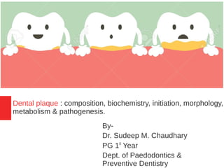
- 1. Dental plaque : composition, biochemistry, initiation, morphology, metabolism & pathogenesis. By- Dr. Sudeep M. Chaudhary PG 1st Year Dept. of Paedodontics & Preventive Dentistry
- 2. Contents ➢Introduction ➢Classification of soft deposits ➢Definitions ➢Classification of dental plaque ➢Composition ➢Formation/development of dental plaque ➢ Plaque as a biofilm ➢Morphology of dental plaque ➢Factors affecting plaque formation ➢Metabolism ➢Pathogenesis ➢Conclusion ➢References
- 3. Dental plaque is a complex community of microorganisms that forms on the surfaces of teeth and restorations and has been implicated as the primary etiological factor in the development of periodontal diseases. So far, more than 700 different bacterial species have been identified from the human oral cavity and the majority of them are associated with dental plaque. Introduction
- 4. ➔Human fetus is sterile. ➔Colonization starts at birth. ➔Within hours – facultative & aerobic bacteria. ➔2nd day – anaerobic bacteria. ➔Within 2 weeks – mature microbiota established in gut. ➔After weaning - 1014 microorganisms with 400 different type of bacteria. ➔There are 10 times more bacteria than human cells.
- 5. ➔ Establishing microbiota - harmony with the host. ➔ Constant renewal microorganisms - Prevents the accumulation of microorganisms. ➔ Teeth provide hard, non-shedding surfaces - accumulation & metabolism of bacteria on hard oral surfaces is considered the primary cause of dental caries, gingivitis, periodontitis and peri-implant infections. ➔ In the oral cavity, the bacterial deposits have been termed dental plaque or bacterial plaque.
- 6. Classification of soft deposits ● A non-cellular thin film ● An organized transparent deposit which is primarily composed of bacteria and their products ● Soft, whitish deposit with no specific architecture, which can be removed by water spray ● Retained food which is usually removed by saliva and oral muscular action Dental Plaque Acquired Pellicle Materia Alba Food Debris
- 7. DEFINITIONS Plaque is a specific but highly variable structural entity resulting from colonization and growth of microorganisms on surfaces of teeth and consisting of numerous microbial species and their products embedded in a extracellular matrix. {WHO (1978)} Dental plaque is defined clinically as a structured, resilient yellow-grayish substance that adheres tenaciously to the intraoral hard surfaces, including removable and fixed restorations. (Carranza, 11th Edition)
- 8. Classification of Dental Plaque 1)Supragingival plaque 2)Subgingival plague a)Attached (a)Tooth associated (b)Tissue associated b)Unattached
- 9. Composition ● Consist of densely packed bacteria which are embedded in an amorphous material called plaque matrix ● 60 – 70% bacterial cells ● 30 – 40% matrix
- 10. Bacteria Approximal surface •Gram positive & gram negative; facultative & obligate anaerobes: 1. Neisseria 2. Streptococcus 3. Prevotella 4. Actinomyces 5. veillonella Fissure •Gram positive •Facultative anaerobes 1. Streptococcus 2. Actinomyces Gingival crevice •Gram positive & gram negative & obligate anaerobes: 1. Streptococcus 2. Prevotella 3. Actinomyces 4. Treponema 5. Eubacterium
- 11. Intercellular matrix - ➔Impart structural integrity to the microbial masses ➔80% water & remaining (20%) solids ➔Bacterial & salivary proteins comprise about one half of the dry weight of plaque ➔Lipids ➔Carbohydrates (25% dry weight) – glucans, fructose, heteropolysaccherides ➢play a role in bacterial attachment & cohesion ➢Reservoir of fermentable substrates which are metabolized by bacteria
- 12. ➔5 - 10% the dry weight of plaque ➔Calcium ➔Phosphate ➔Potassium ➔Sodium ➔Magnesium ➔Copper ➔Lead ➔Iron ➔Strontium ➔Fluoride Inorganic components –
- 13. ●Fluoride 5 – 10 ppm as compared to saliva 0.01 – 0.05 ppm ●Most of fluoride is probably bound on or within bacteria but some may be in form of calcium fluoride or fluorapatite. ●Concentration of Ca & phosphate in plaque is several magnitudes higher than in saliva. (Dawes & Jenkins, 1962) ●Higher concentration is thought to be due to infiltration of salivary proteins which probably includes – statherin.
- 14. Element High DMFS mean Low DMFS mean Fluoride (ppm) 12.4 36.0 Calcium (%) 0.416 2.158 Magnesium (%) 0.156 0.158 Phosphorus (%) 1.58 2.11 Table- The relationship between caries experience & palque mineral concentrarion (Schamschula et al., 1980-82)
- 15. Formation/development of dental plaque 1)Pellicle formation 2)Initial adhesion/attachment of bacteria 3)Colonization & plaque maturation
- 16. 1)Pellicle formation ➔Pellicle is the initial stucture that forms on the surfaces the teeth & artificial prosthesis ➔Involves attachment of positively charged salivary proteins to apetite surface which has negatively charged phosphate group
- 17. 2)Initial adhesion/attachment of bacteria ●Within a few hours, bacteria are found on the dental pellicle. The initial bacteria colonizing the pellicle coated tooth surface are predominantly gram - positive facultative microorganisms such as Actinomyces viscosus and Streptococcus sanguis. ●These initial colonizers adhere to the pellicle, through specific molecules, termed adhesins, on the bacterial surface that interact with receptors in the dental pellicle. Actinomyces spp S.mitis S.oralis S.sanguis S.gordonii S.intermedius V.parvula A.odontolyticus Primary colonizers
- 18. ●There is a transition from the early aerobic environment characterized by gram-positive facultative species to a highly oxygen-deprived environment in which gram-negative anaerobic microorganisms predominate.
- 19. 3)Colonization & plaque maturation Secondary colonizers are the microorganisms that do not initially colonize clean tooth surfaces, including Prevotella intermedia, Prevotella loescheii, Capnocytophaga spp., Fusobacterium nucleatum and Porphyromonas gingivalis. These microorganisms adhere to cells of bacteria already in the plaque mass. C.showae C.rectus E.nodatum P.intermedia P.nigrescens P.micros F.nucleatum E.corrodens Capnocytophaga spp A.actinomycetemcomitans P.gingivalis B.forsythus T.denticola Secondary Colonizers
- 20. ●Extensive laboratory studies have documented the ability of different species and genera of plaque microorganisms to adhere to one another, a process known as coaggregation. This process occurs primarily through the highly specific stereochemical interaction of protein and carbohydrate molecules located on the bacterial cell surfaces, in addition to the less specific interactions resulting from hydrophobic, electrostatic, and van der Waals forces.
- 21. ●Most studies of coaggregation have focused on interactions among different gram-positive species and between gram- positive and gram-negative species. ●In the latter stages of plaque formation, coaggregation between different gram-negative species is likely to predominate. Examples of these types of interactions are the coaggregation of F. nucleatum with P. gingivalis or Treponema denticola.
- 22. ●First 2-8 hours→pioneering streptococci, cover 3-30% of enamel surface ●Next 20 hrs→short period of rapid growth. ●One day, it can be called biofilm. ●As bacterial densities approach 2-6 million bacteria /mm2, a marked increase in growth rate can be observed up to 32 million bacteria/mm2 ●Thickness slowly increases with time to 20- 30 µm after 3 days. ●After 4 days 30% of the tooth crown is covered by plaque.
- 25. Morphology of dental plaque ●Supragingival plaque typically demonstrates a stratified organization of the bacterial morphotypes. Gram-positive cocci and short rods predominate at the tooth surface, whereas Gram-negative rods and filaments as well as spirochetes predominate in the outer surface of the mature plaque mass. ●Highly specific cell-to-cell interactions are also evident from the “corncob” structures often observed. Corncob formations have been observed between rod-shaped bacterial cells (e.g. Bacterionema matruchotii or F. nucleatum) that form the inner core of the structure and coccal cells (e.g., streptococci or P. gingivalis) that attach along the surface of the rodshaped cell.
- 26. Development of dental plaque on a clean enamel surface. Coccal bacteria attach to the enamel pellicle as pioneer species (A) and multiply to form microcolonies (B), eventually resulting in biofilm formation embedded in a matrix of extracellular polymers of bacterial and salivary origin (C). With time, the diversity of the microflora increases and rod and filament-shaped bacteria colonize (D
- 27. Long-standing supragingival plaque near the gingival margin demonstrates “corncob” arrangement. A central gramnegative filamentous core supports the outer coccal cells, which are firmly attached by interbacterial adherence or coaggregation.
- 28. Plaque as a biofilm ●As the bacteria attach to a surface and to each other, they cluster together to form sessile, mushroom-shaped microcolonies that are attached to the surface at a narrow base. ●Each microcolony is a tiny, independent community containing thousands of compatible bacteria. ●Different microcolonies may contain different combinations of bacterial species.
- 29. ●Bacteria in the center of a microcolony may live in a strict anaerobic environment, while other bacteria at the edges of the fluid channels may live in an aerobic environment. ●Thus, the biofilm structure provides a range of customized living environments (with differing pH , nutrient availability and oxygen concentrations) within which bacteria with different physiological needs can survive.
- 30. ●The extracellular slime layer is a protective barrier that surrounds the mushroomshaped bacterial microcolonies. ●The slime layer protects the bacterial microcolonies from antibiotics, antimicrobials and host defense mechanisms. ●A series of fluid channels penetrates the extracellular slime layer.
- 31. ●These fluid channels provide nutrients and oxygen for the bacterial micro colonies and facilitate movement of bacterial metabolites, waste products and enzymes within the biofilm structure. ●Each bacterial microcolony uses chemical signals to create a primitive communication system used to communicate with other bacterial microcolonies Fluid channel
- 32. Quorum sensing ●Involves the regulation of expression of specific genes through the accumulation of signaling compounds that mediate intercellular communication. ●Dependent on cell density and mediated through signaling compounds. ●Quorum sensing gives biofilms their distinct properties Cell – cell communication
- 33. Quorum sensing is involved in the regulation of - a)Genetic competence b)Mating c)Bacteriocin production d)Sporulation e)Stress responses f)Virulence expression g)Biofilm formation
- 34. Factors affecting plaque formation ➢Surface irregularities ➢Restorative materials ➢Erupting teeth ➢Carious lesion ➢Calculus ➢Malocclusion ➢Orthodontic therapy ➢Removable partial denture Plaque retentive factors -
- 35. Plaque formation occurs faster - ➔Lower jaw > upper jaw ➔Molar region > anterior region ➔Buccal surface > palatal surface (especially in upper jaw) ➔Interdental region > buccal/palatal surface Variations in dentition -
- 36. Impact of gingival inflammation - ●Plaque formation is more rapid on tooth surfaces facing inflamed gingival margins, than those facing healthy gingivae. ●Increase in crevicular fluid production enhances plaque formation, it favors initial adhesion & colonization of bacteria.
- 37. Ageing - ●Following tooth eruption the isolation frequency of spirochetes & black pigmented anaerobes increases. ●Increased prevalence of spirochetes & black pigmented anaerobes is found in teenagers, this is due to hormones entering gingival crevice & acting as a novel nutrient source.
- 38. Nutrients Bacteria degrade host proteins to release ammonia which is used by another baceria as a nitrogen source. P. gingivalis - uses hemin iron from the breakdown of Host haemoglobin. Prevotella intermedia - Proportions increases with steroid increase in host.
- 39. Metabolism ●Heterogeneity & complexity of the chemical and the microbial composition of the dental plaque has been emphasized ● A very wide range of metabolic reactions may be detected in plaque ●Degradative reactions whereby bacteria convert organic substances to metabolites & thereby derive energy are readily detectable. ●Opposite biochemical processes also occur which utilize the energy
- 40. Glycolysis ●Anaerobic catabolism of carbohydrates predominates in plaque which have a reduced oxygen tension. ●Bacteria of plaque – capable of using different carbohydrates – starch, disaccherides & monosaccherides – as a substrate. ●1 molecule of glucose →2 molecules of lactic acid + 2 ATP Polyglucose Glucose-1-phosphate Glucose-6-phosphate Glucose-1,6-diphosphate Pyruvic acid Lactic acid ATP ADP ADP
- 41. ●Homorofermentors – some streptococci & many lactobacilli – produced 90% lactic acid ●Heterofermentors – produce mixure of metabolite – propionic acid, butyric acid, succinic acid & ethanol. Polyglucose Pyruvic acid Lactic acid Propionic acid/butyric acid/ succinic acid CO2
- 42. ●The proportion of lactic acid or other organic acids formed by plaque may be markly affected by growth conditions & by the bacterial types present. ●When the concentration of cariogenic bacteria & sugars in plaque is high the pathway leading to lactic acid formation is dominent. On other hand, when carbohydrate is limited the latter reaction is favored. (Yamada & Carlsson, 1975)
- 43. Base production ●pH of plaque is usually highest upon wakening in the morning & it is higher than pH of saliva. ●Due to production of ammonia, amines & other basic components by bacterial degradation of proteins, peptides, urea & other nitrogenous compounds. (Kleinberg & Jenkins, 1964)
- 44. Pathogenesis 1.Nonspecific plaque hypothesis 2.Specific plaque hypothesis 3.Ecologic plaque hypothesis
- 45. 1. Nonspecific plaque hypothesis ●This hypothesid was delineated in the 1976 by Walter Loesche ●The nonspecific plaque hypothesis maintains that periodontal disease results from the “elaboration of noxious products by the entire plaque flora.” ●According to this thinking, when only small amounts of plaque are present, noxious products are neutralized by the host. ●Similarly, large amounts of plaque would produce large amounts of noxious products, which would essentially overwhelm the host's defenses.
- 46. ●Nonspecific plaque hypothesis is the concept that control of periodontal disease depends on control of the amount of plaque accumulation. ●Treatment of periodontitis by debridement (nonsurgical or surgical) and oral hygiene measures focuses on the removal of plaque and its products and is founded in the nonspecific plaque hypothesis.
- 47. 2. Specific plaque hypothesis ●Proposed by Walter Loesche(1976) ●The specific plaque hypothesis states that only certain plaque is pathogenic, and its pathogenicity depends on the presence of or increase in specific microorganisms. ●This concept predicts that plaque harboring specific bacterial pathogens results in periodontal disease because these organisms produce substances that mediate the destruction of host tissues.
- 48. 3. Ecologic plaque hypothesis ●In 1994, Philip D. Marsh proposed a hypothesis that combined key concepts of the earlier hypotheses. ●Disease is the result of an imbalance in the total microflora due to ecological stress, resulting in an enrichment of some oral pathogens or disease-related micro-organisms ●This hypothesis is based on the theory that the unique local microenvironment influences the composition of the oral microflora.
- 49. ●This hypothesis postulated dynamic relationship between environmental cause & ecological shifts within the biofilm. ●It also introduced the concept that the disease can be prevented not only by inhibiting the putative pathogens, but also interfering with the environmental factors driving the selection & enrichment of these bacteria.
- 50. Bacteria associated with health & disese Health ●102 to 103 bacteria/mm2 . ●Certain bacterial species have been proposed to be beneficial to the host, including S. sanguis, Veilonella parvula, and C. ochraceus ●Bacteria associated with periodontal diseases are often found in the subgingival microflora at healthy sites, although they are normally present in small proportions. ●Nonmotile nature.
- 51. Gingivitis ●10 4 to 10 6 bacteria/mm2 . ●Gram-negative bacteria. ●Compared with healthy sites, noticeable increase also occur in the numbers of motile bacteria, including cultivable and uncultivable treponemas (spirochetes). ●Pregnancy associated gingivitis is accompanied by dramatic increases in levels of P. intermedia, which uses the steroid as growth factors.
- 52. Chronic periodontitis ●Campylobacter rectus, Porphyromonas gingivalis, Provella intermedia, Fusobacterium nucleatum and Tannerella forsythia were found to be elevated in the active sites. ●Sites with chronic periodontitis will be populated with greater proportions of gram- negative organisms and motile bacteria. ●Certain gram-negative bacteria with pronounced virulence properties have been strongly implicated as etiologic agents e.g. Porphyromonas gingivalis and Tannerella forsythus.
- 53. Localized aggresive periodontitis ●Gram negative, and anaerobic rods. ●The most numerous isolates are several species from the genera Eubacterium, Actinomyces naeslundii, Fusobacterium nucleatum, Campylobacter rectus and Veillonella parvula. ●In some populations, a strong case can be made for Aggregatibacter actinomycetemcomitans playing a causative role in LAP, especially in cases in which patients harbor highly leukotoxic strains of the organism. ●However, some populations of patients with LAP do not harbor A. actinomycetemcomitans, and in still others Porphyromonas gingivalis may be etiologically more important.
- 54. Generalized aggressive periodontitis ●The sub-gingival flora in patients with generalized aggressive periodontitis resembles that in other forms of periodontitis. ●The predominant subgingival bacteria in patients with generalized aggressive periodontitis are P. gingivalis, T. forsythis, A. actinomycetemcomitans and Campylobacter species.
- 55. Periodontal abscess ●The bacteria isolated from abscesses are similar to those associated with chronic and aggressive forms of periodontitis. ●An average of approximately 70% of the cultivable flora in exudates from periodontal abscesses are gram-negative and about 50% are anaerobic rods. ●Periodontal abscesses revealed a high prevalence of the following putative pathogens: F. nucleatum (70.8%), Peptostreptococcus micros (70.6%), P. intermedia (62.5%), P. gingivalis (50.0%) and T. forsythis (47.1%). ●Enteric bacteria, coagulase-negative staphylococci and Candida albicans have also been detected.
- 56. Necrotizing ulcerative gingivitis & periodontitis ●More than 50% of the isolated species were strict anaerobes with P. gingivalis and F. nucleatum accounting for 7-8% and 3.4%, respectively.
- 57. Conclusion ●Dental plaque biofilm cannot be eliminated permanently. ●Dental plaque is regarded as one of the main etiological factors in the initiation and promotion of periodontal disease i.e. gingivitis and periodontitis & dental caries. ●However, the pathogenic nature of the dental plaque biofilm can be reduced by reducing the bioburden and maintaining a normal flora with appropriate oral hygiene methods that include daily brushing,flossing and rinsing with antimicrobial mouthrinses. ●This can result in the prevention or management of the associated sequelae, including the development of periodontal diseases
- 58. References ● Newman MG, Takei H, Klokkevold PR, Carranza FA. Carranza's clinical periodontology (Vol-1). 11th edition Elsevier health sciences; 2011 Feb 14. ● Reddy S. Essentials of Clinical Periodontology & Periodontics. JP Medical Ltd; 2017 Nov 30. ● Nikiforuk G. Understanding Dental Caries. 1. Etiology and Mechanisms, Basic and Clinical Aspects. 1985:125-7.