cementum seminar
•Als PPTX, PDF herunterladen•
2 gefällt mir•527 views

ultrastructure and function of cementum seminar

Empfohlen
Weitere ähnliche Inhalte
Was ist angesagt?
Was ist angesagt? (20)
Ähnlich wie cementum seminar
Ähnlich wie cementum seminar (20)
Kürzlich hochgeladen
Kürzlich hochgeladen (20)
cementum seminar
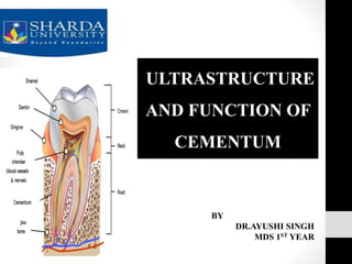
- 1. ULTRASTRUCTURE AND FUNCTION OF CEMENTUM BY DR.AYUSHI SINGH MDS 1ST YEAR
- 2. CONTENTS • Introduction • Physical properties • Chemical composition • Histology of cementum • Classification • Development of cementum • Cemento-dentinal junction • Cemento-enamel junction • Cementum resorption and repair
- 3. CONTENTS • Functions • Effects of ageing on cementum • Cementum in oral environment • Role of cementum in periodontal disease • Developmental anomalies • Conclusion • References
- 4. CEMENTUM • Cementum furnishes a medium for the attachment of collagen fibers that bind the tooth to surrounding structures.1
- 5. DEFINITION • The cementum is the part of the periodontium that attaches the teeth to the alveolar bone by anchoring the periodontal ligament.- GPT • Cementum is the calcified, avascular mesenchymal tissue that forms the outer covering of the anatomic root. Orbans 2
- 6. 1 2 3 The cementum is the surface layer of the tooth root. Rather than being a passive entity like paint on a wall, cementum is a dynamic entity within the periodontium.1 In fully formed and functioning teeth, 1. Cementum is firmly attached to the radicular dentin. 2. Covers the entire surface of the root. 3. Increases in thickness towards the apex and may extend partially into the apical foramen1
- 7. PHYSICAL CHARACTERISTICS • Hardness: less than dentin2 • Color: Light yellow with dull surface and lighter than dentin2 • Thickness: Variable, thinnest at CEJ and thickest at at apex. Apical foramen is surrounded by cementum2 • Its permeable, (as age progresses permeability of cementum diminishes)
- 8. CHEMICAL CHARACTERISTICS On dry weight basis, cementum of fully formed teeth contains: • Inorganic substances- 45-50% 2 • Organic substances and water- 50-55% 2
- 9. INORGANIC PORTION • Hydroxyapatite- calcium and Phosphate1 • Trace elements like - Copper - Fluorine - Iron - Lead - Potassium - Silica • Cementum has highest fluorine content among mineralized tissues
- 10. ORGANIC PORTION 2 • Collagen: Type I predominant (90%), others include type III (5%), V, XII, XIV 1 • Matrix proteins2 • Proteoglycans • Osteonectin • Osteopontin • Osteocalcin • Fibronectin • Bone sialoprotein
- 11. HISTOLOGYOF CEMENTUM • Histology section of cementum shows: • A) Cells, fibers, ground substance • B) Cemento-enamel junction • C) Cemento-dentinal junction Cells of cementum • The cells are:2 • Cementoblast • Cementocytes • Cementoclasts
- 12. CEMENTOBLASTS 1 • Numerous mitochondria • Well defined golgi apparatus • Large number of granular endoplasmic reticulum • Synthesize collagen and protein polysaccharide, which make up the organic matrix of cementum • After some cementum is laid down, its mineralization begins • The cells are found lining the root
- 13. CEMENTOCYTES 1 • Spider shaped cells, characteristic feature of cellular cementum • During the formation of cellular cementum, cementoblasts become entrapped within their own matrix due to rapid deposition and are called cementocytes • Similar to osteocytes, they lie in spaces called as lacunae • Haphazardly arranged and widely dispersed • In deeper layers of cementum, cementocytes shows definite signs of degeneration such as cytoplasmic clumping, empty lacunae, vesiculation
- 14. • CEMENTOCLASTS 1 • Found in howship’s lacunae • Unilocular /multilocular cells • Function: resorption of cementocytes • Major role: resorption and repair
- 15. INCREMENTAL LINES OF CEMENTUM 2 • Referred to as incremental lines of salter • Represent rhythmic periodic deposition of cementum • Appears as dark lines running parallel to root surface • Seen in both cellular and acellular cementum but more prominent in acellular cementum • Best seen in decalcified sections under light microscope • Highly mineralized areas with less collagen and more ground substance
- 16. CLASSIFICATION1 BASED ON LOCATION BASED ON TIME OF FORMATION CORONAL RADICULAR PRIMARY SECONDARY
- 17. BASED ON CELLULARITY BASED ON THE PRESENCE OR ABSENCE OF COLLAGENOUS FIBRILS 1. FIBRILLAR 2. AFIBRILLAR • It lacks embedded cells. • Covers the root adjacent to dentin near crown ACELLULAR • Cells are located within the mineralized matrix. • Seen at apical area and overlying acellular cementum. Also common in interradicular areas CELLULAR
- 18. BASED ON THE ORIGIN OF COLLAGENOUS FIBRILS INTRINSIC FIBERS - Which are formed due to cementoblasts activity. Are found between the Sharpey's fibers. Arranged either randomly or parallel to the surface of the cementum1 EXTRINSIC FIBERS - The first group is made up of the embedded parts of the principal fibers of the periodontal ligament which are known as Sharpey's fibers (extrinsic group).1 They are formed by the fibroblasts of the periodontal ligament.
- 19. SCHRODER & PAGE CLASSIFICATION (1968,1991) 1.ACELLULAR AFIBRILLAR CEMENTUM 2.ACELLULAR EXTRINSIC FIBRE CEMENTUM 3.CELLULAR MIXED FIBRE CEMENTUM 4.CELLULAR INTRINSIC FIBRE CEMENTUM 5.INTERMEDIATE CEMENTUM
- 21. ACELLULARAFIBRILLAR CEMENTUM 1 • Found deposited on mature enamel surfaces (1-15um) as coronal cementum. • It may be deposited during tooth formation, during tooth eruption or after it. • This type of cementum is probably of little significance as it is not involved in fiber insertion and tooth anchorage. • Contains neither cells nor extrinsic or intrinsic collagen fibers, except for a mineralized ground substance. •.
- 22. ACELLULAR EXTRINSIC FIBER CEMENTUM 1 • Is composed almost entirely of densely packed bundles of Sharpey fibers and lacks cells. • Acellular extrinsic fiber cementum is a product of fibroblasts and cementoblasts. • It is found in the cervical third of roots in humans, but it may extend farther apically. • Its thickness is between 30 and 230 µm.
- 23. (PLOS ONE | DOI:10.1371/journal.pone.0167316 December 9, 2016) The results highlighted five main points – 1. The AEFC is mainly composed of carbonated hydroxyapatite and collagen type I, with a Raman spectrum response similar to that of bone and dentin. 2. The spatial distribution of peak and ratio intensities reproduced the alternating bright and dark incremental lines observed in polarized microscopy.
- 24. The alternating pattern of AEFC could thus be partly related to the mineral/organic composition, with higher mineral/ organic ratio in dark lines compared to bright lines. 3. The collagen fibers seemed to be better aligned in dark lines (the dark lines correspond to smaller, more mineralized and better organized incremental layers produced during a winter rest period. ) 4. Mineral crystals were always parallel to collagen fibrils. 5. Based on the results of Raman polarized acquisitions, they proposed an organization of the AEFC consisting of orthogonal or radial fibers in the continuity of periodontal ligament Sharpey’s fibers and “non-orthogonal” fibers (occasionally mistakenly called intrinsic fibers), which in turn may consist of branching and bending radial fibers.
- 25. CELLULAR MIXED STRATIFIED CEMENTUM 1 • Composed of extrinsic & intrinsic fibers & contains cells. • Forms the bulk of the Secondary Cementum. • Co-product of cementoblasts & fibroblasts. • Location: Apical interradicular regions. • Thickness ranges from 100-1000um. • Primary function is adaptation.
- 26. • It has a lower mineral content than Acellular Extrinsic Fiber Cementum. • This difference can in part be accounted for by: • the non mineralized structures present in Cellular Intrinsic Fiber Cementum. • The Sharpey’s fibers present in it generally retain an unmineralized core as these fibers have a coating of type 3 collagen, which may prevent mineralization of the type I collagen in the core.
- 27. CELLULAR INTRINSIC FIBER CEMENTUM 1 • Location: Found in middle and apical third • Contains cells but no extrinsic collagen fibers. • Formed by Cementoblasts. • Only it can repair a resorptive defect of the root in a reasonable time due to its capacity to grow much faster than any other known cementum type. • It has a major role in adaptation and repair • its formation on the developing root commences in closest proximity to the advancing root edge.
- 28. INTERMEDIATE CEMENTUM LAYER • It does not exhibit characterstics of either DENTIN OR CEMENTUM • It is structureless • It mainly observed in apical 2/3rd of molars and premolars , rarely seen in incisors or deciduous teeth. • In this layers hertwig’s epithelial sheath become trapped in rapidly deposited dentin or cementum matrix.1 • It contains no tubules but wide range spaces which are thought to be enlarged terminals of dentinal tubules • Sometimes it found as a continuous layer or in isolated areas • Seen at zone near CDJ
- 29. CEMENTOGENESIS (Development of Cementum)2 Deposition of dentin along the inner aspect of HERS Disintegration of HERS allowing newly formed dentin to come in contact with cells of dental follicle Differentiation of cementoblast along the external surface of the root Protein secretion by cementoblast mainly collagen & proteoglycans which form organic matrix of cementum Phase of matrix maturation which subsequently mineralizes to form cementum.
- 30. FORMATION OFCEMENTOBLASTS • CEMENTOBLASTS FORMATION RESULTS IN 2 WAYS2 FROM HERTWIG’S EPITHELIAL ROOT SHEATH FROM DENTAL FOLLICLE Soon after Hertwig’s root sheath breaks down Epithelial cells of hertwig’s root sheath undergo epithelial mesenchymal transformation CEMENTOBLASTS Ecto- mesenchymal in origin
- 31. • These cells are rich in numerous Mitochondria ,well formed Golgi apparatus, and larger amounts of Granular Endoplasmic Reticulum Electron microscopically cementoblasts show Mitochondria and Endoplasmic reticulum
- 32. • Cementoblasts synthesize collagen and protein polyasaccharides. • Cementoblasts derived from dental follicle have similar phenotype to osteoblasts ,it is shown to be involed in formation of Cellular Intrinsic fiber Cementum.2 • Cementoblasts derived from HERS has different phenotype than osteoblasts and are shown to be involved in Acellular Intrinsic Fiber Cementum.2 • In the differentiation of cementoblasts, growth factors belonging to the TGF beta including BMP, MOLECULE EPIDERMAL GROWTH FACTOR etc. are involved .
- 33. CEMENTO-DENTINALJUNCTION • The terminal apical area of cementum where it joins internal root canal dentin is known as cemento-dentinal junction. • Interface between cementum and dentine • In deciduous teeth- scalloped • In permanent teeth- smooth • Areas of dentine adjacent to CDJ appears granular in ground section due to colaescing and looping of terminal portion of dentinal tubules and is called TOMES GRANULAR LAYER • During RCT obturating material must end near CDJ • No increase or decrease in width with age(2-3 micrometer)1
- 34. CEMENTO-ENAMELJUNCTION 1 It is defined as the area of union of cementum and enamel at the cervical region of the tooth.
- 35. CEMENTUM RESORPTIONAND REPAIR 1 • Permanent teeth doesn’t undergo the process of physiologic resorption as does the counterpart • However the cementum of erupted as well as unerupted teeth is subject to resorptive changes that may be of microscopic proportion or sufficiently extensive to present a radiographically detectable alteration in the root contour • Local conditions1 • 1) Trauma from occlusion • 2) Orthodontic movement • 3) Cysts and Tumors • 4) Pressure from mal-aligned erupted teeth • 5) Peri-apical disease and Periodontal disease
- 36. Systemic Conditions1 1) Paget’s disease 2) Hypothyroidism 3) Heredity fibrous osteodystrophy 4) Calcium deficiency • Cementum resorption appear microscopically as bay like concavities in the root surface1 • Multi-nucleated giant cells and large mononuclear macrophages are generally found adjacent to cementum undergoing active resorption1 • Several sites of resorption coalesce to form a larger area of destruction1 • Resorptive process may extend into underlying dentin and even into pulp, but its usually painless
- 37. • Cementum resorption is not necessarily continuous and may alternate with periods of repair and deposition of new cementum1 • The newly formed cementum is demarcated from the root by a deep stained irregular line, termed as reversal line, which delineates the border of the previous resorption1
- 38. • Repair of cementum is a process to heal the damage caused by resorption or cemental fracture • Cementum repair requires the presence of viable connective tissue • If epithelium proliferates in the area of resorption then repair will not take place • Repair can occur in both vital and non-vital teeth • Repair can be Anatomical Functional
- 39. Anatomic Repair: The root outline is re-established as it was before cemental resorption. It generally occurs when the degree of destruction is low. Cementum resorption is repaired by cellular and acellular cementum. Functional Repair: in cases of large cemental resorption or destruction, repair doesn’t re-establish the same contour as before. To maintain the width of PDL, the adjacent alveolar bone grows and takes the shape of defect, this is done to improve the function of tooth, hence called functional repair.
- 40. FUNCTIONS OF CEMENTUM 2 • ANCHORAGE: Primary function of cementum is to furnish a medium for the attachment of collagen fibers that bind the tooth to alveolar bone. • ADAPTATION: The continuous deposition of cementum is of considerable functional importance. As the superficial layer of cementum ages, a new layer of cementum is deposited to keep the attachment apparatus intact. • REPAIR: Cementum serves as the major reparative tissue for root surfaces. Damage to roots such as fractures and resorption can be repaired by the deposition of new cementum
- 41. AGE CHANGES IN CEMENTUM 5 • Cementum formation continues throughout life • It appears that cementum is deposited at a linear rate • There is tendency for cementum to reduce root surface concavities . Thus, its thicker layers are found in root surface grooves and furcation areas • Tooth position may exert temporal and spatial variation in pressure and tension on root surface and these stimuli influence the rate as well as pattern of cementum formation • Non- functioning impacted teeth have thicker cementum • Sharpey’s fibers may nearly completely absent
- 43. (2018 International Journal of Oral Health Sciences) The relationship between chronological age and estimated age using light microscope, phase contrast and polarized microscope showed stronger correlation while stereo microscope showed a weaker correlation. The aim of this study was to examine the correlation between chronologic age and estimated age and to find the most accurate method of calculating cemental annulations using different types of microscopes (light microscope, phase contrast microscope, polarized, and stereomicroscope).
- 44. EXPOSURE OF CEMENTUM TO ORALENVIRONMENT 5 • Cementum becomes exposed to oral cavity in cases of gingival recession and a consequence of loss of attachment in pocket formation • Cementum is sufficiently permeable to be penetrated by micro- organisms, organic and inorganic substances in such cases • Bacterial invasion of the cementum occurs commonly in periodontal disease • Caries of cementum can also develop.
- 45. (Journal of Indian Society of Periodontology - Vol 18, Issue 5, Sep-Oct 2014) The results of this study indicated that the hardness and modulus of elasticity of the cervical third cementum decreases significantly in chronic periodontitis. This study aims to analyze the nanomechanical properties of the cervical third of the cementum (transverse section) in health and chronic periodontitis Twenty teeth (10 healthy and 10 periodontally diseased) were collected and the nanomechanical properties of the transverse section of the cervical third cementum were evaluated
- 46. CHANGES IN CEMENTUMASSOCIATED WITH PERIODONTALDISEASE • Structural Changes • Partial demineralization • Decrease or loss of cross banding of collagen • Sub-surface condensation of organic material of exogenous origin • Chemical Changes • Increase in calcium and phosphate levels • Increase in fluoride content • Decrease in sodium levels
- 47. • Cytotoxic changes • Effects on cell proliferation • Hatfield and Bomhammers- inhibitory substance penetrates the exposed cementum that prevents growth of epithelial cells in tissue cultures • Presence of endotoxins - limits fibroblast proliferation – detrimental to the arrest of disease • Cementum bound endotoxins – 50 times more toxic • Destructive physical changes – cavitation, partial demineralization • Effect on cell attachment • Cultured human gingival fibroblasts do not attach to diseased tooth (Aleo et al 1975)
- 48. Inhibitory principle of matrix- Morris (1975) • Disease inhibited development of implanted marrow whereas demineralized healthy tooth did not • Demineralized diseased roots showed more inhibition-toxins must have seeped into root matrix during pocket formation and demineralization removed the toxin allowing development of marrow • According to inhibitory principle of matrix-phenol extraction usually required to remove toxins from bacterial cell wall is not necessary to make diseased cementum receptive cell attachment
- 49. • In early and moderate periodontitis- acellular cementum is affected (coronal half of the root) • Damage extends to cellular cementum in most advanced periodontal conditions and furcally positioned lesions • These surfaces are almost always covered by cellular cementum during successful regeneration, whether this is adequate is unclear (McNeil and Sommerman-1999)
- 50. DEVELOPMENTALANOMALIES OF CEMENTUM Enamel Projections If amelogenesis does not stop before the start of root formation, enamel may continue to form over portions normally covered by cementum3 Enamel Pearls This consist of globules of enamel on root surface in cervical regeneration (act as plaque retentive areas)3
- 51. Hypercementosis • Refers to prominent thickening of cementum • It may be localised to one tooth or may involve the entire dentition3 (Paget’s disease) • Occurs as generalised thickening of the cementum, with nodular enlargement of cementum in apical third of root • Etiology of Hypercementosis Spike-like type of Hypercementosis : results from excessive forces from orthodontic appliances or occlusal forces Generalised type-occurs in teeth without antagonist
- 52. (2015 Journal of Indian Academy of Oral Medicine and Radiology) A very unusual case of mammoth, dumbbell-shaped hypercementosis in a 49-year-old male patient is reported. Literature on hypercementosis is reviewed. Etiology, radiographic appearances, and differential diagnosis are discussed. Importance of pre-extraction radiographs in all cases is stressed.
- 53. Cementoblastoma • Only neoplasm of cementum • Cementum like tissue is deposited around the roots of teeth as irregular or rounded mass • Age less than 25 • Involves commonly- mandibular molars and pre- molars • Tooth usually as vital pulp • Attached to roots and causes its resorption, involves pulp canal and also causes slow expansion of cortical plate • Enlargement produced is usually asymptomatic
- 54. Cementicles • Are small, globular masses of cementum found in approx 35% of human roots. • May not be always attached to the cementum surface but may be located free in Pdl. • These may result from microtrauma, when extra stress on sharpey’s fibers causes a tear in the cementum. 3 • Are more commonly found in apical & middle third of root and in root furcation areas
- 55. • May develop from calcified epithelial rests; around small spicules of cementum or alveolar bone traumatically displaced into the periodontal ligament; from calcified Sharpey's fibers; and from calcified, thrombossed vessels within the periodontal ligament
- 56. Cementoma • Benign cementoblastoma /cemental dysplasia2 • Represents an unusual reaction of bone • Caused due to occlusal trauma • Present usually at apex of mandibular incisor • Almost exclusively found in black persons • Age-20 to 40 years • Causes expansion of jaw
- 57. Concrescence • Form of fusion which occurs root formation has been completed • Thought to arise as a result of crowding or traumatic injury of the teeth with resorption of interdental bone so that the two roots are in approximate contact and fused by deposition of cementum between them2 • May occur before or after tooth has erupted
- 58. Hypophosphatasia • This is a hereditary disease characterized by total absence of cementum • It results in early loss of teeth • It occurs due to deficiency of enzyme ALP in serum and tissues Acromegaly • The tooth crowns are usually longer in size and the roots often enlarge as a result of secondary cemental hyperplasia. • This hypercementosis may be the result of functional and structural demands on teeth.
- 59. Ankylosis • Fusion of alveolar bone and cementum with obliteration of PDL space is termed as ankylosis2 • Occurs in cases of cemental resorption, may represent a form of abnormal repair • May develop after chronic apical inflammation, tooth re-plantation and occlusal trauma • Results in resorption of root and gradual replacement with bone tissue
- 60. Cemental tears • The detachment of a fragment of cementum is described as a cemental tear. • Cemental tears have been reported in the periodontal literature associated with localized, rapid periodontal breakdown.
- 61. CONCLUSION A thorough knowledge of the structure and function of cementum is essential to understand its involvement in disease & also about its treatment. Cementum by virtue of its structural and dynamic qualities, provides tooth attachment & maintenance of occlusal relationship. Fibers in cementum helps in tooth anchorage. Certain pathologic factors may cause irreversible damage to cementum else it is self reparative to small disturbance .
- 62. REFERENCES 1. Carranza’s clinical periodontology (10th , 11th & 13th edition) 2. Orban’s –Text Book Of Oral Histology And Embryology 11th edition 3. Shafers textbook of oral pathology – 7th edition 4. Mallar KB, Girish HC, Murgod S, Yathindra Kumar BN. Age estimation using annulations in root cementum of human teeth: A comparison between longitudinal and cross sections. J Oral Maxillofac Pathol 2015;19:396-404 5. Boscchardt D, Selvig K. Dental Cementum: The dynamic tissue covering the root surface. Periodontology 2000. Vol. 13, 1997, 41-75
- 63. 6. Colard T et al. (2016) New Insights on the Composition and the Structure of the Acellular Extrinsic Fiber Cementum by Raman Analysis. PLoS ONE 11(12):e0167316. 7. Geetha HL, Baghisath VP, Vinay HB, Sudheer B, Kumar VJ, Gayathri CH. Age estimation using tooth cementum annulations method by different types of microscope: A comparative study. Int J Oral Health Sci 2018;8:73-80. 8. Sundaram, et al.: Nanostructure of cervical third cementum in health and chronic periodontitis JISP -2014 ;18( 5). 9. Raghavan V, Singh C. Hypercementosis: Review of literature and report of a case of mammoth, dumbbell-shaped hypercementosis. J Indian Acad Oral Med Radiol 2015;27:160-3.