SlideShare ist ein Scribd-Unternehmen logo
1 von 31
Introduction to Diffusion
Tensor Imaging
 Why DTI?
 Diffusion – what it is, how it affects MR signal
 Tensor – how we represent diffusion
 Imaging – how we measure it in MRI
 Stroke/ischemia
 Alzheimer’s Disease
 Multiple Sclerosis
 Brain maturation studies
 Ischemia and stroke
 Neoplasm
 Preoperative planning
 Traumatic brain injury
 Congenital anomalies and diseases of white
matter
 Encephalopathies
 Neurodegenerative diseases
 Spinal Cord Injury
 Epilepsy
 Dementia, schizophrenia, depression
 Developmental disorders
 Autism
 Aging
Why diffusion?
http://www.vh.org/Providers/Textbooks/
BrainAnatomy/Ch5Text/Section18.html
http://eclipse.nichd.nih.gov/nichd/DTMRI/mri/
Conceptually: in vivo histology
Why diffusion?
 Diffusion is EXTREMELY SENSITIVE to
differences and changes in tissue microstructure
 Myelination/Demyelination
 Axon damage/loss
 Inflammation/Edema
 Necrosis
 It is NOT a biomarker of white matter integrity
 It is NOT just about white matter
 Gray matter
 Cardiac tissue
Example DTI image
 “Fractional Anisotropy”
map
 “map” is a computed
parameter, unlike an
“image” which is acquired
signal
 Also called a “tractogram”
since it clearly shows
major white matter fiber
tracts
What is Diffusion?
 stochastic movement of particles in a solvent,
driven by the thermal molecular motion of the
solvent…
 … and also applies to motion of the solvent
itself (Einstein, 1905)
time τ∆
NOTE: In the limit N→∞, use the
Central Limit Theorem to assume
“step size” ∆ is fixed and equal to
the average of individual
displacements ∆i.
1D Fick’s Law - what the flux?
t = t0 + τ
x
t = t0
0 2x − ∆ 0x − ∆ 0x + ∆0x 0 2x + ∆
t = t0 + τ
x
t = t0
0 2x − ∆ 0x − ∆ 0x + ∆0x 0 2x + ∆
What is the flux (J) through x0 after one time interval τ ?
C1(x)C2(x)
dx
dC
J
τ
2
2
1 ∆
−=
Adolf Fick, 1855: Flux is proportional to
the particle concentration gradient
(conservation of mass)
The Diffusion Coefficient
 3D Fick’s Law
 Note the minus sign: flux goes
from high to low concentration
 del operator replaces partial
derivative
 factor of 6, not 2 (why?)
 D is the diffusion coefficient
 This is the expression for
isotropic diffusion
τ62
∆=D
CDJ ∇−=

CJ ∇
∆
−=
τ6
2
Isotropic Diffusion (water)
water
ink
Dtr 6=
r1
t1
r2
t2
τ62
∆=D
Diffusion in Tissue (Anisotropic)
t
ink
r2
r3
r1
diffusion
ellipsoid
tDr 11 2=
tDr 22 2=
tDr 33 2=
x
y
z
laboratory
frame
DON’T
try this at
lab!!!!!
The Diffusion Tensor










zzyzxz
yzyyxy
xzxyxx
DDD
DDD
DDD
x
y
z
r2
r3
r1










3
2
1
00
00
00
D
D
D
diagonalization
Lab frame Intrinsic frame
Tensor Invariants
 Eigenvalues:
diagonalization
(iterative QR
factorization)
 Eigenvectors
xx xy xz
xy yy yz
xz yz zz
D D D
D D D
D D D
 
 
 
 
 
{ }1 2 3D D D
{ }321 eee

Tensor Invariants
 Shape invariants:
analytical calculation
directly from tensor
coeffs
xx xy xz
xy yy yz
xz yz zz
D D D
D D D
D D D
 
 
 
 
 
( )1
3
av xx yy zzD D D D= + +
1
2
2 2 2
1
3
xx yy xx zz yy zz
surf
xy xz yz
D D D D D D
D
D D D
+ + 
=  
 − − − 
1
2 3
2 2
2
xx yy zz xx yz
vol
xy xz yz zz xy yy xz
D D D D D
D
D D D D D D D
 −
 =
 + − − 
Scalar Anisotropy Indices
( )
avND
D
DADC
DDDADC
=
++= 321
3
1
( ) ( ) ( )
2
2
2
3
2
2
2
1
2
3
2
2
2
1
1
2
3
mag
surf
ND
D
D
D
FA
DDD
DDDDDD
FA
−=
++
−+−+−
=
FA vs. ADC
FA (x 10
-4
)
0 1000 2000 3000 4000 5000 6000 7000 8000 9000 10000
Probability
0.0000
0.0002
0.0004
0.0006
0.0008
0.0010
WM
GM
CSF
MD (x 10 -3
mm2
/s)
0 500 1000 1500 2000 2500 3000 3500 4000
Probability
0.000
0.001
0.002
0.003
0.004
0.005
WM
GM
CSF
FA and ADC are very
useful clinically, but are
very different.
Tensor has a LOT of
information!
Q. Which metric would you use to detect brain cancer?
Vector anisotropy measures
 We can use eigenvector
information from the
tensor as well
 Represent direction of
primary eigenvector as
color on a scalar map
 Or render the primary
eigenvectors as “fibers”
for astonishing* 3D
visualizations
Red = R/L
Green = A/P
Blue = S/I
*but how “real” is it? Many PhD
theses have asked….
Diffusion tensor coefficients Diffusion tensor invariants
Scalar anisotropy indices
Vector anisotropy indices
Effect of Diffusion on MRI signal
Signal attenuation!
Diffusion term
Diffusion weighted MRI
∆ δ
G G
echoπα
( ) ( )( )2
0
δexpδ
3
M
G D
M
γ= − × ∆ − ×
∆ δ
( ) ( )2
δδ
3
b Gγ= × ∆ −
(boxcar gradients)
“b-value”
Consider
simplified diffusion
experiment…
MR Measurement of Diffusion Tensor
j
T
j j
j
p
G G q
r
 
 
= × 
 
 
ur
0
xx xy xz j
j j j xy yy yz j
xz yz zz j
D D D p
p q r D D D q
D D D r
j
b
S S e
   
   
 − × × ×    
   
   
=
( )
22 2
γ δ Δ δ 3b G= −
( )
1
6
j N
N
=
≥
Kjth
diffusion-
weighted
image
Diffusion
magnitude
Diffusion
direction
Gz
Gy
Gx
...
...
...
Solving for D
20
0
xx xy xz j
j j j xy yy yz j
xz yz zz j
D D D p
p q r D D D q
D D D r
j
b
S S e
   
   
 − × × ×    
   
   
=
1. Acquire T2W image (b = 0 s/mm2
)
3. Choose a diffusion gradient orientation2. Choose a b-value
4. Acquire image (Sj)
5. Repeat steps 1 – 3, j = 1 … N times
6. Solve for D…. How?
Let’s do some linear algebra…
[ ]










⋅










⋅⋅−=
zj
yj
xj
zzzyzx
yzyyyx
xzxyxx
zjyjxjj
DDD
DDD
DDD
bSS
α
α
α
αααexp0
( )
( )
( )
( )
( )
( ) 



















⋅




















⋅=
yz
xz
xy
xx
yy
xx
T
jxy
jxy
jxy
jzz
jyy
jxx
j
D
D
D
D
D
D
b
S
S
α
α
α
α
α
α
2
2
2
ln
2
2
2
0
1661 xNxNx xAY ⋅=
B-matrix formalism
22
( )yzyzxzxzxyxyzzzzyyyyxxxx DDDDDDb αααααα 222222
+++++⋅
( )yzyzxzxzxyxyzzzzyyyyxxxx DbDbDbDbDbDb 222 +++++=
∑∑= =
=
3
1
3
1i j
ijij Db
The “b-matrix”
The b-matrix formalism
summarizes total
attenuating effect of all
gradient waveforms in
all directions (including
imaging gradients)
T2W
(b = 0 s/mm2
)
Y, -ZY, Z-X, Y
X, Y-X, Z+X, Z
24
SVD
DIAG
T2W
(b = 0 s/mm2
)
…
DWI
(j = 1, 2, 3 … N)
Dij
N=27 N=55
N=13N=6
N NEX # DWI
6 8 56
13 4 56
27 2 56
55 1 56
#DWI = (N + 1) x NEX
If TR = 4 sec, then acq time
= 56*4sec = 3.7 minutes
Tradeoff: N vs NEX
Rotational invariance
Hasan et al, JMRI 2001
Jones MRM 2004
27
Empirical Image Quality
increasing N, decreasing NEX
increasingb-value
How low can you go?
 High b-values mean more
attenuation, lower SNR
 Lower b-values mean higher
SNR, room for more N
 At very low b-values, imaging
gradients’ diffusion effects
are no longer negligible
 Lower b-values also do not
probe same diffusion scale,
less clinically interesting
b=100 s/mm2
b=500 s/mm2
(N = 6, 8 NEX)
Echo-Planar Imaging (EPI)
 Advantages
 Minimal motion artifacts
 NEX
 N
 Disadvantages
 Eddy current artifacts
 T2* limits spatial
resolution
 Geometric distortion
(susceptibility)
29
DT-MRI Alexander
SLFSLF CRCR CCCC CINGCING
Partial Volume Effects on Anisotropy
DT-MRI Alexander
Mapping Complex Diffusion
Based Upon Q-Space Theory – Model Independent
 ODF – orientation density function (Tuch et al., Neuron 2003)
 Diffusion Spectrum Imaging (DSI)
(Tuch et al. Neuron 2003, Wedeen et al. 2005)
 High Angular Diffusion Imaging (HARDI), Q-Ball
(Frank 2002; Tuch et al. Neuron 2003)

Weitere ähnliche Inhalte

Andere mochten auch

Flow Cytometry - basics, principles and applications
Flow Cytometry - basics, principles and applicationsFlow Cytometry - basics, principles and applications
Flow Cytometry - basics, principles and applicationsAnkit Raiyani
 
Diffusion Weighted MRI (2011-09-29 이정원)
Diffusion Weighted MRI (2011-09-29 이정원)Diffusion Weighted MRI (2011-09-29 이정원)
Diffusion Weighted MRI (2011-09-29 이정원)이정원 JeongwonLee
 
Diffusion MRI, Tractography,and Connectivity: what machine learning can do?
Diffusion MRI, Tractography,and Connectivity: what machine learning can do?Diffusion MRI, Tractography,and Connectivity: what machine learning can do?
Diffusion MRI, Tractography,and Connectivity: what machine learning can do?Ting-Shuo Yo
 
Week 5. Basics and clinical uses of MR spectroscopy.
Week 5. Basics and clinical uses of MR spectroscopy.Week 5. Basics and clinical uses of MR spectroscopy.
Week 5. Basics and clinical uses of MR spectroscopy.Dr. Jakab András
 
Perfusion cerebral EN TC
Perfusion cerebral EN TCPerfusion cerebral EN TC
Perfusion cerebral EN TCcristiancg2005
 
Presentation1.pptx, perfusiona and specroscopy imaging in brain tumour.
Presentation1.pptx, perfusiona and specroscopy imaging in brain tumour.Presentation1.pptx, perfusiona and specroscopy imaging in brain tumour.
Presentation1.pptx, perfusiona and specroscopy imaging in brain tumour.Abdellah Nazeer
 
DWI/ ADC MRI principles/ applications in veterinary medicine
DWI/ ADC MRI principles/ applications  in veterinary medicineDWI/ ADC MRI principles/ applications  in veterinary medicine
DWI/ ADC MRI principles/ applications in veterinary medicineRobert Cruz
 
Diffusion-weighted and Perfusion MR Imaging for Brain Tumor Characterization ...
Diffusion-weighted and Perfusion MR Imaging for Brain Tumor Characterization ...Diffusion-weighted and Perfusion MR Imaging for Brain Tumor Characterization ...
Diffusion-weighted and Perfusion MR Imaging for Brain Tumor Characterization ...Arif S
 
Imaging in pediatric brain tumors
Imaging in pediatric brain tumorsImaging in pediatric brain tumors
Imaging in pediatric brain tumorsDr.Suhas Basavaiah
 
MRI imaging of brain tumors. A practical approach.
MRI imaging of brain tumors. A practical approach. MRI imaging of brain tumors. A practical approach.
MRI imaging of brain tumors. A practical approach. hazem youssef
 
Helpful radiological signs in cxr25 11-91
Helpful radiological signs in cxr25 11-91Helpful radiological signs in cxr25 11-91
Helpful radiological signs in cxr25 11-91aalmasi1970
 

Andere mochten auch (16)

Flow Cytometry - basics, principles and applications
Flow Cytometry - basics, principles and applicationsFlow Cytometry - basics, principles and applications
Flow Cytometry - basics, principles and applications
 
Diffusion Weighted MRI (2011-09-29 이정원)
Diffusion Weighted MRI (2011-09-29 이정원)Diffusion Weighted MRI (2011-09-29 이정원)
Diffusion Weighted MRI (2011-09-29 이정원)
 
Diffusion MRI, Tractography,and Connectivity: what machine learning can do?
Diffusion MRI, Tractography,and Connectivity: what machine learning can do?Diffusion MRI, Tractography,and Connectivity: what machine learning can do?
Diffusion MRI, Tractography,and Connectivity: what machine learning can do?
 
Week 5. Basics and clinical uses of MR spectroscopy.
Week 5. Basics and clinical uses of MR spectroscopy.Week 5. Basics and clinical uses of MR spectroscopy.
Week 5. Basics and clinical uses of MR spectroscopy.
 
Perfusion cerebral EN TC
Perfusion cerebral EN TCPerfusion cerebral EN TC
Perfusion cerebral EN TC
 
Presentation1.pptx, perfusiona and specroscopy imaging in brain tumour.
Presentation1.pptx, perfusiona and specroscopy imaging in brain tumour.Presentation1.pptx, perfusiona and specroscopy imaging in brain tumour.
Presentation1.pptx, perfusiona and specroscopy imaging in brain tumour.
 
DWI/ ADC MRI principles/ applications in veterinary medicine
DWI/ ADC MRI principles/ applications  in veterinary medicineDWI/ ADC MRI principles/ applications  in veterinary medicine
DWI/ ADC MRI principles/ applications in veterinary medicine
 
Diffusion-weighted and Perfusion MR Imaging for Brain Tumor Characterization ...
Diffusion-weighted and Perfusion MR Imaging for Brain Tumor Characterization ...Diffusion-weighted and Perfusion MR Imaging for Brain Tumor Characterization ...
Diffusion-weighted and Perfusion MR Imaging for Brain Tumor Characterization ...
 
Git signs
Git signsGit signs
Git signs
 
Imaging in pediatric brain tumors
Imaging in pediatric brain tumorsImaging in pediatric brain tumors
Imaging in pediatric brain tumors
 
MRI imaging of brain tumors. A practical approach.
MRI imaging of brain tumors. A practical approach. MRI imaging of brain tumors. A practical approach.
MRI imaging of brain tumors. A practical approach.
 
Helpful radiological signs in cxr25 11-91
Helpful radiological signs in cxr25 11-91Helpful radiological signs in cxr25 11-91
Helpful radiological signs in cxr25 11-91
 
Basics Of MRI
Basics Of MRIBasics Of MRI
Basics Of MRI
 
Dual energy case study
Dual energy case studyDual energy case study
Dual energy case study
 
Tensor de difusão e tratografia
Tensor de difusão e tratografiaTensor de difusão e tratografia
Tensor de difusão e tratografia
 
Copy of NCSU Workshop - DTI.1
Copy of NCSU Workshop - DTI.1Copy of NCSU Workshop - DTI.1
Copy of NCSU Workshop - DTI.1
 

Ähnlich wie DTI lecture 100710

Computational Information Geometry on Matrix Manifolds (ICTP 2013)
Computational Information Geometry on Matrix Manifolds (ICTP 2013)Computational Information Geometry on Matrix Manifolds (ICTP 2013)
Computational Information Geometry on Matrix Manifolds (ICTP 2013)Frank Nielsen
 
Principal Component Analysis for Tensor Analysis and EEG classification
Principal Component Analysis for Tensor Analysis and EEG classificationPrincipal Component Analysis for Tensor Analysis and EEG classification
Principal Component Analysis for Tensor Analysis and EEG classificationTatsuya Yokota
 
CVPR2010: Advanced ITinCVPR in a Nutshell: part 2: Interest Points
CVPR2010: Advanced ITinCVPR in a Nutshell: part 2: Interest PointsCVPR2010: Advanced ITinCVPR in a Nutshell: part 2: Interest Points
CVPR2010: Advanced ITinCVPR in a Nutshell: part 2: Interest Pointszukun
 
On optimization ofON OPTIMIZATION OF DOPING OF A HETEROSTRUCTURE DURING MANUF...
On optimization ofON OPTIMIZATION OF DOPING OF A HETEROSTRUCTURE DURING MANUF...On optimization ofON OPTIMIZATION OF DOPING OF A HETEROSTRUCTURE DURING MANUF...
On optimization ofON OPTIMIZATION OF DOPING OF A HETEROSTRUCTURE DURING MANUF...ijcsitcejournal
 
Thesis seminar
Thesis seminarThesis seminar
Thesis seminargvesom
 
INFLUENCE OF OVERLAYERS ON DEPTH OF IMPLANTED-HETEROJUNCTION RECTIFIERS
INFLUENCE OF OVERLAYERS ON DEPTH OF IMPLANTED-HETEROJUNCTION RECTIFIERSINFLUENCE OF OVERLAYERS ON DEPTH OF IMPLANTED-HETEROJUNCTION RECTIFIERS
INFLUENCE OF OVERLAYERS ON DEPTH OF IMPLANTED-HETEROJUNCTION RECTIFIERSZac Darcy
 
Distributed Architecture of Subspace Clustering and Related
Distributed Architecture of Subspace Clustering and RelatedDistributed Architecture of Subspace Clustering and Related
Distributed Architecture of Subspace Clustering and RelatedPei-Che Chang
 
Pattern learning and recognition on statistical manifolds: An information-geo...
Pattern learning and recognition on statistical manifolds: An information-geo...Pattern learning and recognition on statistical manifolds: An information-geo...
Pattern learning and recognition on statistical manifolds: An information-geo...Frank Nielsen
 
ON INCREASING OF DENSITY OF ELEMENTS IN A MULTIVIBRATOR ON BIPOLAR TRANSISTORS
ON INCREASING OF DENSITY OF ELEMENTS IN A MULTIVIBRATOR ON BIPOLAR TRANSISTORSON INCREASING OF DENSITY OF ELEMENTS IN A MULTIVIBRATOR ON BIPOLAR TRANSISTORS
ON INCREASING OF DENSITY OF ELEMENTS IN A MULTIVIBRATOR ON BIPOLAR TRANSISTORSijcsitcejournal
 
Vector Distance Transform Maps for Autonomous Mobile Robot Navigation
Vector Distance Transform Maps for Autonomous Mobile Robot NavigationVector Distance Transform Maps for Autonomous Mobile Robot Navigation
Vector Distance Transform Maps for Autonomous Mobile Robot NavigationJanindu Arukgoda
 
QMC Error SAMSI Tutorial Aug 2017
QMC Error SAMSI Tutorial Aug 2017QMC Error SAMSI Tutorial Aug 2017
QMC Error SAMSI Tutorial Aug 2017Fred J. Hickernell
 

Ähnlich wie DTI lecture 100710 (20)

Computational Information Geometry on Matrix Manifolds (ICTP 2013)
Computational Information Geometry on Matrix Manifolds (ICTP 2013)Computational Information Geometry on Matrix Manifolds (ICTP 2013)
Computational Information Geometry on Matrix Manifolds (ICTP 2013)
 
main
mainmain
main
 
Principal Component Analysis for Tensor Analysis and EEG classification
Principal Component Analysis for Tensor Analysis and EEG classificationPrincipal Component Analysis for Tensor Analysis and EEG classification
Principal Component Analysis for Tensor Analysis and EEG classification
 
Interactive High-Dimensional Visualization of Social Graphs
Interactive High-Dimensional Visualization of Social GraphsInteractive High-Dimensional Visualization of Social Graphs
Interactive High-Dimensional Visualization of Social Graphs
 
CVPR2010: Advanced ITinCVPR in a Nutshell: part 2: Interest Points
CVPR2010: Advanced ITinCVPR in a Nutshell: part 2: Interest PointsCVPR2010: Advanced ITinCVPR in a Nutshell: part 2: Interest Points
CVPR2010: Advanced ITinCVPR in a Nutshell: part 2: Interest Points
 
On optimization ofON OPTIMIZATION OF DOPING OF A HETEROSTRUCTURE DURING MANUF...
On optimization ofON OPTIMIZATION OF DOPING OF A HETEROSTRUCTURE DURING MANUF...On optimization ofON OPTIMIZATION OF DOPING OF A HETEROSTRUCTURE DURING MANUF...
On optimization ofON OPTIMIZATION OF DOPING OF A HETEROSTRUCTURE DURING MANUF...
 
CLIM Fall 2017 Course: Statistics for Climate Research, Statistics of Climate...
CLIM Fall 2017 Course: Statistics for Climate Research, Statistics of Climate...CLIM Fall 2017 Course: Statistics for Climate Research, Statistics of Climate...
CLIM Fall 2017 Course: Statistics for Climate Research, Statistics of Climate...
 
Thesis seminar
Thesis seminarThesis seminar
Thesis seminar
 
Cit112010
Cit112010Cit112010
Cit112010
 
Oaxaca-Blinder type Decomposition Methods for Duration Outcomes
Oaxaca-Blinder type Decomposition Methods for Duration OutcomesOaxaca-Blinder type Decomposition Methods for Duration Outcomes
Oaxaca-Blinder type Decomposition Methods for Duration Outcomes
 
QMC: Operator Splitting Workshop, Structured Decomposition of Multi-view Data...
QMC: Operator Splitting Workshop, Structured Decomposition of Multi-view Data...QMC: Operator Splitting Workshop, Structured Decomposition of Multi-view Data...
QMC: Operator Splitting Workshop, Structured Decomposition of Multi-view Data...
 
Program on Quasi-Monte Carlo and High-Dimensional Sampling Methods for Applie...
Program on Quasi-Monte Carlo and High-Dimensional Sampling Methods for Applie...Program on Quasi-Monte Carlo and High-Dimensional Sampling Methods for Applie...
Program on Quasi-Monte Carlo and High-Dimensional Sampling Methods for Applie...
 
INFLUENCE OF OVERLAYERS ON DEPTH OF IMPLANTED-HETEROJUNCTION RECTIFIERS
INFLUENCE OF OVERLAYERS ON DEPTH OF IMPLANTED-HETEROJUNCTION RECTIFIERSINFLUENCE OF OVERLAYERS ON DEPTH OF IMPLANTED-HETEROJUNCTION RECTIFIERS
INFLUENCE OF OVERLAYERS ON DEPTH OF IMPLANTED-HETEROJUNCTION RECTIFIERS
 
Distributed Architecture of Subspace Clustering and Related
Distributed Architecture of Subspace Clustering and RelatedDistributed Architecture of Subspace Clustering and Related
Distributed Architecture of Subspace Clustering and Related
 
Pattern learning and recognition on statistical manifolds: An information-geo...
Pattern learning and recognition on statistical manifolds: An information-geo...Pattern learning and recognition on statistical manifolds: An information-geo...
Pattern learning and recognition on statistical manifolds: An information-geo...
 
ON INCREASING OF DENSITY OF ELEMENTS IN A MULTIVIBRATOR ON BIPOLAR TRANSISTORS
ON INCREASING OF DENSITY OF ELEMENTS IN A MULTIVIBRATOR ON BIPOLAR TRANSISTORSON INCREASING OF DENSITY OF ELEMENTS IN A MULTIVIBRATOR ON BIPOLAR TRANSISTORS
ON INCREASING OF DENSITY OF ELEMENTS IN A MULTIVIBRATOR ON BIPOLAR TRANSISTORS
 
Vector Distance Transform Maps for Autonomous Mobile Robot Navigation
Vector Distance Transform Maps for Autonomous Mobile Robot NavigationVector Distance Transform Maps for Autonomous Mobile Robot Navigation
Vector Distance Transform Maps for Autonomous Mobile Robot Navigation
 
QMC Error SAMSI Tutorial Aug 2017
QMC Error SAMSI Tutorial Aug 2017QMC Error SAMSI Tutorial Aug 2017
QMC Error SAMSI Tutorial Aug 2017
 
KAUST_talk_short.pdf
KAUST_talk_short.pdfKAUST_talk_short.pdf
KAUST_talk_short.pdf
 
Program on Quasi-Monte Carlo and High-Dimensional Sampling Methods for Applie...
Program on Quasi-Monte Carlo and High-Dimensional Sampling Methods for Applie...Program on Quasi-Monte Carlo and High-Dimensional Sampling Methods for Applie...
Program on Quasi-Monte Carlo and High-Dimensional Sampling Methods for Applie...
 

DTI lecture 100710

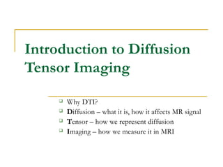
  • 1. Introduction to Diffusion Tensor Imaging  Why DTI?  Diffusion – what it is, how it affects MR signal  Tensor – how we represent diffusion  Imaging – how we measure it in MRI
  • 2.  Stroke/ischemia  Alzheimer’s Disease  Multiple Sclerosis  Brain maturation studies  Ischemia and stroke  Neoplasm  Preoperative planning  Traumatic brain injury  Congenital anomalies and diseases of white matter  Encephalopathies  Neurodegenerative diseases  Spinal Cord Injury  Epilepsy  Dementia, schizophrenia, depression  Developmental disorders  Autism  Aging Why diffusion? http://www.vh.org/Providers/Textbooks/ BrainAnatomy/Ch5Text/Section18.html http://eclipse.nichd.nih.gov/nichd/DTMRI/mri/ Conceptually: in vivo histology
  • 3. Why diffusion?  Diffusion is EXTREMELY SENSITIVE to differences and changes in tissue microstructure  Myelination/Demyelination  Axon damage/loss  Inflammation/Edema  Necrosis  It is NOT a biomarker of white matter integrity  It is NOT just about white matter  Gray matter  Cardiac tissue
  • 4. Example DTI image  “Fractional Anisotropy” map  “map” is a computed parameter, unlike an “image” which is acquired signal  Also called a “tractogram” since it clearly shows major white matter fiber tracts
  • 5. What is Diffusion?  stochastic movement of particles in a solvent, driven by the thermal molecular motion of the solvent…  … and also applies to motion of the solvent itself (Einstein, 1905) time τ∆ NOTE: In the limit N→∞, use the Central Limit Theorem to assume “step size” ∆ is fixed and equal to the average of individual displacements ∆i.
  • 6. 1D Fick’s Law - what the flux? t = t0 + τ x t = t0 0 2x − ∆ 0x − ∆ 0x + ∆0x 0 2x + ∆ t = t0 + τ x t = t0 0 2x − ∆ 0x − ∆ 0x + ∆0x 0 2x + ∆ What is the flux (J) through x0 after one time interval τ ? C1(x)C2(x) dx dC J τ 2 2 1 ∆ −= Adolf Fick, 1855: Flux is proportional to the particle concentration gradient (conservation of mass)
  • 7. The Diffusion Coefficient  3D Fick’s Law  Note the minus sign: flux goes from high to low concentration  del operator replaces partial derivative  factor of 6, not 2 (why?)  D is the diffusion coefficient  This is the expression for isotropic diffusion τ62 ∆=D CDJ ∇−=  CJ ∇ ∆ −= τ6 2
  • 8. Isotropic Diffusion (water) water ink Dtr 6= r1 t1 r2 t2 τ62 ∆=D
  • 9. Diffusion in Tissue (Anisotropic) t ink r2 r3 r1 diffusion ellipsoid tDr 11 2= tDr 22 2= tDr 33 2= x y z laboratory frame DON’T try this at lab!!!!!
  • 11. Tensor Invariants  Eigenvalues: diagonalization (iterative QR factorization)  Eigenvectors xx xy xz xy yy yz xz yz zz D D D D D D D D D           { }1 2 3D D D { }321 eee 
  • 12. Tensor Invariants  Shape invariants: analytical calculation directly from tensor coeffs xx xy xz xy yy yz xz yz zz D D D D D D D D D           ( )1 3 av xx yy zzD D D D= + + 1 2 2 2 2 1 3 xx yy xx zz yy zz surf xy xz yz D D D D D D D D D D + +  =    − − −  1 2 3 2 2 2 xx yy zz xx yz vol xy xz yz zz xy yy xz D D D D D D D D D D D D D  −  =  + − − 
  • 13. Scalar Anisotropy Indices ( ) avND D DADC DDDADC = ++= 321 3 1 ( ) ( ) ( ) 2 2 2 3 2 2 2 1 2 3 2 2 2 1 1 2 3 mag surf ND D D D FA DDD DDDDDD FA −= ++ −+−+− =
  • 14. FA vs. ADC FA (x 10 -4 ) 0 1000 2000 3000 4000 5000 6000 7000 8000 9000 10000 Probability 0.0000 0.0002 0.0004 0.0006 0.0008 0.0010 WM GM CSF MD (x 10 -3 mm2 /s) 0 500 1000 1500 2000 2500 3000 3500 4000 Probability 0.000 0.001 0.002 0.003 0.004 0.005 WM GM CSF FA and ADC are very useful clinically, but are very different. Tensor has a LOT of information! Q. Which metric would you use to detect brain cancer?
  • 15. Vector anisotropy measures  We can use eigenvector information from the tensor as well  Represent direction of primary eigenvector as color on a scalar map  Or render the primary eigenvectors as “fibers” for astonishing* 3D visualizations Red = R/L Green = A/P Blue = S/I *but how “real” is it? Many PhD theses have asked….
  • 16. Diffusion tensor coefficients Diffusion tensor invariants Scalar anisotropy indices Vector anisotropy indices
  • 17. Effect of Diffusion on MRI signal Signal attenuation! Diffusion term
  • 18. Diffusion weighted MRI ∆ δ G G echoπα ( ) ( )( )2 0 δexpδ 3 M G D M γ= − × ∆ − × ∆ δ ( ) ( )2 δδ 3 b Gγ= × ∆ − (boxcar gradients) “b-value” Consider simplified diffusion experiment…
  • 19. MR Measurement of Diffusion Tensor j T j j j p G G q r     = ×      ur 0 xx xy xz j j j j xy yy yz j xz yz zz j D D D p p q r D D D q D D D r j b S S e          − × × ×             = ( ) 22 2 γ δ Δ δ 3b G= − ( ) 1 6 j N N = ≥ Kjth diffusion- weighted image Diffusion magnitude Diffusion direction Gz Gy Gx ... ... ...
  • 20. Solving for D 20 0 xx xy xz j j j j xy yy yz j xz yz zz j D D D p p q r D D D q D D D r j b S S e          − × × ×             = 1. Acquire T2W image (b = 0 s/mm2 ) 3. Choose a diffusion gradient orientation2. Choose a b-value 4. Acquire image (Sj) 5. Repeat steps 1 – 3, j = 1 … N times 6. Solve for D…. How?
  • 21. Let’s do some linear algebra… [ ]           ⋅           ⋅⋅−= zj yj xj zzzyzx yzyyyx xzxyxx zjyjxjj DDD DDD DDD bSS α α α αααexp0 ( ) ( ) ( ) ( ) ( ) ( )                     ⋅                     ⋅= yz xz xy xx yy xx T jxy jxy jxy jzz jyy jxx j D D D D D D b S S α α α α α α 2 2 2 ln 2 2 2 0 1661 xNxNx xAY ⋅=
  • 22. B-matrix formalism 22 ( )yzyzxzxzxyxyzzzzyyyyxxxx DDDDDDb αααααα 222222 +++++⋅ ( )yzyzxzxzxyxyzzzzyyyyxxxx DbDbDbDbDbDb 222 +++++= ∑∑= = = 3 1 3 1i j ijij Db The “b-matrix” The b-matrix formalism summarizes total attenuating effect of all gradient waveforms in all directions (including imaging gradients)
  • 23. T2W (b = 0 s/mm2 ) Y, -ZY, Z-X, Y X, Y-X, Z+X, Z
  • 24. 24 SVD DIAG T2W (b = 0 s/mm2 ) … DWI (j = 1, 2, 3 … N) Dij
  • 25. N=27 N=55 N=13N=6 N NEX # DWI 6 8 56 13 4 56 27 2 56 55 1 56 #DWI = (N + 1) x NEX If TR = 4 sec, then acq time = 56*4sec = 3.7 minutes Tradeoff: N vs NEX
  • 26. Rotational invariance Hasan et al, JMRI 2001 Jones MRM 2004
  • 27. 27 Empirical Image Quality increasing N, decreasing NEX increasingb-value
  • 28. How low can you go?  High b-values mean more attenuation, lower SNR  Lower b-values mean higher SNR, room for more N  At very low b-values, imaging gradients’ diffusion effects are no longer negligible  Lower b-values also do not probe same diffusion scale, less clinically interesting b=100 s/mm2 b=500 s/mm2 (N = 6, 8 NEX)
  • 29. Echo-Planar Imaging (EPI)  Advantages  Minimal motion artifacts  NEX  N  Disadvantages  Eddy current artifacts  T2* limits spatial resolution  Geometric distortion (susceptibility) 29
  • 30. DT-MRI Alexander SLFSLF CRCR CCCC CINGCING Partial Volume Effects on Anisotropy
  • 31. DT-MRI Alexander Mapping Complex Diffusion Based Upon Q-Space Theory – Model Independent  ODF – orientation density function (Tuch et al., Neuron 2003)  Diffusion Spectrum Imaging (DSI) (Tuch et al. Neuron 2003, Wedeen et al. 2005)  High Angular Diffusion Imaging (HARDI), Q-Ball (Frank 2002; Tuch et al. Neuron 2003)

Hinweis der Redaktion

  1. Before I go over the diffusion enhancements, let me give a bit of background on diffusion imaging and why it is desirable Heres a highly invasive technique for imaging the corpus callosum And heres diffusion fiber tractography of the structure
  2. The particles will move forward or backwards with equal probability, so half the particles undergo displacement -D and the other half undergo displacement +D . After a single time interval tau we only need consider particles within distance D of a given point x0 to calculate the flux half are displaced left and the other half are displaced right. Therefore, only half the particles between x0 and x0  D will cross x0 in one step.
  3. THIS IS ONLY A CARTOON! demonstrate concept injecting ink not part of methods and materials if we repeat thought experiment in brain tissue, diffusing molecules will encounter obstructions from tissue microstructure (unlike free water) isotropic: degenerate case uynder this formalism where D1 = D2 = D3 asymmetric diffusion called “anisotropic”, Diffusion is strongly anisotropic in WMFT NOTE: - ellipsoid true in limit of infinitesimal point - oblique diffusion has components along three axes
  4. we actually measure directly in DT-MRI : diffusion tensor coefficients, averaged over entire voxel. (volume element) Diagonalize -> reduce to intrinsic reference frame of ellipsoid (lose orientation)
  5. The shape invariants have the advantage that they can be constructed analytically from the tensor coefficients, unlike the eigenvalues which are usually obtained using an iterative computation.
  6. The shape invariants have the advantage that they can be constructed analytically from the tensor coefficients, unlike the eigenvalues which are usually obtained using an iterative computation.
  7. to visualize-> construct vector (2d) and scalar (1d) indices to characterize the anisotropy of tissue in that voxel focus on scalar indices in this research RA : VISUALIZATION gold standard, linear, 0-sqrt(2) FA : more sensitive to low aniso, high aniso saturates (0-1) ND/D definitions are equivalent - index VALUE is the same --- EXTRA INFO -- - vector indices sensitive to noise, therefore formidable acquisition requirements (time, hardware). - don’t need vectors : construct scalar indices from any set of tensor invariants
  8. MR signal is attenuated by diffusion effects: apply gradient For every diffusion-weighted gradient we apply, we obtain one such equation. 6 unknowns in the diffusion tensor (due to symmetry), need at least N = 6 diffusion weighted gradients. more than 6 gradients -> use least-squares method like SVD to find the best fit -- EXTRA info -- [ typically maximize Gd for shortest TE] static spins see no net dephasing - 1800 pulse reverses polarity of first gradient. Diffusing spins, undergo net dephasing - experience different local magnetic susceptibility as sensitize any MR pulse sequence by inserting diffusion-weighting magnetic field gradients
  9. example of data set in Experiment 1 - N = 6, b = 500 s/mm2 diffusion-weighted images are attenuated from T2 baseline signal varies with orientation of the gradient
  10. fiber tracts’ orientation not known a-priori equal weighting of anisotropy in all directions needed for max sensitivity -> even spacing of diffusion-weighting gradients (“acquisition scheme”) for this project. we developed acquisition schemes for N = 6, 13, 27, and 55 [ shown from same angle for consistency ] N = 6: standard isocahedral encoding [X, Z], [Y, Z], and [X, Y] N = 27, 55: Perl algorithm tiling unit hemisphere by equal solid angle disks uses circular tiles as an approximation, cannot cover the surface precisely algorithm performs poorly for N < 27 N = 13: modeled by electrostatic repulsion each gradient treated as point charge on surface of hemisphere iterative computation - solve for lowest energy configuration
  11. all images are RA, gold standard commonly used
  12. experiment 14 (N = 6) exp. 15 - similar results < 500 are worse, interference with imaging gradients