Spinal Dural Arteriovenous Fistula Case
•
1 gefällt mir•364 views

Case record...Spinal dural arteriovenous fistula withn congestive myelopathy http://yassermetwally.com http://yassermetwally.net

Empfohlen
Empfohlen
Weitere ähnliche Inhalte
Was ist angesagt?
Was ist angesagt? (20)
Andere mochten auch
Andere mochten auch (20)
Ähnlich wie Spinal Dural Arteriovenous Fistula Case
Ähnlich wie Spinal Dural Arteriovenous Fistula Case (20)
Mehr von Professor Yasser Metwally
Mehr von Professor Yasser Metwally (20)
Kürzlich hochgeladen
Kürzlich hochgeladen (20)
Spinal Dural Arteriovenous Fistula Case
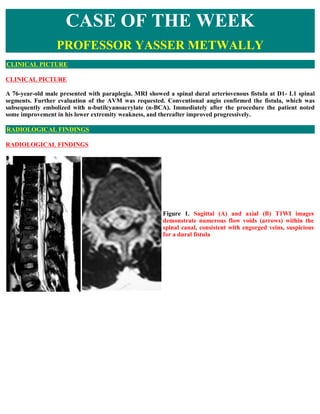
- 1. CASE OF THE WEEK PROFESSOR YASSER METWALLY CLINICAL PICTURE CLINICAL PICTURE A 76-year-old male presented with paraplegia. MRI showed a spinal dural arteriovenous fistula at D1- L1 spinal segments. Further evaluation of the AVM was requested. Conventional angio confirmed the fistula, which was subsequently embolized with n-butilcyanoacrylate (n-BCA). Immediately after the procedure the patient noted some improvement in his lower extremity weakness, and thereafter improved progressively. RADIOLOGICAL FINDINGS RADIOLOGICAL FINDINGS Figure 1. Sagittal (A) and axial (B) T1WI images demonstrate numerous flow voids (arrows) within the spinal canal, consistent with engorged veins, suspicious for a dural fistula
- 2. Figure 2. Central cord T2 hyperintensity representing cord edema. Figure 3. Sagittal (A) and coronal (B) MRA images show enlarged venous channels at the T10-T11 levels, confirming a dural fistula.
- 3. Figure 4. Conventional Angio: a Simmons 2 catheter was used to catheterize the right sided T10 posterior intercostal artery. The fistula and enlarged venous channels are well demonstrated. B, A microcatheter (arrow) was coaxially advanced into the segmental branch, adjacent to the dural fistula, 25% n-BCA (Glue) was injected and the fistula was embolized. The fistula and enlarged venous channels are no longer seen. DIAGNOSIS: DIAGNOSIS: DURAL SPINAL ARTERIOVENOUS FISTULA DISCUSSION DISCUSSION Spinal Dural AVFs, most common type of spinal vascular malformation constituting 80% of spinal AVMs [1]. There are three basic types of AVFs - extradural, dorsal intradural, and ventral intradural [3]. Dorsal intradural AVFs have been given a number of alternate names including long dorsal, angioma racemosum, dorsal extramedullary, angioma racemosum venosum, and Type I. The current literature supports use of term Type I [3]. Type I is further classified into subtypes A and B to designate AVMs with single or multiple feeding arteries, respectively [3]. These dural AVFs are malformations that most commonly occur in the thoracic region [1]. The dural AVFs consist of an abnormal communication between a radicular artery and a radicular vein within the dural sleeve of a nerve root [1]. The vein communicates with the coronal venous plexus along the surface of the spinal cord. The clinical manifestation of patients with this miscommunication is the insidious development of venous hypertension induced-progressive myelopathy [1]. Venous congestion produces progressive neurological deterioration that can manifest as sensory disturbances, paraparesis, and sphincter dysfunction [2,3]. The pathognomonic MR imaging studies is a pencil-like hyperintensity in the spinal cord on a T2-weighted image [2]. Aminoff and Logue detailed the clinical prognosis of 60 cases of dural AVFs [3]. Seven patients with acute onset symptoms had no progression of neurological dysfunction. The 53 other patients had symptom progression with acute neurological episodes, and lower extremity impairment. Within three years of developing impairment the
- 4. patients became severely disabled [3]. This and similar outcomes has led to the rule that elimination of dural AVFs will halt the progressive neurological deterioration [1,3]. MR imaging findings of Type I AVMs include enlargement of the spinal cord, cord hypointensity on Tl-weighted images, central cord hyperintensity on T2-weighted images, scalloping of the surface of the cord on sagittal images, serpentine flow voids, and enhancement of the cord and dilated perimedullary veins after administration of gadopentetate dimeglumine. Hyperintensity of the cord on T2-weighted images is believed to be the most sensitive finding. In a review of 91 spinal vascular malformations, which included 31 Type I AVMs visualized by MR imaging, only increased hyperintensity in the center of the cord on T2-weighted images was seen in all cases. [6] Increased T2 signal intensity is thought to result from cord edema. Isu et al, 50 studied two patients with Type I AVMs in the lumbar spine with MR imaging and angiography. They noted that the intramedullary high-signal- intensity changes on T2-weighted images corresponded with the level at which delay was seen in the venous drainage on spinal angiography. In both cases, the intramedullary high-intensity T2 signal disappeared after therapy, leading them to conclude that these MR imaging findings are caused by edema of the cord and are reversible. [7] Although high signal intensity on T2-weighted images is the most sensitive sign, it is nonspecific and can be seen in inflammatory, neoplastic, degenerative, demyelinating, and posttraumatic conditions. In the study by Gilbertson et al, no patient with a dural fistula had increased T2 signal abnormality as the only finding. [6] The eliminations of dural AVFs can be performed through endovascular or surgical therapy. Endovascular therapy consists of embolization with liquid acrylic embolic material into the AVF and/or the proximal draining vein [1]. Surgical therapy involves microsurgical ligation of AVF draining vein and excision of affected dura1. Literature has supported the use of endovascular therapy as initial treatment with awareness of a 39% failure rate that should be followed up with definitive surgical therapy [1,5,8]. Addendum A new version of case record of the week publication is uploaded in my web site every week (every Saturday and remains available till Friday.) To download the current PDF version of case record of the week publication follow the link "http://pdf.yassermetwally.com/case.pdf". To download the current software version of case record of the week publication (crow.exe) follow the link: http://neurology.yassermetwally.com/crow.zip You can also download the current version from my web site at "http://yassermetwally.com". The case is also presented as a short case in PDF format, to download the short case follow the link: http://pdf.yassermetwally.com/short.pdf At the end of each year, all the publications are compiled on a single CD-ROM, please contact the author to know more details. Screen resolution is better set at 1024*768 pixel screen area for optimum display Click here for an archive of the previously reported cases in downloadable PDF files. For an archive of the previously reported cases go to www.yassermetwally.net, then under pages in the right
- 5. panel, scroll down and click on the text entry "Downloadable case records in PDF format" and "Downloadable short cases in PDF format" REFERENCES References 1. Eskandar EN, Borges LF, et al. Spinal dural arteriovenous fistulas: Experience with endovascular and surgical therapy. J. Neurosurg (Spine 2) 96: 162-167, 2002. 2. Kataoka H, Miyamoto S, et al. Venous congestion is a major cause of neurological deterioration in spinal arteriovenous malformations. 3. Neurosurgery 48:1224-1230, 2001. 4. Spetzler RF, Detwiler PW, et al. Modified classification of spinal cord vascular lesions. J. Neurosurg (Spine 2) 96:145-156, 2002. 5. Doaa Abdulla: Venous disorders of the CNS. MS thesis, Ain Shams university school of medicine, Cairo, Egypt, 2005 6. Gilbertson JR, Miller GM, Goldman MS, et al: Spinal dural arteriovenous fistulas: MR and myelographic findings. AJNR Am j Neuroradiol 16:2049-2057,1995 7. Isu T, Iwasaki Y, Akino M, et al: Magnetic resonance imaging in cases of spinal dural arteriovenous malformation. Neurosurgery 24:919-923,1989 8. Metwally, MYM: Textbook of Neuroimaging, a CD-ROM based publication in Metwally MYM (Ed), WEB- CD agency for electronic publishing, version 8.4a October 2007