
Empfohlen
Weitere ähnliche Inhalte
Was ist angesagt?
Was ist angesagt? (20)
Ähnlich wie Mastigomycotina
Ähnlich wie Mastigomycotina (20)
Mehr von vaishalidandge3
Mehr von vaishalidandge3 (20)
Kürzlich hochgeladen
Kürzlich hochgeladen (20)
Mastigomycotina
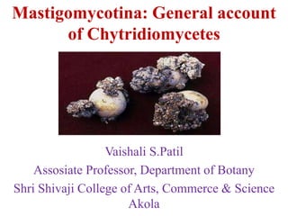
- 1. Mastigomycotina: General account of Chytridiomycetes Vaishali S.Patil Assosiate Professor, Department of Botany Shri Shivaji College of Arts, Commerce & Science Akola
- 2. General account of Mastigomycotina: • Mastigomycotina is former polyphyletic taxonomic grouping, a subdivision, of fungi, similar to Phycomycetes, and that includedthe zoosporic classes Chytridiomycetes, Hyphochytrio mycetes, Plasmodiophoromycetes and Oomycetes. General features of Mastigomycotina: • They produce flagellated cells during their lifetime. • May bear rhizoids. • Mostly, filamentous and having coenocytic mycelium. • Show centric nuclear division. • Perfect state of spores is typically oospores.
- 3. General account of Chytridiomycetes- • The members of the class Chytridiomycetes, commonly called chytrids, are mostly aquatic, but a few species occur on the soil as saprophytes and some as parasites on many land plants. The aquatic habitats including peat, bogs, rivers, ponds, springs, and ditches, and terrestrial habitats such as acidic soils, alkaline soils, temperate forest soils, rainforest soils, arctic and Antarctic soils. • Most of the members are unicellular, but some advanced taxa form short chains of cells which are attached to the substratum with the help of rhizoids. Some forms possess undeveloped mycelium. Chitin and glycan are the main constituents of the cell wall. •In unicellular forms the thallus is holocarpic (whole vegetative thallus transforms into one or more reproductive
- 4. structures), whereas in filamentous forms it is eucarpic (some part of the vegetative thallus transforms into reproductive structure, while the rest remains vegetative). •The members of the class may be epibiotic (reproductive bodies present on the host’s surface) or endobiotic (live completely within the cells of the host) and monocentric(having only a single reproductive structure) or polycentric (having more than one reproductive structures). •The thallus is coenocytic but sex organs are separated from vegetative part by a septum. • Asexual reproduction takes place with the help of zoospores which are posteriorly uniflagellate. The
- 5. flagellum is of whiplash type. Zoosporangia are spherical or pear-shaped and inoperculate or operculate. The zoospore with a posteriorly inserted flagellum is called opisthocont. •The flagellum is attached to the blepharoplast within the cell. The motile cells of some species possess a nuclear cap which consists of RNA. It shields the nucleus at the anterior end of the cell. Majority of the members occur in water. •Planogametes are also posteriorly uniflagellate. • The zygote is formed by the fusion of planogametes and it is transformed into a resting spore which produces zoospores on germination.
- 6. Significance of Chytridiomycetes: (i) Some of the soil-inhabiting Chytridiomycetes attack the underground as well as aerial parts of the higher plants and cause diseases which are of great economic significance. For example, Synchytrium endobioticum causes black wart disease of potato; Urophlyctis alfalfae causes crown wart of alfalfa (Medicago); and Physoderma maydis causes brown spot disease of maize (Zea mays). (ii) Many chytrids indirectly harm humans and animals. They parasitize and destroy the phytoplanktonic forms of algae that form an important link in food chain of aquatic ecosystems. (iii) Various species of Allomyces and Blastocladiella have been found to be valuable research tools in studying morphogenesis. (iv) Species of Coelomomyces (C. anophelescia) are endoparasites on mosquito larvae and can be utilized for the biological control of the mosquito (Anopheles spp.), which is an important vector for the spread of malaria in human beings.
- 7. Classification- It includes 10 orders, 700 species &90 genera. Chytridiales Spizellomycetales Cladochytriales Rhizophydiales Polychytriales Rhizophlctidales Lobulomycetales Synchytriales Gromochytriales Mesochytriales.
- 8. Life cycle of Synchytrium endobioticum- wart disease or black wart of potato It is one of the chytrid fungi which causes black scab or the wart disease of potato. Solanum are also infected by it. Synchytrium is represented by about 200 species reported from all over the world, occurs as parasite on aquatic alga, bryophytes (mosses), pteridophytes (ferns) and mostly on flowering plants. Classification Kingdom: Fungi Division: Chytridiomycota Class: Chytridiomycetes Order: Synchytriales Family: Synchytriaceae Genus: Synchytrium Species: S. Endobioticum Sexual reproduction is by junction of isogametes, resulting in the formation of thick walled resting sporangia.
- 9. Symptoms of Black Wart Disease of Potato The disease is characterised by cauliflower-like black warty growth on tubers (Fig. 4.16), stolons and stem bases (Fig. 4.15). Sometimes, the size of the warts are more than the size of the tuber. •Usually the disease affects the underground parts of the host. •Diseased potato tubers appear as brown or black cauliflower like outgrowths. •The fungus cause the enlargement of the surface cells (hypertrophy) as well as increased the numbers of cells (hyperplasia) in the infected potato tuber, converting them into useless masses of watery tissue. •Most of the host cells contain resting sporangia. •Galls or tumors may be formed on aerial parts (stems and leaves). Vegetative Structure of Synchytrium: The vegetative body of Synchytrium consists of minute endobiotic holocarpic thallus, represented by naked uniflagellate zoospore with whiplash flagellum.
- 11. Reproduction - During reproduction, the entire thallus transforms into a reproductive unit i.e., holocarpic. 1. Asexual Reproduction: Asexual reproduction generally occurs during favourable condition, i.e., in spring season. At reproduction the thallus body may be converted directly into a group (or sorus) of sporangia. During this period, minute naked uninucleate and uniflagellate zoospores (Fig. 4.17U) are released from the resting sporangium (which turns during unfavourable condition i.e., in winter season) after water soaking (Fig. 4.17T). The zoospores are capable of swimming for about two hours. After coming in contact either with the potato ‘eye’ or stolon or young tuber (Fig. 4.17B), they come to rest and withdraw their flagella (Fig. 4.17C). The content of the zoospore cyst enters into the host cell through the wall by minute pore in amoeboid movement, keeping the cyst membrane outside (Fig. 4.17D).
- 12. The protoplast of zoospore, after entry in the host epidermal cell, absorbs food and becomes spherical in shape. The infected host cell also enlarges in volume. The host cell surrounding the infected cell becomes stimulated and starts swelling (hypertrophy) resulting into the formation of tumour or wart-like structure. The infected cell dies and remains in the middle of the wart. The pathogen along with its nucleus enlarges considerably, rounds off and develops two layered walls, consisting of a thick golden brown exospore and a thin hyaline endospore. This is the summer spore (Fig. 4.17E). The summer spore germinates within the infected host cell. Before germination, the nucleus enlarges and the inner wall protrudes out through a minute pore on the outer wall and forms a vesicle towards the upper portion of the infected host cell. The total content of the summer spore is transferred to the vesicle. The nucleus of summer spore then undergoes repeated mitotic divisions and forms about 32 nuclei. This multinucleate vesicle is known as prosorus (Fig. 4.17G). The protoplast of the vesicle becomes cleaved into 4-9 segments, covered by thin hyaline wall. Each segment is known
- 13. as summer sporangium or zoosporangium (Fig. 4.1 7H). The total aggregated structure of the zoosporangia is known as Sorus (Fig. 4.1 71). The nuclei of each zoosporangium undergo repeated mitotic divisions and form generally 200 to 300 nuclei (their number may go up to 500 or more in large sporangium). The protoplast then divides into many uninucleate segments (Fig. 4.17J). The mature sporangium swells up by absorbing water and creates pressure on the host wall to burst. After bursting, the zoospores get released through a small slit on the sporangial wall. The zoospores are uninucleate and uniflagellate (Fig. 4.17A). They swim actively in water and infect again the new host or different regions of the same host. The behavior of the zoospore varies with the environmental condition. If the condition is favourable (i.e., summer continues), the zoospore causes infection to a new host or different region of the same host and continues the asexual cycle again. During unfavourable condition they behave as gamete and undergo sexual reproduction.
- 14. Sexual Reproduction: During unfavourable condition (if winter comes), the multinucleate segment of prosorus instead of behaving as zoosporangium behaves as gametangium (Fig. 4.17K) which produces many gametes (Fig. 4.1 7L), those are smaller in size than the zoospores. The gametes coming from different gametangia of a same or different sorus may fuse, but not from same gametangia of a sorus. The planogametes after union form diploid biflagellate zygote (Fig. 4.17N). The planogametes are similar in size and shape therefore, copulation is isogamous. The zygote swims for sometime and encysts on the surface of the host epidermis and penetrates the host cell by a process similar to zoospore penetration (Fig. 4.170). The surrounding host cell then undergoes hyperplasia i.e., repeated cell division. The infected cell is then buried into the deeper layer of host cells (Fig. 4.17P). The effect on the surrounding tissue varies between zoospore and zygote infection. Hypertrophy (i.e., enlargement of cells) takes place on zoospore infection, but the zygote infection causes Hyperplasia (i.e.,
- 15. repeated cell division). During this development, the zygote enlarges and becomes surrounded by two layered wall formed by itself (Fig. 4.17Q) and then a third wall is developed from the host content after its death. It is now called winter sporangium or resting sporangium (Fig. 4.14R). The resting sporangium remains dormant throughout the winter season. The resting sporangia are released into the soil after decaying the host tissue and are capable to germinate within about two months. With the onset of favourable condition i.e., in spring season, the resting sporangium becomes active and its nucleus undergoes repeated nuclear division of which first one is meiotic, followed by many mitotic divisions. The protoplast, along with a single nucleus, divides into many uninucleate segments (Fig. 4.14S). After absorbing water, the wall of resting sporangium bursts open (Fig. 4.14T) and releases the zoospores. The zoospores are like the asexual zoospores (Fig. 4.14U), which on coming in contact with a suitable host cause infection and repeat the cycle again.