
Empfohlen
Weitere ähnliche Inhalte
Was ist angesagt?
Was ist angesagt? (20)
Ähnlich wie Cutaneous receptors
Ähnlich wie Cutaneous receptors (20)
Kürzlich hochgeladen
Kürzlich hochgeladen (20)
Cutaneous receptors
- 1. CUTANEOUS RECEPTORS PRESENTOR: Dr ROHINI MODERATOR: Dr PASCHAL D’SOUZA
- 2. SHERRINGTON CLASSIFICATION OF RECEPTORS Depending upon the source of stimulus • Exteroreceptors • Interoreceptors • Proprioreceptors
- 3. Exteroreceptors • Receptors responsible for sensing stimulus coming from external environment. • Present at or near the surface of the body. • Includes: 1. Touch 2. Temperature 3. Nociception
- 4. Interoreceptors • Respond to stretch ,volume and pressure in the wall of viscera, excessive muscle contraction and to overstretching in the visceral organs. • Essential in regulating blood flow ,pressure in the CVS ,maintaining respiration etc.
- 5. Proprioceptors • Responds to stimulus arising in muscles ,tendons, joints etc. • Essential for coordination of movements and maintenance of posture. • Includes 1. Neuromuscular 2. Neurotendinous spindles
- 6. Depending upon type of stimulus energy • Mechanoreceptors 1. Cutaneous receptors 2. Muscle and joint receptors 3. Hair cells 4. Baroreceptors of carotid sinus and aortic arch • Thermoreceptors • Nociceptors: receptors responding to pain • Photorecepetors: rods and cones • Chemoreceptors
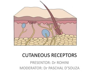
- 7. Cutaneous receptors • Sensory receptors • Located in epidermis as well as dermis of skin • Exteroreceptors :informs us about touch , temperature ,pain and pressure. • Densly present over face ,finger tips. • Includes: 1. Mechanoreceptors 2. Thermoreceptors 3. Nociceptors
- 8. Mechanoreceptors • Touch ,pressure , softness, texture of the stimulus. • Includes 1. Merkels disc and Meissners corpuscles 2. Pacinian corpuscles 3. Ruffini ‘s end organs 4. Krause end bulbs 5. Free nerve endings
- 10. Meissner's corpuscles • Meissner's corpuscles localize in the dermis between epidermal ridges. • They contain an unmyelinated nerve ending surrounded by Schwann cells. • The center of the capsule contains one or more afferent nerve fibers that generate rapidly adapting action potentials following minimal skin depression.
- 11. • Present in glabrous (smooth, hairless) skin (the fingertips) • Their afferent fibers account for about 40% of the sensory innervation of the human hand. • Efficient in transducing information about the relatively low-frequency vibrations (30–50 Hz) that occur when textured objects are moved across the skin.
- 13. Pacinian corpuscles • Large encapsulated endings located in the subcutaneous tissue . • Has an onion like capsule in which the inner core of membrane lamellae is separated from an outer lamella by a fluid-filled space. • one or more rapidly adapting afferent axons lie at the center of this structure. • The capsule again acts as a filter, allowing only transient disturbances at high frequencies (250–350 Hz) to activate the nerve endings.
- 14. • Rapidly adapting • Involved in the discrimination of fine surface textures or high- frequency vibrations. • Provide information primarily about the dynamic qualities of mechanical stimuli.
- 16. Merkel's disks • Located in the epidermis, where they are precisely aligned with the papillae that lie beneath the dermal ridges. • Account for about 25% of the mechanoreceptors of the hand and are particularly dense in the fingertips, lips, and external genitalia.
- 17. • The slowly adapting nerve fiber associated with each Merkel's disk enlarges into a saucer-shaped ending that is closely applied to another specialized cell containing vesicles that apparently release peptides that modulate the nerve terminal. • Selective stimulation of these receptors in humans produces a sensation of light pressure.
- 19. IGGO DOME RECEPTORS • Merkel's discs are often grouped together in a receptor organ called the Iggo dome receptor, which projects upward against the underside of the epithelium of the skin. • This causes the epithelium at this point to protrude outward, thus creating a dome and constituting an extremely sensitive receptor.
- 20. RUFFINI CORPUSCLES • These elongated, spindle-shaped capsular specializations are located deep in the skin, as well as in ligaments and tendons. • The long axis of the corpuscle is usually oriented parallel to the stretch lines in skin. • Particularly sensitive to the cutaneous stretching produced by digit or limb movements.
- 21. Ruffini corpuscle from original slide sent by Ruffini to Sir Charles Sherrington
- 22. HAIR FOLLICLE RECEPTOR • Unencapsulated • Primary afferent spiral around hair follicle base. • Runs parallel to the hair shaft to form a lattice like pattern. • Signals the direction and velocity of movement. • Rapidly adaptating
- 24. Krause end bulb • Type of meissners receptor • Mainly present in glabrous skin such as skin of genitalia, papillae of lips,conjunctiva etc • Afferent fibres belong to A delta group • Detects fine touch and pressure
- 26. Adaptation in cutaneous receptors • Rapid adaptation means that there is no response to sustained pressure, only to changes in pressure (either an increase or a decrease). • Slow adaptation means that the receptor continues to respond to pressure for as long as it is sustained, within some reasonable time frame. ADAPTATION RAPIDLY SLOWLY
- 28. RAPIDLY ADAPTING PACINIAN CORPUSCELS MEISSNER'S CORPUSCLES High frequency vibration , pressure Light touch SLOWLY ADAPTING MERKELS DISC RUFFINI Low frequency vibration and texture Skin stretching
- 29. Receptive fields of cutaneous receptors • Portion of the skin which, when stimulated, affects the activity or state of the receptor.
- 30. • Receptive Fields — the typically oval areas of skin along which a mechanical stimulus (e.g., touch) leads to a change in that neurons action potential firing rates. • In particular, Pacinian corpuscles have fairly large receptive fields, making them more sensitive (if you consider the goal of a mechanoreceptor to sense touch anywhere on the body) • Meissner’s corpuscles have fairly small receptive fields, making them more specific
- 32. The A-beta fibers are large, fast, myelinated afferents. The Meisnner's corpuscles and Pacinian corpuscles belong to this class. These are rapidly adapting touch afferents.
- 33. Pathways from Skin to Cortex • Two major pathways in the spinal cord: – • Medial lemniscal pathway consists of large fibers that carry proprioceptive and touch information . • Spinothalamic pathway consists of smaller fibers that carry temperature and pain information . • These cross over to the opposite side of the body and synapse in the thalamus, and then on to the Somatosensory cortex
- 35. • Signals travel from the thalamus to the somatosensory receiving area (S1) and the secondary receiving area (S2) in the parietal lobe . • Body map (homunculus) on the cortex shows more cortical space allocated to parts of the body that are responsible for detail.
- 36. THERMORECEPTORS • Thermoreceptors are free dendrite endings in skin, and thus are primary sensory organs. They have no specialized epithelial cells or supporting cells. • The nerves of skin branch from musculocutaneous nerves that arise segmentally from spinal nerves. • The pattern of nerve fibers in skin is similar to the vascular patterns—nerve fibers form a deep plexus, then ascend to a superficial, subpapillary plexus.
- 37. • The penicillate fibers are the primary nerve fibers found subepidermally in haired skin. • Rapidly adapting receptors that function in the perception of touch, temperature, pain, and itch. • Overlapping innervation,leads discrimination to be generalized. FREE NERVE ENDING Penicillate Papillary
- 38. • Papillary nerve endings are found at the orifice of a follicle and are thought to be particularly receptive to cold sensation. • Hair follicles also contain , slow-adapting receptors that respond to the bending or movement of hairs.
- 40. • A very high concentration of thermosensitive TRP (Transient Receptor Potential )ion channels are found in keratinocytes in the epidermis. • Each TRP has a unique temperature threshold of firing.
- 42. THERMORECEPTIVE NEURONS C-FIBRES 1.Heat sense neurons 2.Unmyelinated 3.Less velocity of transduction 4.Innervated epidermis 5.Fewer dendrites per neuron ,small receptive fied A DELTA FIBRES 1.Cold sense neuron 2.Myelinated 3.More velocity of transduction 4.Innervated layers between epidermis and dermis 5.High receptive field
- 44. • Cell bodies of the afferent fibers of cutaneous thermoreceptors reside in the dorsal root ganglion (DRG) or the trigeminal ganglion on the dorsal horn of the spinal cord. • The trigeminal neuron is particularily sensitive to cold due to its high expression of cold- activated TRP ion channels.
- 46. NOCICEPTORS • Mediate pain • Terminal branches of thin myelinated A delta and unmyleniated C fibres 1. Somatic nociceptors: free nerve endings in skin 2. Visceral nociceptors: not well known • Nociceptors are generally electrically silent and transmit all-or-none action potentials only when stimulated.
- 47. • Nociceptive fibers have been classified on the basis of their conduction velocity and sensitivity and threshold to noxious mechanical (M), heat (H), and cold (C) . • C fibres:(C-MH, C-MC, C-MHC) • A-fiber nociceptors are predominately heat- and or mechanosensitive (A-MH, A-H, A-M)
- 48. • Excitatory neurons and release glutamate as their primary neurotransmitter as well as other components including peptides (e.g., substance P, calcitonin gene-related peptide [CGRP], somatostatin.)