Acute Calculous Cholecystitis
•Als PPT, PDF herunterladen•
72 gefällt mir•20,011 views

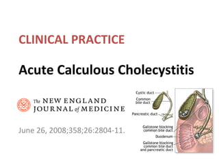
Acute Calculous Cholecystitis NEJM June 26, 2008;358;26:2804-11

Empfohlen
Weitere ähnliche Inhalte
Was ist angesagt?
Was ist angesagt? (20)
Andere mochten auch
Andere mochten auch (20)
Ähnlich wie Acute Calculous Cholecystitis
Ähnlich wie Acute Calculous Cholecystitis (20)
Mehr von Sun Yai-Cheng
Mehr von Sun Yai-Cheng (20)
Kürzlich hochgeladen
Kürzlich hochgeladen (20)
Acute Calculous Cholecystitis
- 1. Acute Calculous Cholecystitis June 26, 2008;358;26:2804-11. CLINICAL PRACTICE
- 2. Clinical Problem • Biliary colic develops in 1 to 4% annually, and acute cholecystitisdevelops in about 20% of these symptomatic patientsif they are left untreated. • Acute cholecystitis may coexist with choledocholithiasis,cholangitis, or gallstone pancreatitis.
- 3. Pathogenesis • Obstruction of the cysticduct in the presence of bile supersaturated with cholesterol. • Brief impaction may cause pain only, but if impaction is prolongedover many hours, inflammation can result. • With inflammation,the gallbladder becomes enlarged, tense, and reddened, and wall thickening and an exudate of peri-cholecystic fluid may develop.
- 4. • Enterobacteriaceae familyor with enterococci or anerobes occurs in the majority of patients. • The wall of the gallbladder may undergo necrosis and gangrene (gangrenous cholecystitis). • Bacterial super-infection with gas-forming organisms may lead to gas in the wall or lumen of the gallbladder(emphysematous cholecystitis).
- 5. Diagnosis • The main symptom of uncomplicated cholelithiasis is biliarycolic, caused by the obstruction of the gallbladder neck bya stone. • The pain is characteristically episodic, severe, andlocated in the epigastrium or RUQ. • It frequentlyfollows food intake or comes on at night. • Patients commonlyhave pain that radiates into the back, accompanied by nauseaand vomiting.
- 6. Diagnosis • Murphy's sign — the arrest of inspirationwhile palpating the gallbladder during a deep breath. • Systemic sepsis and organ failure gangrenous or emphysematous cholecystitis. • Fever, elevation in the WBC and CRP. • Elevated serum amylase level concomitant gallstonepancreatitis or gangrenous cholecystitis. • In elderly patients,delays in diagnosis are common, the only symptoms maybe a change in mental status or decreased food intake, and physicalexamination and laboratory indexes may be normal.
- 7. Imaging • Ultrasonographydetects cholelithiasis in about 98% of patients. • Acute calculous cholecystitis is diagnosed radiologically bythe concomitant presence of thickening of the gallbladder wall( >5 mm), peri-cholecystic fluid, or direct tenderness when the probe is pushed against the gallbladder (ultrasonographicMurphy's sign).
- 8. Ultrasonographic Images of Three Gallbladders. A normal, sonolucent gallbladder (Panel A) is characterized by a thin wall and an absence of acoustic shadows. In a patient with symptomatic gallstones (Panel B), the gallbladder contains small echogenic objects with posterior acoustic shadows that are typical of gallstones (arrow), with a normal wall thickness. In a patient with acute calculous cholecystitis (Panel C), thickening is visible in the gallbladder wall (arrow), along with a large gallstone (arrowhead).
- 9. Imaging • Hepatobiliary scintigraphy involves intravenous injection oftechnetium-labeled analogues of iminodiacetic acid, which areexcreted into bile. The absence of gallbladder filling within60 minutes after the administration of tracer indicates obstructionof the cystic duct and has a sensitivity of 80 to 90% for acutecholecystitis. • The "rim sign" isa blush of increased pericholecystic radioactivity, which ispresent in about 30% of patients with acute cholecystitis andin about 60% with acute gangrenous cholecystitis.
- 10. Hepatobiliary Scintigraphy In Panel A, a normal liver is visible 10 minutes after the intravenous injection of a technetium-labeled analogue of iminodiacetic acid. In Panel B, at 55 minutes after tracer injection, filling of the bile duct (arrow) and gallbladder (arrowhead) can be seen. In Panel C, at 1 hour after tracer injection in a patient with acute cholecystitis and obstruction of the cystic duct, there is filling of the bile duct (arrow) but no filling of the gallbladder.
- 13. Treatment • Timing of Cholecystectomy • Antibiotic Therapy • Percutaneous Cholecystostomy
- 14. Timing of Cholecystectomy • Cholecystectomy can be performed by laparotomy or by laparoscopy,either at the time of the initial attack (early treatment) or2 to 3 months after the initial attack has subsided (delayedtreatment). • “Early" has been variably definedas anywhere from 24 hours to 7 days after either the onset of symptoms or the time of diagnosis. • If delayed, or "conservative,"treatment is selected, patients are treated during the acutephase with antibiotics and intravenous fluids and NPO.
- 15. • Early laparoscopic cholecystectomy is considered the treatmentof choice for most patients. • The rate of conversion to open cholecystectomy is higher whenlaparoscopic cholecystectomy is performed for acute cholecystitisthan for uncomplicated cholelithiasis. • Predictors of theneed for conversion include – WBC > 18000/mm3 – duration of symptoms of more than a range of 72 to 96 hours – age over 60 years
- 16. Antibiotic Therapy • The guidelinesof the Infectious Diseases Society of America recommend that antimicrobial therapy be instituted if infection is suspectedon the basis of laboratory and clinical findings (WBC > 12500/mm3 or temperature > 38.5°C) and radiographic findings (e.g., air inthe gallbladder or gallbladder wall).
- 17. • Antibiotics coverage against microorganisms in the Enterobacteriaceae family(e.g., second- generation cephalosporin or a combination ofa quinolone and metronidazole); activity against enterococciis not required. • Antibiotics are also recommended for routineuse in patients who are elderly or have diabetes or immunodeficiencyand for prophylaxis in patients undergoing cholecystectomy toreduce septic complications even when infection is not suspected.
- 18. Percutaneous Cholecystostomy • Percutaneous cholecystostomy is often used when the patient presentswith sepsis (severe acute cholecystitis, according to the Tokyo guidelines) and in cases in which conservative treatment alonefails, especially in patients who are poor candidates for surgery.
- 19. Guidelines 1. Mild acute cholecystitis: early laparoscopic cholecystectomy is recommended. 2. Moderate acute cholecystitis: either early or delayed cholecystectomy may be selected butthat early laparoscopic cholecystectomy should be performedonly by a highly experienced surgeon and promptly terminated by conversion to open cholecystostomy if operative conditionsmake anatomical identification difficult. 3. Severe acute cholecystitis: initial conservative management with antibiotics is recommended, preferably in ahigh-acuity setting, with the use of percutaneous cholecystostomyas needed; surgery is reserved for patients in whom this treatmentfails.