Immunohistochemistry Antibody Validation Report for Anti-Caspase-8 Antibody (STJ97394)

Most upstream protease of the activation cascade of caspases responsible for the TNFRSF6/FAS mediated and TNFRSF1A induced cell death. Binding to the adapter molecule FADD recruits it to either receptor. The resulting aggregate called death-inducing signaling complex (DISC) performs CASP8 proteolytic activation. The active dimeric enzyme is then liberated from the DISC and free to activate downstream apoptotic proteases. Proteolytic fragments of the N-terminal propeptide (termed CAP3, CAP5 and CAP6) are likely retained in the DISC. Cleaves and activates CASP3, CASP4, CASP6, CASP7, CASP9 and CASP10. May participate in the GZMB apoptotic pathways. Cleaves ADPRT. Hydrolyzes the small-molecule substrate, Ac-Asp-Glu-Val-Asp-|-AMC. Likely target for the cowpox virus CRMA death inhibitory protein. Isoform 5, isoform 6, isoform 7 and isoform 8 lack the catalytic site and may interfere with the pro-apoptotic activity of the complex. / Strict requirement for Asp at position P1 and has a preferred cleavage sequence of (Leu/Asp/Val)-Glu-Thr-Asp-|-(Gly/Ser/Ala). / Inhibited by the effector protein NleF that is produced by pathogenic E.coli; this inhibits apoptosis. Anti-Caspase-8-http://www.stjohnslabs.com/caspase-8-antibody-p-99045 Join our Antibody Validation Project - http://www.stjohnslabs.com/services/antibody-validation

Empfohlen
Empfohlen
Weitere ähnliche Inhalte
Was ist angesagt?
Was ist angesagt? (20)
Andere mochten auch
Andere mochten auch (20)
Ähnlich wie Immunohistochemistry Antibody Validation Report for Anti-Caspase-8 Antibody (STJ97394)
Ähnlich wie Immunohistochemistry Antibody Validation Report for Anti-Caspase-8 Antibody (STJ97394) (20)
Mehr von St John's Laboratory Ltd
Mehr von St John's Laboratory Ltd (20)
Kürzlich hochgeladen
Kürzlich hochgeladen (20)
Immunohistochemistry Antibody Validation Report for Anti-Caspase-8 Antibody (STJ97394)
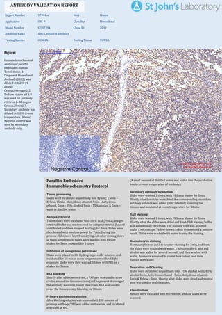
- 1. Figure: Immunohistochemical analysis of paraffin embedded Human Tonsil tissue. 1: Caspase-8 Monoclonal Antibody(2G12) was diluted at 1:200 (4 degree Celsius,overnight). 2: Sodium citrate pH 6.0 was used for antibody retrieval (>98 degree Celsius,20min). 3: Secondary antibody was diluted at 1:200 (room temperature, 30min). Negative control was used by secondary antibody only. Report Number 97394-a Host Mouse Application IHC-P Clonality Monoclonal Model Number STJ97394 Clone ID 2G12 Antibody Name Anti-Caspase-8 antibody Testing Species HUMAN Testing Tissue TONSIL ANTIBODY VALIDATION REPORT b. (A small amount of distilled water was added into the incubation box to prevent evaporation of antibody). 51. Secondary antibody incubation a. Slides were washed 3 times, with PBS on a shaker for 5min. Shortly after the slides were dried the corresponding secondary antibody solution was added (HRP labelled), covering the tissues, and incubated at room temperature for 30min. b. 52. DAB staining a. Slides were washed 3 times, with PBS on a shaker for 5min. b. Shortly after, the slides were dried and fresh DAB staining buffer was added inside the circles. The staining time was adjusted under a microscope. Yellow-brown colour represented a positive result. Slides were washed with water to stop the staining. c. 53. Haematoxylin staining a. Haematoxylin was used to counter-staining for 1min, and then the slides were washed with water. 1% Hydrochloric acid and alcohol was added for several seconds and then washed with water. Ammonia was used to reveal blue colour, and then flushed with water. b. 54. Desolation and Clearing i. Slides were incubated sequentially into: 75% alcohol 5min, 85% alcohol 5min, Anhydrous ethanol - 5min, Anhydrous ethanol - 5min & Xylene - 5min. Shortly after slides were dried and neutral gum was used to seal the slides. ii. 55. Visualization a. Results were validated with microscope, and the slides were scanned. Paraffin-Embedded Immunohistochemistry Protocol 45. 46. Tissue processing a. Slides were incubated sequentially into Xylene; 15min – Xylene, 15min - Anhydrous ethanol, 5min - Anhydrous ethanol, 5min - 85% alcohol, 5min - 75% alcohol & 5min – wash in distilled water. b. 47. Antigen retrieval a. Tissue slides were incubated with citric acid (PH6.0) antigen retrieval buffer and microwaved for antigen retrieval (heated until boiled and then stopped heating) for 8min. Slides were then heated with medium power for 7min. During this process slides were kept from drying out. After cooling down at room temperature, slides were washed with PBS on shaker for 5min, repeated for 3 times. b. 48. Inhibition of endogenous peroxidase a. Slides were placed in 3% Hydrogen peroxide solution, and incubated for 10 min at room temperature without light exposure. Slides were then washed 3 times with PBS on a shaker for 5mins. b. 49. BSA Blocking a. Shortly after slides were dried, a PAP pen was used to draw circles around the tissue sections (and to prevent draining of the antibody solution). Inside the circles, BSA was used to cover the tissue evenly, blocking for 30min. b. 50. Primary antibody incubation After blocking solution was removed a 1:200 solution of primary antibody/PBS was added on the slide, and incubated overnight at 4°C. St John's Laboratory Ltd. www.stjohnslabs.com
- 2. Figure: Immunohistochemical analysis of paraffin embedded Human colon tissue. 1: Caspase-8 Monoclonal Antibody(2G12) was diluted at 1:200 (4 degree Celsius,overnight). 2: Sodium citrate pH 6.0 was used for antibody retrieval (>98 degree Celsius,20min). 3: Secondary antibody was diluted at 1:200 (room temperature, 30min). Negative control was used by secondary antibody only. Report Number 97394-b Host Mouse Application IHC-P Clonality Monoclonal Model Number STJ97394 Clone ID 2G12 Antibody Name Anti-Caspase-8 antibody Testing Species HUMAN Testing Tissue COLON ANTIBODY VALIDATION REPORT b. (A small amount of distilled water was added into the incubation box to prevent evaporation of antibody). 40. Secondary antibody incubation a. Slides were washed 3 times, with PBS on a shaker for 5min. Shortly after the slides were dried the corresponding secondary antibody solution was added (HRP labelled), covering the tissues, and incubated at room temperature for 30min. b. 41. DAB staining a. Slides were washed 3 times, with PBS on a shaker for 5min. b. Shortly after, the slides were dried and fresh DAB staining buffer was added inside the circles. The staining time was adjusted under a microscope. Yellow-brown colour represented a positive result. Slides were washed with water to stop the staining. c. 42. Haematoxylin staining a. Haematoxylin was used to counter-staining for 1min, and then the slides were washed with water. 1% Hydrochloric acid and alcohol was added for several seconds and then washed with water. Ammonia was used to reveal blue colour, and then flushed with water. b. 43. Desolation and Clearing i. Slides were incubated sequentially into: 75% alcohol 5min, 85% alcohol 5min, Anhydrous ethanol - 5min, Anhydrous ethanol - 5min & Xylene - 5min. Shortly after slides were dried and neutral gum was used to seal the slides. ii. 44. Visualization a. Results were validated with microscope, and the slides were scanned. Paraffin-Embedded Immunohistochemistry Protocol 34. 35. Tissue processing a. Slides were incubated sequentially into Xylene; 15min – Xylene, 15min - Anhydrous ethanol, 5min - Anhydrous ethanol, 5min - 85% alcohol, 5min - 75% alcohol & 5min – wash in distilled water. b. 36. Antigen retrieval a. Tissue slides were incubated with citric acid (PH6.0) antigen retrieval buffer and microwaved for antigen retrieval (heated until boiled and then stopped heating) for 8min. Slides were then heated with medium power for 7min. During this process slides were kept from drying out. After cooling down at room temperature, slides were washed with PBS on shaker for 5min, repeated for 3 times. b. 37. Inhibition of endogenous peroxidase a. Slides were placed in 3% Hydrogen peroxide solution, and incubated for 10 min at room temperature without light exposure. Slides were then washed 3 times with PBS on a shaker for 5mins. b. 38. BSA Blocking a. Shortly after slides were dried, a PAP pen was used to draw circles around the tissue sections (and to prevent draining of the antibody solution). Inside the circles, BSA was used to cover the tissue evenly, blocking for 30min. b. 39. Primary antibody incubation After blocking solution was removed a 1:200 solution of primary antibody/PBS was added on the slide, and incubated overnight at 4°C. St John's Laboratory Ltd. www.stjohnslabs.com
- 3. Figure: Immunohistochemical analysis of paraffin embedded Human liver tissue. 1: Caspase-8 Monoclonal Antibody(2G12) was diluted at 1:200 (4 degree Celsius,overnight). 2: Sodium citrate pH 6.0 was used for antibody retrieval (>98 degree Celsius,20min). 3: Secondary antibody was diluted at 1:200 (room temperature, 30min). Negative control was used by secondary antibody only. Report Number 97394-c Host Mouse Application IHC-P Clonality Monoclonal Model Number STJ97394 Clone ID 2G12 Antibody Name Anti-Caspase-8 antibody Testing Species HUMAN Testing Tissue LIVER ANTIBODY VALIDATION REPORT b. (A small amount of distilled water was added into the incubation box to prevent evaporation of antibody). 29. Secondary antibody incubation a. Slides were washed 3 times, with PBS on a shaker for 5min. Shortly after the slides were dried the corresponding secondary antibody solution was added (HRP labelled), covering the tissues, and incubated at room temperature for 30min. b. 30. DAB staining a. Slides were washed 3 times, with PBS on a shaker for 5min. b. Shortly after, the slides were dried and fresh DAB staining buffer was added inside the circles. The staining time was adjusted under a microscope. Yellow-brown colour represented a positive result. Slides were washed with water to stop the staining. c. 31. Haematoxylin staining a. Haematoxylin was used to counter-staining for 1min, and then the slides were washed with water. 1% Hydrochloric acid and alcohol was added for several seconds and then washed with water. Ammonia was used to reveal blue colour, and then flushed with water. b. 32. Desolation and Clearing i. Slides were incubated sequentially into: 75% alcohol 5min, 85% alcohol 5min, Anhydrous ethanol - 5min, Anhydrous ethanol - 5min & Xylene - 5min. Shortly after slides were dried and neutral gum was used to seal the slides. ii. 33. Visualization a. Results were validated with microscope, and the slides were scanned. Paraffin-Embedded Immunohistochemistry Protocol 23. 24. Tissue processing a. Slides were incubated sequentially into Xylene; 15min – Xylene, 15min - Anhydrous ethanol, 5min - Anhydrous ethanol, 5min - 85% alcohol, 5min - 75% alcohol & 5min – wash in distilled water. b. 25. Antigen retrieval a. Tissue slides were incubated with citric acid (PH6.0) antigen retrieval buffer and microwaved for antigen retrieval (heated until boiled and then stopped heating) for 8min. Slides were then heated with medium power for 7min. During this process slides were kept from drying out. After cooling down at room temperature, slides were washed with PBS on shaker for 5min, repeated for 3 times. b. 26. Inhibition of endogenous peroxidase a. Slides were placed in 3% Hydrogen peroxide solution, and incubated for 10 min at room temperature without light exposure. Slides were then washed 3 times with PBS on a shaker for 5mins. b. 27. BSA Blocking a. Shortly after slides were dried, a PAP pen was used to draw circles around the tissue sections (and to prevent draining of the antibody solution). Inside the circles, BSA was used to cover the tissue evenly, blocking for 30min. b. 28. Primary antibody incubation After blocking solution was removed a 1:200 solution of primary antibody/PBS was added on the slide, and incubated overnight at 4°C. St John's Laboratory Ltd. www.stjohnslabs.com
- 4. Figure: Immunohistochemical analysis of paraffin embedded Human liver cancer tissue. 1: Caspase-8 Monoclonal Antibody(2G12) was diluted at 1:200 (4 degree Celsius,overnight). 2: Sodium citrate pH 6.0 was used for antibody retrieval (>98 degree Celsius,20min). 3: Secondary antibody was diluted at 1:200 (room temperature, 30min). Negative control was used by secondary antibody only. Report Number 97394-d Host Mouse Application IHC-P Clonality Monoclonal Model Number STJ97394 Clone ID 2G12 Antibody Name Anti-Caspase-8 antibody Testing Species HUMAN Testing Tissue LIVER CANCER ANTIBODY VALIDATION REPORT b. (A small amount of distilled water was added into the incubation box to prevent evaporation of antibody). 18. Secondary antibody incubation a. Slides were washed 3 times, with PBS on a shaker for 5min. Shortly after the slides were dried the corresponding secondary antibody solution was added (HRP labelled), covering the tissues, and incubated at room temperature for 30min. b. 19. DAB staining a. Slides were washed 3 times, with PBS on a shaker for 5min. b. Shortly after, the slides were dried and fresh DAB staining buffer was added inside the circles. The staining time was adjusted under a microscope. Yellow-brown colour represented a positive result. Slides were washed with water to stop the staining. c. 20. Haematoxylin staining a. Haematoxylin was used to counter-staining for 1min, and then the slides were washed with water. 1% Hydrochloric acid and alcohol was added for several seconds and then washed with water. Ammonia was used to reveal blue colour, and then flushed with water. b. 21. Desolation and Clearing i. Slides were incubated sequentially into: 75% alcohol 5min, 85% alcohol 5min, Anhydrous ethanol - 5min, Anhydrous ethanol - 5min & Xylene - 5min. Shortly after slides were dried and neutral gum was used to seal the slides. ii. 22. Visualization a. Results were validated with microscope, and the slides were scanned. Paraffin-Embedded Immunohistochemistry Protocol 12. 13. Tissue processing a. Slides were incubated sequentially into Xylene; 15min – Xylene, 15min - Anhydrous ethanol, 5min - Anhydrous ethanol, 5min - 85% alcohol, 5min - 75% alcohol & 5min – wash in distilled water. b. 14. Antigen retrieval a. Tissue slides were incubated with citric acid (PH6.0) antigen retrieval buffer and microwaved for antigen retrieval (heated until boiled and then stopped heating) for 8min. Slides were then heated with medium power for 7min. During this process slides were kept from drying out. After cooling down at room temperature, slides were washed with PBS on shaker for 5min, repeated for 3 times. b. 15. Inhibition of endogenous peroxidase a. Slides were placed in 3% Hydrogen peroxide solution, and incubated for 10 min at room temperature without light exposure. Slides were then washed 3 times with PBS on a shaker for 5mins. b. 16. BSA Blocking a. Shortly after slides were dried, a PAP pen was used to draw circles around the tissue sections (and to prevent draining of the antibody solution). Inside the circles, BSA was used to cover the tissue evenly, blocking for 30min. b. 17. Primary antibody incubation After blocking solution was removed a 1:200 solution of primary antibody/PBS was added on the slide, and incubated overnight at 4°C. St John's Laboratory Ltd. www.stjohnslabs.com
- 5. Figure: Immunohistochemical analysis of paraffin embedded Human lung cancer tissue. 1: Caspase-8 Monoclonal Antibody(2G12) was diluted at 1:200 (4 degree Celsius,overnight). 2: Sodium citrate pH 6.0 was used for antibody retrieval (>98 degree Celsius,20min). 3: Secondary antibody was diluted at 1:200 (room temperature, 30min). Negative control was used by secondary antibody only. Report Number 97394-e Host Mouse Application IHC-P Clonality Monoclonal Model Number STJ97394 Clone ID 2G12 Antibody Name Anti-Caspase-8 antibody Testing Species HUMAN Testing Tissue LUNG CANCER ANTIBODY VALIDATION REPORT b. (A small amount of distilled water was added into the incubation box to prevent evaporation of antibody). 7. Secondary antibody incubation a. Slides were washed 3 times, with PBS on a shaker for 5min. Shortly after the slides were dried the corresponding secondary antibody solution was added (HRP labelled), covering the tissues, and incubated at room temperature for 30min. b. 8. DAB staining a. Slides were washed 3 times, with PBS on a shaker for 5min. b. Shortly after, the slides were dried and fresh DAB staining buffer was added inside the circles. The staining time was adjusted under a microscope. Yellow-brown colour represented a positive result. Slides were washed with water to stop the staining. c. 9. Haematoxylin staining a. Haematoxylin was used to counter-staining for 1min, and then the slides were washed with water. 1% Hydrochloric acid and alcohol was added for several seconds and then washed with water. Ammonia was used to reveal blue colour, and then flushed with water. b. 10. Desolation and Clearing i. Slides were incubated sequentially into: 75% alcohol 5min, 85% alcohol 5min, Anhydrous ethanol - 5min, Anhydrous ethanol - 5min & Xylene - 5min. Shortly after slides were dried and neutral gum was used to seal the slides. ii. 11. Visualization a. Results were validated with microscope, and the slides were scanned. Paraffin-Embedded Immunohistochemistry Protocol 1. 2. Tissue processing a. Slides were incubated sequentially into Xylene; 15min – Xylene, 15min - Anhydrous ethanol, 5min - Anhydrous ethanol, 5min - 85% alcohol, 5min - 75% alcohol & 5min – wash in distilled water. b. 3. Antigen retrieval a. Tissue slides were incubated with citric acid (PH6.0) antigen retrieval buffer and microwaved for antigen retrieval (heated until boiled and then stopped heating) for 8min. Slides were then heated with medium power for 7min. During this process slides were kept from drying out. After cooling down at room temperature, slides were washed with PBS on shaker for 5min, repeated for 3 times. b. 4. Inhibition of endogenous peroxidase a. Slides were placed in 3% Hydrogen peroxide solution, and incubated for 10 min at room temperature without light exposure. Slides were then washed 3 times with PBS on a shaker for 5mins. b. 5. BSA Blocking a. Shortly after slides were dried, a PAP pen was used to draw circles around the tissue sections (and to prevent draining of the antibody solution). Inside the circles, BSA was used to cover the tissue evenly, blocking for 30min. b. 6. Primary antibody incubation After blocking solution was removed a 1:200 solution of primary antibody/PBS was added on the slide, and incubated overnight at 4°C. St John's Laboratory Ltd. www.stjohnslabs.com
- 6. Figure: Immunohistochemical analysis of paraffin embedded Human stomach cancer tissue. 1: Caspase-8 Monoclonal Antibody(2G12) was diluted at 1:200 (4 degree Celsius,overnight). 2: Sodium citrate pH 6.0 was used for antibody retrieval (>98 degree Celsius,20min). 3: Secondary antibody was diluted at 1:200 (room temperature, 30min). Negative control was used by secondary antibody only. Report Number 97394-f Host Mouse Application IHC-P Clonality Monoclonal Model Number STJ97394 Clone ID 2G12 Antibody Name Anti-Caspase-8 antibody Testing Species HUMAN Testing Tissue STOMACH CANCER ANTIBODY VALIDATION REPORT b. (A small amount of distilled water was added into the incubation box to prevent evaporation of antibody). 56. Secondary antibody incubation a. Slides were washed 3 times, with PBS on a shaker for 5min. Shortly after the slides were dried the corresponding secondary antibody solution was added (HRP labelled), covering the tissues, and incubated at room temperature for 30min. b. 57. DAB staining a. Slides were washed 3 times, with PBS on a shaker for 5min. b. Shortly after, the slides were dried and fresh DAB staining buffer was added inside the circles. The staining time was adjusted under a microscope. Yellow-brown colour represented a positive result. Slides were washed with water to stop the staining. c. 58. Haematoxylin staining a. Haematoxylin was used to counter-staining for 1min, and then the slides were washed with water. 1% Hydrochloric acid and alcohol was added for several seconds and then washed with water. Ammonia was used to reveal blue colour, and then flushed with water. b. 59. Desolation and Clearing i. Slides were incubated sequentially into: 75% alcohol 5min, 85% alcohol 5min, Anhydrous ethanol - 5min, Anhydrous ethanol - 5min & Xylene - 5min. Shortly after slides were dried and neutral gum was used to seal the slides. ii. 60. Visualization a. Results were validated with microscope, and the slides were scanned. Paraffin-Embedded Immunohistochemistry Protocol 61. 62. Tissue processing a. Slides were incubated sequentially into Xylene; 15min – Xylene, 15min - Anhydrous ethanol, 5min - Anhydrous ethanol, 5min - 85% alcohol, 5min - 75% alcohol & 5min – wash in distilled water. b. 63. Antigen retrieval a. Tissue slides were incubated with citric acid (PH6.0) antigen retrieval buffer and microwaved for antigen retrieval (heated until boiled and then stopped heating) for 8min. Slides were then heated with medium power for 7min. During this process slides were kept from drying out. After cooling down at room temperature, slides were washed with PBS on shaker for 5min, repeated for 3 times. b. 64. Inhibition of endogenous peroxidase a. Slides were placed in 3% Hydrogen peroxide solution, and incubated for 10 min at room temperature without light exposure. Slides were then washed 3 times with PBS on a shaker for 5mins. b. 65. BSA Blocking a. Shortly after slides were dried, a PAP pen was used to draw circles around the tissue sections (and to prevent draining of the antibody solution). Inside the circles, BSA was used to cover the tissue evenly, blocking for 30min. b. 66. Primary antibody incubation After blocking solution was removed a 1:200 solution of primary antibody/PBS was added on the slide, and incubated overnight at 4°C. St John's Laboratory Ltd. www.stjohnslabs.com
- 7. Figure: Immunohistochemical analysis of paraffin embedded Mouse brain tissue. 1: Caspase-8 Monoclonal Antibody(2G12) was diluted at 1:200 (4 degree Celsius,overnight). 2: Sodium citrate pH 6.0 was used for antibody retrieval (>98 degree Celsius,20min). 3: Secondary antibody was diluted at 1:200 (room temperature, 30min). Negative control was used by secondary antibody only. Report Number 97394-g Host Mouse Application IHC-P Clonality Monoclonal Model Number STJ97394 Clone ID 2G12 Antibody Name Anti-Caspase-8 antibody Testing Species MOUSE Testing Tissue BRAIN ANTIBODY VALIDATION REPORT b. (A small amount of distilled water was added into the incubation box to prevent evaporation of antibody). 67. Secondary antibody incubation a. Slides were washed 3 times, with PBS on a shaker for 5min. Shortly after the slides were dried the corresponding secondary antibody solution was added (HRP labelled), covering the tissues, and incubated at room temperature for 30min. b. 68. DAB staining a. Slides were washed 3 times, with PBS on a shaker for 5min. b. Shortly after, the slides were dried and fresh DAB staining buffer was added inside the circles. The staining time was adjusted under a microscope. Yellow-brown colour represented a positive result. Slides were washed with water to stop the staining. c. 69. Haematoxylin staining a. Haematoxylin was used to counter-staining for 1min, and then the slides were washed with water. 1% Hydrochloric acid and alcohol was added for several seconds and then washed with water. Ammonia was used to reveal blue colour, and then flushed with water. b. 70. Desolation and Clearing i. Slides were incubated sequentially into: 75% alcohol 5min, 85% alcohol 5min, Anhydrous ethanol - 5min, Anhydrous ethanol - 5min & Xylene - 5min. Shortly after slides were dried and neutral gum was used to seal the slides. ii. 71. Visualization a. Results were validated with microscope, and the slides were scanned. Paraffin-Embedded Immunohistochemistry Protocol 72. 73. Tissue processing a. Slides were incubated sequentially into Xylene; 15min – Xylene, 15min - Anhydrous ethanol, 5min - Anhydrous ethanol, 5min - 85% alcohol, 5min - 75% alcohol & 5min – wash in distilled water. b. 74. Antigen retrieval a. Tissue slides were incubated with citric acid (PH6.0) antigen retrieval buffer and microwaved for antigen retrieval (heated until boiled and then stopped heating) for 8min. Slides were then heated with medium power for 7min. During this process slides were kept from drying out. After cooling down at room temperature, slides were washed with PBS on shaker for 5min, repeated for 3 times. b. 75. Inhibition of endogenous peroxidase a. Slides were placed in 3% Hydrogen peroxide solution, and incubated for 10 min at room temperature without light exposure. Slides were then washed 3 times with PBS on a shaker for 5mins. b. 76. BSA Blocking a. Shortly after slides were dried, a PAP pen was used to draw circles around the tissue sections (and to prevent draining of the antibody solution). Inside the circles, BSA was used to cover the tissue evenly, blocking for 30min. b. 77. Primary antibody incubation After blocking solution was removed a 1:200 solution of primary antibody/PBS was added on the slide, and incubated overnight at 4°C. St John's Laboratory Ltd. www.stjohnslabs.com