IB Biology 3.4 inheritance
•
46 gefällt mir•32,216 views

IB Biology 2015 Curriculum Genetics
![Understandings
Statement
3.4.U1
Mendel discovered the principles of inheritance with experiments in which large numbers
of pea plants were crossed.
3.4 U2 Gametes are haploid so contain only one allele of each gene.
3.4 U3
The two alleles of each gene separate into different haploid daughter nuclei during
meiosis.
3.4 U4
Fusion of gametes results in diploid zygotes with two alleles of each gene that may be the
same allele or different alleles.
3.4 U5
Dominant alleles mask the effects of recessive alleles but co-dominant alleles have joint
effects.
3.4 U6
Many genetic diseases in humans are due to recessive alleles of autosomal genes,
although some genetic diseases are due to dominant or co-dominant alleles.
3.4 U7
Some genetic diseases are sex-linked. The pattern of inheritance is different with sex-
linked genes due to their location on sex chromosomes. [Alleles carried on X
chromosomes should be shown as superscript letters on an upper case X, such as Xh.]
3.4 U8 Many genetic diseases have been identified in humans but most are very rare.
3.4 U9
Radiation and mutagenic chemicals increase the mutation rate and can cause genetic
diseases and cancer.](data:image/gif;base64,R0lGODlhAQABAIAAAAAAAP///yH5BAEAAAAALAAAAAABAAEAAAIBRAA7)
Empfohlen
Weitere ähnliche Inhalte
Was ist angesagt?
Was ist angesagt? (20)
Andere mochten auch
Andere mochten auch (12)
Ähnlich wie IB Biology 3.4 inheritance
Ähnlich wie IB Biology 3.4 inheritance (20)
Mehr von Bob Smullen
Mehr von Bob Smullen (20)
Kürzlich hochgeladen
Kürzlich hochgeladen (20)
IB Biology 3.4 inheritance
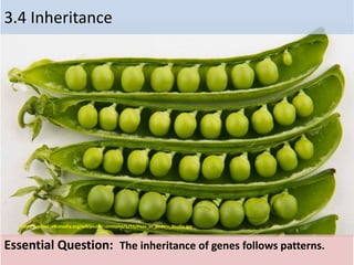
- 1. 3.4 Inheritance Essential Question: The inheritance of genes follows patterns. http://upload.wikimedia.org/wikipedia/commons/1/11/Peas_in_pods_-_Studio.jpg
- 2. Understandings Statement 3.4.U1 Mendel discovered the principles of inheritance with experiments in which large numbers of pea plants were crossed. 3.4 U2 Gametes are haploid so contain only one allele of each gene. 3.4 U3 The two alleles of each gene separate into different haploid daughter nuclei during meiosis. 3.4 U4 Fusion of gametes results in diploid zygotes with two alleles of each gene that may be the same allele or different alleles. 3.4 U5 Dominant alleles mask the effects of recessive alleles but co-dominant alleles have joint effects. 3.4 U6 Many genetic diseases in humans are due to recessive alleles of autosomal genes, although some genetic diseases are due to dominant or co-dominant alleles. 3.4 U7 Some genetic diseases are sex-linked. The pattern of inheritance is different with sex- linked genes due to their location on sex chromosomes. [Alleles carried on X chromosomes should be shown as superscript letters on an upper case X, such as Xh.] 3.4 U8 Many genetic diseases have been identified in humans but most are very rare. 3.4 U9 Radiation and mutagenic chemicals increase the mutation rate and can cause genetic diseases and cancer.
- 3. Applications and Skills Statement Guidance 3.4 A1 Inheritance of ABO blood groups. [The expected notation for ABO blood group alleles: O = i, A=IA, B = IB.] 3.4 A2 Red-green color blindness and hemophilia as examples of sex- linked inheritance. 3.4 A3 Inheritance of cystic fibrosis and Huntington’s disease. 3.4 A4 Consequences of radiation after nuclear bombing of Hiroshima and accident at Chernobyl. 3.4 S1 Construction of Punnett grids for predicting the outcomes of monohybrid genetic crosses. 3.4 S2 Comparison of predicted and actual outcomes of genetic crosses using real data. (Penny Lab) 3.4 S3 Analysis of pedigree charts to deduce the pattern of inheritance of genetic diseases.
- 4. Gregor Mendel • Austrian monk who published results of garden pea plants inheritance in 1865 • Used artificial pollination in a series of experiments by using a small brush to place the pollen on the reproductive parts of the flowers 3.4 U.1 Mendel discovered the principles of inheritance with experiments in which large numbers of pea plants were crossed.
- 5. Key terminology 1. Genotype – symbolic representation of pair of alleles possessed by an organism, typically represented by two letters • Ex: Bb, GG, tt 2. Phenotype – characteristics or traits of an organism • Ex: five fingers on each hand, color blindness, type O blood 3. Dominant allele – an allele that has the same effect on the phenotype whether it is paired with the same allele or a different one; always expressed in phenotype • Ex: Aa give dominant trait A because the a allele is masked; the a allele is not transcribed and translated during protein synthesis 3.4 U.1 Mendel discovered the principles of inheritance with experiments in which large numbers of pea plants were crossed.
- 6. 4. Recessive allele – an allele that has an effect on the phenotype only when present in the homozygous state • Ex: aa gives rise to the recessive trait because no dominant allele is there to mask it 5. Codominant allele – pairs of alleles that both affect the phenotype when present in a heterozygote • Ex: parent with curly hair and parent with straight hair can have children with different degrees of curliness as both alleles influence hair condition when both are present in the genotype 6. Locus – particular position on homologous chromosomes of a gene 3.4 U.1 Mendel discovered the principles of inheritance with experiments in which large numbers of pea plants were crossed.
- 7. 7. Homozygous – having two identical alleles of a gene Ex: AA is a genotype which is homozygous dominant whereas aa is the genotype which is homozygous recessive 8. Heterozygous – having two different alleles of a gene Ex: Aa is a heterozygous genotype 9. Carrier – an individual who has a recessive allele of a gene that does not have an effect on their phenotype 3.4 U.1 Mendel discovered the principles of inheritance with experiments in which large numbers of pea plants were crossed.
- 8. 10. Test cross – testing a suspected heterozygote plant or animal by crossing it with a known homozygous recessive (aa). Since a recessive allele can be masked, it is often impossible to tell if an organism is AA or Aa until they produce offspring which have the recessive trait. 3.4 U.1 Mendel discovered the principles of inheritance with experiments in which large numbers of pea plants were crossed.
- 9. 3.4 U.1 Mendel discovered the principles of inheritance with experiments in which large numbers of pea plants were crossed.
- 10. 3.4 U.1 Mendel discovered the principles of inheritance with experiments in which large numbers of pea plants were crossed.
- 11. Mendel’s Law of Segregation Four parts • Alternative versions of genes account for variations in inherited characteristics. • For each characteristic, an organism inherits two alleles, one from each parent. • If the two alleles differ, then one, the allele that encodes the dominant trait, is fully expressed in the organism's appearance; the other, the allele encoding the recessive trait, has no noticeable effect on the organism's appearance. • The two alleles for each characteristic segregate during gamete production 3.4 S.1 Construction of Punnett grids for predicting the outcomes of monohybrid genetic crosses.
- 12. Principle of Segregation: Each Parent or Gamete Contributes One Allele to Offspring 3.4 S.1 Construction of Punnett grids for predicting the outcomes of monohybrid genetic crosses.
- 13. Punnet Square: Used to determine the outcome of a cross between two individuals. In the example we have two parents that are heterozygous dominant for a trait Offspring: Genotype: 1/4 PP, 1/2 Pp, and 1/4 pp Phenotype: 3/4 Purple and 1/4 white 3.4 S.1 Construction of Punnett grids for predicting the outcomes of monohybrid genetic crosses.
- 14. 3.4 U.2 Gametes are haploid so contain only one allele of each gene. • Gametes are sex cell. • Sex cells contain one chromosome of each type, as an example Humans have 23 types. • Parents pass information in the form of genes in gametes (sex cell) • These cell will fuse together with the cell of the opposite sex to create a zygote.
- 15. Meiosis = reduction division • Cells divide twice • Result: 4 daughter cells, each with half as many chromosomes as parent cell 3.4 U.3 The two alleles of each gene separate into different haploid daughter nuclei during meiosis.
- 16. Life cycle: reproductive history of organism, from conception production of own offspring • Fertilization and meiosis alternate in sexual life cycles • Meiosis: cell division that reduces # of chromosomes (2n n), creates gametes • Fertilization: combine gametes (sperm + egg) – Fertilized egg = zygote (2n) • Zygote divides by mitosis to make multicellular diploid organism 3.4 U.4 Fusion of gametes results in diploid zygotes with two alleles of each gene that may be the same allele or different alleles. .
- 17. Human Life Cycle 3.4 U.4 Fusion of gametes results in diploid zygotes with two alleles of each gene that may be the same allele or different alleles. .
- 18. 3.4 U.4 Fusion of gametes results in diploid zygotes with two alleles of each gene that may be the same allele or different alleles. .
- 19. 3.4 U.5 Dominant alleles mask the effects of recessive alleles but co- dominant alleles have joint effects. . http://gestblog.scientopia.org/wp-content/uploads/sites/35/2012/07/10-04.gif
- 20. Problems with predictions I. Codominance II. Multiple alleles III. Sex linked genes
- 21. http://image.slidesharecdn.com/incompletecodominancemultiplealleles-120127095123-phpapp02/95/incomplete-codominance-multiplealleles-7-728.jpg 3.4 U.6 Many genetic diseases in humans are due to recessive alleles of autosomal genes, although some genetic diseases are due to dominant or co-dominant alleles
- 22. 3.4 U.6 Many genetic diseases in humans are due to recessive alleles of autosomal genes, although some genetic diseases are due to dominant or co-dominant alleles Inheritance characterized by full expression of both alleles in the heterozygote. Seen in: • Roan Cattle • Tay Sacs disease • Blood Types You have a brown Bull and a white Cow. You cross them and get a mix of the two colors. BB = Brown Bull/Cow WW = white Bull/Cow BW = Mixture of the two colors
- 23. 3.4 U.6 Many genetic diseases in humans are due to recessive alleles of autosomal genes, although some genetic diseases are due to dominant or co-dominant alleles https://jeanurquharthighlandsandislandsmsp.files.wordpress.com/2014/06/cavans-bourbon-orkney-by-robert_scarth-on-flickr.jpg
- 24. Sickle Cell Anemia: (example of a codomainant gene mutation and its consequences through protein synthesis) The Genetics of Sickle Cell Anemia •HBA HBA Suceptible to malaria with anemia • HBA HBs Increase resistance to malaria with mild anemia • HBs HBs Sickle cell shaped cell Suceptible to malaria with severe anemia 3.4 U.6 Many genetic diseases in humans are due to recessive alleles of autosomal genes, although some genetic diseases are due to dominant or co-dominant alleles
- 25. 3.4 U.6 Many genetic diseases in humans are due to recessive alleles of autosomal genes, although some genetic diseases are due to dominant or co-dominant alleles
- 26. 3.4 U.6 Many genetic diseases in humans are due to recessive alleles of autosomal genes, although some genetic diseases are due to dominant or co-dominant alleles
- 27. 3.4 U.6 Many genetic diseases in humans are due to recessive alleles of autosomal genes, although some genetic diseases are due to dominant or co-dominant alleles
- 28. 3.4 U.6 Many genetic diseases in humans are due to recessive alleles of autosomal genes, although some genetic diseases are due to dominant or co-dominant alleles
- 29. 3.4 U.6 Many genetic diseases in humans are due to recessive alleles of autosomal genes, although some genetic diseases are due to dominant or co-dominant alleles
- 30. 3.4 A.1 Inheritance of ABO blood groups. [The expected notation for ABO blood group alleles: O = i, A=IA, B = IB.] There are 4: A, B, AB and O A & B refer to 2 genetically inherited A and B antigens on the surface of red blood cells. IA – codes for A IB – codes for B i - codes for no antigen = type O blood
- 31. Multiple Alleles: ABO Blood Groups Blood type O: Universal donor. Blood type AB: Universal acceptor 3.4 A.1 Inheritance of ABO blood groups. [The expected notation for ABO blood group alleles: O = i, A=IA, B = IB.]
- 32. 3.4 A.1 Inheritance of ABO blood groups. [The expected notation for ABO blood group alleles: O = i, A=IA, B = IB.]
- 33. Sex Chromosomes 3.4 U.7 Some genetic diseases are sex-linked. The pattern of inheritance is different with sex-linked genes due to their location on sex chromosomes. [Alleles carried on X chromosomes should be shown as superscript letters on an upper case X, such as Xh.] • The X chromosome in humans spans more than 153 million base pairs (the building material of DNA). It represents about 2000 out of 20,000 - 25,000 genes. • The Y chromosome containing 78genes, out of the estimated 20,000 to 25,000 total genes in the human genome. Genetic disorders that are due to mutations in genes on the X chromosome are described as X linked.
- 34. 3.4 U.7 Some genetic diseases are sex-linked. The pattern of inheritance is different with sex-linked genes due to their location on sex chromosomes. [Alleles carried on X chromosomes should be shown as superscript letters on an upper case X, such as Xh.] Male sex chromosomes • There are non-homologous region males in which there is only one allele per gene and that is inherited from the female on the X- chromosome • In the homologous region the male inherited two copies of an allele per gene.
- 35. Female sex chromosomes • All regions of the X chromosome are homologous. • There are two alleles per gene as with all other genes on all other chromosomes This difference in x and y chromosomes plays a large role in determining rates of genetic inherited defects 3.4 U.7 Some genetic diseases are sex-linked. The pattern of inheritance is different with sex-linked genes due to their location on sex chromosomes. [Alleles carried on X chromosomes should be shown as superscript letters on an upper case X, such as Xh.]
- 36. Sex Linkage Alleles on the non-homologous region of the X chromosome are more common in females than in males • A gene with two alleles where one is dominant and one is recessive. • Female has three possible genotypes and one is the homozygous recessive. • In a population the chance of being homozygous recessive is 33.3 %. • Males have two possible genotypes. • There is a 50% chance of the homozygous recessive condition in the population. • In sex linked conditions the recessive condition is more common in males than females. 3.4 U.7 Some genetic diseases are sex-linked. The pattern of inheritance is different with sex-linked genes due to their location on sex chromosomes. [Alleles carried on X chromosomes should be shown as superscript letters on an upper case X, such as Xh.]
- 37. 3.4 U.7 Some genetic diseases are sex-linked. The pattern of inheritance is different with sex-linked genes due to their location on sex chromosomes. [Alleles carried on X chromosomes should be shown as superscript letters on an upper case X, such as Xh.]
- 38. Female carriers of sex linked alleles • Female heterozygote's for sex linked alleles e.g. Hemophilia XHXh or Color Blindness XBXb are carriers of the allele. • They are unaffected by the condition. • They do pass on the allele which may result in a homozygous female or a male with the sex linked recessive allele. 3.4 A.2 Red-green color blindness and hemophilia as examples of sex-linked inheritance.
- 39. Sex Linkage Examples: Hemophilia • Hemophilia is an example of a sex linkage condition. • The hemophilia allele is recessive to the normal allele. • The gene is located on the non- homologous region of the X chromosome. • The disease is associated with an inability to produce a clotting factor in blood. • Internal bleeding takes longer to stop. 3.4 A.2 Red-green color blindness and hemophilia as examples of sex-linked inheritance. http://blog.nz-online.de/lieb/wp-content/uploads/sites/8/2010/07/blut.jpg
- 40. • The homozygous genotype(*) in females has a high mortality. • The genotype XnY in males has a high mortality. 3.4 A.2 Red-green color blindness and hemophilia as examples of sex-linked inheritance. Hemophilia Hemophilia
- 41. • Red Green Color Blindness is an example of a sex linked condition. • Red Green Color blindness is a recessive condition. • The color blind allele is recessive to the normal allele. • Female homozygous recessives XbXb are color blind. • Males with the genotype XbY are color blind. • Notice that in a population the probability of having a Red Green color blind genotype in males is higher. 3.4 A.2 Red-green color blindness and hemophilia as examples of sex-linked inheritance. http://en.wikipedia.org/wiki/Color_blindness#/media/File:Ishihara_9.png Above is a color test plate.[The numeral "74" should be clearly visible to viewers with normal color vision.
- 42. 3.4 A.2 Red-green color blindness and hemophilia as examples of sex-linked inheritance. http://upload.wikimedia.org/wikipedia/commons/a/a3/XlinkRecessive.jpg Normal color vision Red/green color blindness
- 43. Pedigree Chart • Another way to visualize a monohybrid crosses or determining a genotype is by using a pedigree chart • Knowing the phenotype of individuals in a family will sometimes allow genotypes to be determined. • In genetic counseling this enables probabilities to be determined for the inheritance of characteristics in children. 3.4 S.3 Analysis of pedigree charts to deduce the pattern of inheritance of genetic diseases.
- 44. Pedigree Chart • White circle : Normal female • White Square: Normal male • Black Circle: affected female • Black square: affected male • (1) and (2)..Normal Parents • (3) affected female • (4),(5) and (6) normal 3.4 S.3 Analysis of pedigree charts to deduce the pattern of inheritance of genetic diseases.
- 45. 3.4 S.3 Analysis of pedigree charts to deduce the pattern of inheritance of genetic diseases.
- 46. 3.4 S.3 Analysis of pedigree charts to deduce the pattern of inheritance of genetic diseases.
- 47. 1. Phenylketonuria (Pku) • Using the allele key provided state the genotype of parents 1 and 2? • Give the genotype and phenotype of individual 5 ? • Is it possible that the condition is sex linked ? • What is the genotype and phenotype of individuals 7 and 8? • Which two individuals have the incorrect pedigree 3.4 S.3 Analysis of pedigree charts to deduce the pattern of inheritance of genetic diseases.
- 48. 2. Muscular Dystrophy • What type of genetic disease is muscular dystrophy? • Give the genotype and phenotype of 1? • Give the genotype and phenotype of 2? • Give the genotype and phenotype of 8 ? • Give the genotype and phenotype of 5 and 6 ? 3.4 S.3 Analysis of pedigree charts to deduce the pattern of inheritance of genetic diseases.
- 49. Cystic fibrosis (CF) Non Sex link recessive genetic trait found on Chromosome 7 Example: Cross The couple below are heterozygous for CF 3.4 A.3 Inheritance of cystic fibrosis and Huntington’s disease. http://www.bbc.co.uk/staticarchive/088e5fc50b3c51cfb49ebc4b6eaf203b18b93bbc.gif
- 50. Huntington’s Disease Non Sex link dominant genetic trait The couple two couples below are examples • Couple 1: 1 heterozygous (has trait) with 1 homozygous (without the trait) • Couple 2: Both parents are heterozygous with Huntington's 3.4 A.3 Inheritance of cystic fibrosis and Huntington’s disease. http://vanhornhuntingtonsdisease.weebly.com/uploads/1/3/7/4/13740905/4993818.jpg?1347964948 Couple 1 Couple 2
- 51. 3.4 U.8 Many genetic diseases have been identified in humans but most are very rare. • Medical research has identified over 4,000 genetic diseases, however many individuals do not suffer from one. • Most genetic diseases are caused by rare recessive alleles. Making the chance of inheritance very small. • Genetic sequencing of the human genome current estimates are that there maybe as little as 75-200 genes out of over 20,000 genes in the genome that contain these traits.
- 52. 3.4 U.9 Radiation and mutagenic chemicals increase the mutation rate and can cause genetic diseases and cancer. A mutagen is a physical (radiation) or chemical agent like Nitrosamines, found in tobacco. These mutagens change the genetic material, usually DNA, of an organism and increases the frequency of mutations above the natural background level. Many mutations cause cancer, mutagens are therefore also likely to be carcinogens.
- 53. • Radiation-induced cancers do not appear until at least 10 years after exposure (for tumors) or 2 years after exposure (for leukemia). • The risk of cancer after exposure can extend beyond this latent period for the rest of a person’s life for tumors or about 30 years for leukemia. • Risk is calculations are based on: – The type of radiation. • Each type of radiation is different and affects tissues differently. – The energy that it leaves in the body. • More energy means a higher probability of an effect. – Where in the body the energy remains. • Radiation exposure to a non-sensitive area of the body (i.e., wrist) really has no actual effect. Radiation exposure to a sensitive area of the body (i.e., blood-forming organs) can have an effect if the amount of energy left is high enough. 3.4 U.9 Radiation and mutagenic chemicals increase the mutation rate and can cause genetic diseases and cancer.
- 54. • Indirect damage – Water molecule is ionized, breaks apart, and forms OH free radical. – OH free radical contains an unpaired electron in the outer shell and is highly reactive: Reacts with DNA. – 75 percent of radiation-caused DNA damage is due to OH free radical. • Direct damage – DNA molecule is struck by radiation, ionized, resulting in damage. 3.4 U.9 Radiation and mutagenic chemicals increase the mutation rate and can cause genetic diseases and cancer.
- 55. Chromosome Damage Formation of a ring and fragments followed by replication of chromosomes. 3.4 U.9 Radiation and mutagenic chemicals increase the mutation rate and can cause genetic diseases and cancer.
- 56. Chromosome Damage Interchange between two chromosomes forms a chromosome with two centromeres and fragment, followed by replication. 3.4 U.9 Radiation and mutagenic chemicals increase the mutation rate and can cause genetic diseases and cancer.
- 57. Commonly Encountered Radiation Doses Effective Dose Radiation Source <= 0.01 rem annual dose living at nuclear power plant panoramic, or full-mouth dental x rays; skull or chest x ray <=0.1 rem single spine x ray; abdominal or pelvic x ray; hip x ray; mammogram <=0.5 rem kidney series of x rays; most barium-related x rays; head CT; any spine x-ray series; annual natural background radiation dose; most nuclear medicine brain, liver, kidney, bone, or lung scans <=1.0 rem barium enema (x rays of the large intestine); chest, abdomen, or pelvic CT <=5.0 rem cardiac catheterization (heart x rays); coronary angiogram (heart x rays); other heart x-ray studies; most nuclear medicine heart scans CT = computerized tomography; a specialized x-ray exam. 3.4 A.4 Consequences of radiation after nuclear bombing of Hiroshima and accident at Chernobyl.
- 58. Radiation Doses and Expected Effects (cont.) General radiation doses to the entire body and expected effects: • 100-200 rem received in a short time will cause nausea and fatigue. • 100-200 rem received over a long period will increase a person’s chances of getting cancer. • 200-300 rem received in a short time will cause nausea and vomiting within 24-48 hours. Medical attention should be sought. • 300-500 rem received in a short time will cause nausea, vomiting, and diarrhea within hours. Loss of hair and appetite occurs within a week. Medical attention must be sought for survival; half of the people exposed to radiation at this high level will die if they receive no medical attention. • 500-1,200 rem in a short time will likely lead to death within a few days. • Greater than 10,000 rem in a short time will lead to death within a few hours. 3.4 A.4 Consequences of radiation after nuclear bombing of Hiroshima and accident at Chernobyl.
- 59. 3.4 A.4 Consequences of radiation after nuclear bombing of Hiroshima and accident at Chernobyl. http://inapcache.boston.com/universal/site_graphics/blogs/bigpicture/hiroshima_08_05/h17_04.jpg
- 60. 3.4 A.4 Consequences of radiation after nuclear bombing of Hiroshima and accident at Chernobyl. http://www.nucleardarkness.org/include/nucleardarkness/images/cityonfire/hiroshima_after_02_full.jpg
- 61. 3.4 A.4 Consequences of radiation after nuclear bombing of Hiroshima and accident at Chernobyl. http://www.zap-actu.fr/wp-content/uploads/2013/11/pripyat-une-des-villes-fantomes-pres-de-tchernobyl-01.jpg
- 62. 3.4 A.4 Consequences of radiation after nuclear bombing of Hiroshima and accident at Chernobyl.