Primary structure of protein
Secondary structure of protein
Tertiary structure of protein
Quaternary structure of protein
Methods to determine protein structure
Conclusion
References
METHODS TO DETERMINE PROTEIN STRUCTURE
Each protein has a unique sequence of amino acids.
The amino acids are held together in a protein by
covalent peptide bonds or linkages.
A peptide bond are formed when amino group of an
amino acid combines with the carboxyl group of another.
The conformation of polypeptide chain by twisting or folding is referred to as secondary structure.
Two types of secondary structures α-helix and β-sheet are mainly identified.
α-Helical structure was proposed by Pauling and Corey in 1951.
It occurs when the sequence of amino acids are linked by hydrogen bonds.
Each turn of α-helix contains 3.6 amino acids.
β-pleated sheets are composed of two or more segments of fully extended peptide chains.
β-Sheets may be arranged either in parallel or anti-parallel direction.
Many globular proteins contain combinations of α-helix and β-pleated sheet secondary structure, these patterns are called supersecondary structures also called motifs.
The three dimensional arrangement of protein structure is referred to as tertiary structure.
It is a compact structure with hydrophobic side chains held interior while the hydrophilic groups are on the surface.
This type of arrangement provide stability of the molecule.
Besides the H-bongs, disulfide bonds, ionic interactions, hydrophobic interactions also contribute to the tertiary structure.
2. Introduction
Primary structure of protein
Secondary structure of protein
Tertiary structure of protein
Quaternary structure of protein
Methods to determine protein structure
Conclusion
References
3. Proteins (Greek: Proteios ‘Holding the first place’) are the most abundant
organic molecules of the living system.
They constitute about 50% of the cellular dry weight.
They are the polymers of Lα-amino acids.
The structures of proteins can be divided into 4 levels of organization:
Primary structure
Secondary structure
Tertiary structure
Quaternary structure
4. Each protein has a unique sequence of amino acids.
The amino acids are held together in a protein by
covalent peptide bonds or linkages.
A peptide bond are formed when amino group of an
amino acid combines with the carboxyl group of another.
5. The conformation of polypeptide chain by twisting or folding is referred to as secondary structure.
Two types of secondary structures α-helix and β-sheet are mainly identified.
α-Helical structure was proposed by Pauling and Corey in 1951.
It occurs when the sequence of amino acids are linked by hydrogen bonds.
Each turn of α-helix contains 3.6 amino acids.
β-pleated sheets are composed of two or more segments of fully extended peptide chains.
β-Sheets may be arranged either in parallel or anti-parallel direction.
Many globular proteins contain combinations of α-helix and β-pleated sheet secondary structure,
these patterns are called supersecondary structures also called motifs.
7. The three dimensional arrangement of protein structure is referred to as tertiary
structure.
It is a compact structure with hydrophobic side chains held interior while the
hydrophilic groups are on the surface.
This type of arrangement provide stability of the molecule.
Besides the H-bongs, disulfide bonds, ionic interactions, hydrophobic interactions
also contribute to the tertiary structure.
9. Proteins consisting of two or more polypeptides which may be identical or unrelated,
such proteins are termed as oligomers and possess quaternary structure.
The individual polypeptide chains are known as monomers.
The monomeric subunits are held together by H-bonds, hydrophobic interactions and
ionic bonds.
These proteins play a significant role in the regulation of metabolism and cellular
functions. E.g., Hemoglobin.
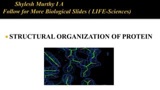
11. The first protein structure was determined by protein crystallography was of Myoglobin by Max Perutz and
John Kendrew in 1962.
Currently used techniques including
X-ray crystallography, NMR spectroscopy and Electron microscopy.
Fig: X-Ray Crystallography
13. Proteins are the most abundant organic molecules of life, performing structural and
dynamic functions in the living cells. The dynamic functions of proteins are highly
diversified such as enzymes, hormones, clotting factors, immunoglobulins, etc.
14. M.C. Michael, L.N. David; Principles of Biochemistry; 5th edi; P:92-138; W.H.
Freeman and Company.
U. Satyanarayan, U. Chakrapani; Biochemistry; 3rd edi; P:43-68; Books and allied
(P) Ltd.
K. Pranav, M. Usha; Life Sciences; 5th edi; P:12-20; Pathfinder Publication.