
Empfohlen
Weitere ähnliche Inhalte
Was ist angesagt?
Was ist angesagt? (20)
Ähnlich wie Ancient Parasitic Disease: The Global Burden of Malaria
Ähnlich wie Ancient Parasitic Disease: The Global Burden of Malaria (20)
Mehr von shiv chaudhary
Kürzlich hochgeladen
Kürzlich hochgeladen (20)
Ancient Parasitic Disease: The Global Burden of Malaria
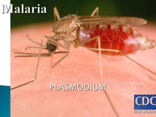
- 1. PLASMODIUM
- 2. An ancient disease : substantial toll of human life and sufferings Originated from Italian word Mala (bad) and ayia(air) Latin word : Marshy So , malaria : disease caused by heat humidity and marshy areas 1st discovered : Alphonse Laveran : 1880 in RBC of patient (Algeria)
- 3. Transmitted by female Anopheles mosquito Common insect borne infection Most deadly vector borne disease in the world Life threatening parasitic problem : global problem worldwide 40% of world’s population : (2.4) billion risk 400-900 million people are affected
- 4. Kingdom: Animalia Phylum: Apicomplexa Class: Coccidia Order: Haemosporidia Genus: Plasmodium Species: vivax, falciparum, ovale , malariae
- 6. P.vivax and P.falciparum : account 95% of infection Some estimate : P.vivax : accounts 80% of infections : widely distributed in tropics, subtropics and temperate zones
- 10. Different stages: 1.Pre-Erythryocytic Schizogony 2. Erythrocytic Schizogony 3. Gametogony 4. Exo-Erythryocytic Schizogony (P.vivax, P.ovale)
- 11. 1.Pre-erythrocytic Schizogony 1st stage of human cycle Sporozoites : doesn’t directly enter into RBC : so k/as PES occurs : inside parenchyma of cells Fully developed schizont measures 42µm : contains large no of merozoites Smaller micromerozoites : enter into circulation : to start ES Larger macromerozoites : Re-enter liver cells : to start Exo ES Some sporozoites : remain dormant in liver : Hypnozoites : cause relapse
- 12. Duration of this phase : P.falciparum : 6 days P.vivax : 8 days P.ovale : 9days P. malariae :13-16days
- 13. 2. Erythrocytic schizogony Infected liver ruptures : merozoites release : invade RBC’s Parasite reside the RBC and passes through : RBC Trophozoite Schizont Merozoite P.vivax : greater tendency for younger erythrocytes and reticulocytes P.falciparum : any age P.malariae : old P.ovale : Young
- 14. Parasitised red cells : enlarged : cells mature with parasites : show stippling(formation of small dot) P.vivax : Schuffner’s dot P.falciparum : Maurer’s dot (large red spots) P.malariae : Ziemann’s dot (few tiny dots) P.ovale : Schuffner’s dot
- 15. Trophozoite : Have active amoeboid 2 forms : 1. Ring form (early trophozoite) Nucleus : thinner side of ring 2. Amoeboid form (late trophozoite) Presence of pseudopodia Contains malarial pigment
- 16. Schizont Appears after a period of 36-40 hours Full grown trophozoite : ready to divide Round in shape Lost all amoeboid activities Nucleus is large and lie at periphery 2 form : Immature schizont (Nucleus not divided) Mature schizont : Nucleus divided
- 17. 3.Gametogony After ES : some merozoites : give rise to gametocytes : sexual function after leaving man host Occurs inside capillaries of bone marrow and spleen Mature gametocytes : appears in peripheral blood Microgametocyte (male) : boarder, shorter with blunt ends Macrogametocyte (female): longer, narrower, pointed ends Changes in infected RBC’s (increase in size, pallor and different dots )
- 18. 4.Exo-erythrocytic Schizogony Resembles PE form in morphology Maintained upto 3 yrs and independent of ES Short term and long term relapses (deteriorate after a period of improvement) Sporozoite PES Development of hypnozoites ES EES primary malaria Relapses
- 19. Relapse : in case of P.vivax and P.ovale due to the presence of hypnozoites Recrudescence : situation : RBC infection is not eliminated by the immune system or by therapy No of RBCs begin to increase again with subsequent clinical symptoms All species may cause
- 20. Sexual cycle of malarial parasite Starts in human body : formation of gametocytes Mosquito : blood meal : ingests both sexual and asexual forms Asexual forms : digested Sexual forms (gametocyte) : undergo further development Blood of human carrier : must contain 12 gametocytes/mm3
- 21. No of female gametocytes more than male gametocytes 1st phase : mid-gut of stomach Nucleus of each male gametocyte : 8 long flagellates(microgamete) : highly motile Process : observed : outside mosquito : thick film: exflagellation Female gametocyte : don't divide : Macrogamete Fertilizes : Zygote : motion less: later becomes motile : Ookinite
- 22. Ookinite : migrates to stomach wall : oocyst Large no of sporozoites inside oocyst When fully mature : oocyst ruptures : liberates sporozoites : spread all parts : salivary gland Ready to be transmitted : when it takes blood meal
- 23. MOT : bite of Anopheles mosquito Extrinsic Incubation period : different periods for the development of sexual cycle at given temp Varies : 8 to 21 days Incubation period P. falciparum : 12 days(9-14 days) P. vivax : 14 days (8-17 days) P. malariae : 28 days (18-40 days) P. ovale :17 days (16-18days)
- 24. Main features : fever peaks followed by anemia and splenomegaly Mild to severe and complicated : According to species of parasite present Patient’s state of immunity Certain disease like : malnutrition and other disease Severe in children and pregnancy
- 25. Main clinical features 1.Prodromal period Malarial paroxysm :preceed by prodromal period Non-specific symptoms : malaise, myalgia, headache and fatigue Some localized symptoms :chest pain, abdominal pain and arthalgia 2. Malarial paroxysm Classical manifestation of acute malaria Characterised by fever chills and rigors
- 26. Primary fever Typical attack 3 distinct stages: cold stage, hot stage and sweating stage a. Cold stage : Onset with lassitude (lethargy), headache, nausea and chilly sensations followed in an hour or so by rigors b. Hot stage : Patient feels hot and the skin is hot and dry to touch Headache intense Lasts for 30 min to 6 hrs
- 27. c. Sweating stage Profuse sweating follows the hot stage Continues for hour or so Temp drops rapidly to normal Skin is cool and moist So, primary attack follows a febrile interval of 48-72hrs
- 28. 3.Anemia Normocytic normochromic anemia Severe in falciparum malaria 4. Hepatospleenomegaly Spleen : palpable after 2nd weeks of fever Severe in P.falciparum : so K/as malignat malaria 5. Malaria in pregnancy Miscarriage or abortion 6.Malaria in children More severe than as in adults May develop convulsion (muscular contarction) during malarial attack Dehydration: as a result of vomiting and sweating.
- 29. P.vivax : Benign tertiary malaria : 48hrs P.falciparum : Malignant tertiary malaria:36-48hrs P.malariae : Benign Quartan Malaria : 72 hours P.ovale : Benign Tertian Malaria : 48 hrs
- 30. 1.Black water fever Repeated infection of P.falciparum: inadequately treated with quinine Massive hemolysis followed by fever and haemoglobinuria(black coloured urine),hyperbilirubenemia Complication : uraemia (blood poisoning), renal failure ,anemia, pigment calculi
- 31. 2.Pernicious anemia (Cerebral malaria or algid malaria) May be different forms : a. Pernicious malaria affecting nervous system : cerebral malaria b. Pernicious malaria affecting GIT system (algid malaria) c. Pernicious types affecting cardiovascular, respiratory and genitourinary tract
- 32. Specimen : Blood (before antimalarial drug) Earlobe or finger in adults Toe in infants Collected : peak fever More imp : frequently examination of blood smear
- 33. 1.Light microscopy 2. Fluorescence microscopy 3. Quantitative buffy coat 1.Light microscope Blood smear Gold standard method Most commonly used Depends upon : demonstration of parasite in stained PBS Ring forms and gametocyte : commonly seen in PBS
- 34. 1.Thick smear Smear preparation Dehaemoglobinisation with d/w Dried and stained with Romanowsky’s stain : Leishman stain. Geimsa stain Uses To detect parasite Demonstrating malarial pigment
- 36. P. falciparum : (only ring and crescent form) Many ring forms Crescent forms gametocyte Malarial pigments : inside the blood P. vivax Trophozoites, Schizont and Gametocytes can be seen in PBS Ring form : nucleus more thicker Gametocyte : spherical or globular Schufnner’s dot
- 41. Thin smear Rapidly dried Fixed in alcohol and stained Uses Detecting parasites Identify species P. falciparum Ring form alone or along with gametocytes Multiple rings in individual RBC’s Presence of Maurer’s dot Banana shaped gametocytes
- 44. The rings are small: 1/3 or less of the rbc
- 46. Ameboid trophozoite, enlarged RBC , Schüffner’s dots Gametocyte
- 47. More than 20 merozoites
- 49. 2. Fluorescence microscope Kawamoto technique : fluorescent staining methods : malarial parasite Blood smear : slide : stain with Acridine orange Nuclear DNA : green Cytoplasmic RNA : Red Examined under fluorescence microscope
- 50. 3. Quantitative buffy coat (QBC)method Sensitive method Based on : ability of acridine orange to stain nucleic acid containing parasites Blood : capillary tube : coated with fluorescent dye : microhaematocrit centrifugation Buffy coat : observed under fluorescent microscope Acridine orange stained malarial parasite : brilliant green
- 51. 4.Serodiagnosis IHA ELISA IFA 5. Rapid diagnostic kit 6. Dipstick method An enzyme immunoassay : which detects histidine rich protein 2 Pf(HRP-2) : metabolic product produced by P.falciparum 7. Molecular diagnosis DNA and RNA : PCR
- 53. Chloroquine Amodiquine Proguanil Quinine
- 55. THANK YOU