SlideShare ist ein Scribd-Unternehmen logo
1 von 13
DNA Fingerprinting
How DNA Fingerprinting Works
How to produce a genetic fingerprint:
The first step to making a genetic fingerprint requires getting a sample of DNA. This sample can come from blood,
semen, hair or saliva, and may be an extremely small sample. The root from a single strand of hair is enough for
researchers to work with. This sample contains white blood cells which are broken open using detergent, and all the
useable DNA is separated from the extra cellular material. Next the restriction enzymes are used to cut the DNA into
smaller pieces. Restriction enzymes work by cutting the DNA at a specific sequence, which produces either blunt ends or
sticky ends, and results in many fragments of different lengths. These fragments are called restriction fragments length
polymorphisms, or RFLPs.
These RFLPs are then put into an agarose gel. Using gel electrophoresis, the fragments are sorted according to size.
When the current of the electric field is turned on, the negative RFLPs will start to move across the gel towards the
positive end. The smaller fragments move farther across the gel than the larger ones. Also, alkali is responsible for
causing the hydrogen bonds to break, and the DNA to become single-stranded. When the DNA becomes single-
stranded, it causes nucleotides to become free, and they will later be used to pair up with probes. The gel is then covered
by a piece of nylon and thin paper towels, which are used to absorb moisture from the gel. The DNA fragments get gently
transferred from the gel to the surface of the nylon. This process is called blotting.
Finally, radioactive probes get washed over the nylon surface. These probes will join to any DNA fragments that share
the same composition. The final step to making a genetic fingerprint is to place a photographic film on top of the nylon
surface. The probes leave marks on the film wherever they attached to the RFLPs. Dark bands will then show up when
the film is developed, which marks the length of the RFLPs that were hybridized. Researchers are then able to read the
fingerprint and match it to others. They do this by placing the xray on a light background, and comparing the RFLP
lengths in the DNA from the crime scene, to the DNA of the suspect.
This picture shows DNA being sorted by an electric field. When the current is turned on, it attracts the shortest
and lightest particles the farthest.
When fingerprinting was a fairly new process, during 1984-1990, it could take as long as four to six weeks of lab work to
complete and compare DNA fingerprinting evidence. Jeffreys was responsible for coming up with the original DNA
fingerprinting technique and in November of 1991, he also was responsible for creating a better test that could obtain
results in as little as two days.
Polymerase Chain Reaction (PCR)
PCR can be used in DNA fingerprinting as a way to make numerous copies of isolated DNA. The process can selectively
amplify a single copy of a desired sequence.
First Cycle:
-Dna molecules are melted by raising the temperature to 95 degrees Celcius.
-Strands separate, temperature is lowered to 60 degrees Celcius.
-Each primer binds specifically to the 3 prime end of the target sequence on the appropriate strands of DNA.
-Primers direct taq polymerase to synthesize complementary nucleotides.
-Only DNA containing target sequence are copied by taq polymerase.
-At the end of cycle 1, both strands have been copied to form 2 partially double-stranded molecules.
-These steps are repeated during the next cycles. After two cycles, four partially double-stranded molecules are
produced, all containing the target sequence.
-After many cycles, millions of copies of the DNA can be produced.
-This is useful in DNA fingerprinting, as researchers can do many tests on the same DNA sample.
What is DNA Fingerprinting?
DNA fingerprinting is a process
through which the unique genetic
code of a person is identified and
codified. DNA fingerprint is called
so because just like the
technology of fingerprint became
very popular in the 1930s as
means of crime detection,
beginning in the 80s, DNA
testing achieved a similar level of
popularity in forensic science.
Reasons Why It is Conducted
Though fingerprints can be
changed through surgical means
or may be tampered with, the
DNA code of every individual is
completely unique and it
therefore serves as a distinct
marker of a person or evidence
of his/her presence at a certain
place. Even very small amounts
of body fluids like sweat, mucus,
blood and semen that the
criminal inadvertently leaves
behind on the scene of the crime can be later traced, and successfully reveal his/her identity conclusively, since DNA is
present uniformly in each cell of an individual’s body. Thus, forensic DNA fingerprinting has become extremely popular as
a method of crime detection. However, this method can also be used for medical purposes like detecting and predicting
the chances of rare hereditary diseases that can be passed on from parents to their unborn children as well as in newborn
babies.
DNA fingerprinting is also extremely useful in research and development of cures for genetic disorders, as it helps locate
and isolate the genetic patterns that cause certain diseases in particular families or racial groups, and thereby eventually
shows the way to curing the condition. In addition, maintaining official records of the DNA fingerprints of each citizen in a
state can help identify missing persons and casualties of accidents, as well as serve as a device of personal identification
in important commercial transactions.
DNA Fingerprinting
DNA Fingerprinting is widely used in investigating and solving criminal crimes. Below are the steps involved for
completing DNA fingerprinting.
1. Steps in DNA Fingerprinting
A.-DNA is cut into fragments of specific sizes by certain enzymes called restriction enzymes.
- Restriction enzymes are taken from bacteria
- Each restriction enzymes recognizes and cuts DNA at a specific sequence
- The DNA fragments can vary in number and size
- Gel electrophoresis determine the numbers and sizes of DNA fragments procuduced by restriction enzyme
treatment
* If DNA is too small, the PCR is used to increase the quantity of DNA for analysis. The PCR makes many copies of the
DNA in a small sample. Example; blood, saliva, hair, urine, or muscle.
2. The value of PCR (polymerase chain reaction) is that it makes many copies of the DNA in a small sample.
3. Restriction enzymes are taken from bacteria that use them to stop infection by viruses.
4. DNA probes are known sequences of DNA that will bind to their coomplementary DNA sequences wherever they are.
It's used with the specific sequence that to the DNA fragement on a membrane with another sequence. 5'-
TATATAGCTC-3' ---> 3'-ATATATCGAG-5'
5.ID criminals, ID remains, and genetic disorders are examples of uses of DNA fingerprinting
Introduction
Like the fingerprints that came into use by detectives and police labs during the 1930s, each person has a unique DNA
fingerprint (Figure 1). Unlike a conventional fingerprint that occurs only on the fingertips and can be altered by surgery, a
DNA fingerprint is the same for every cell, tissue, and organ of a person. It cannot be altered by any known treatment.
Consequently, DNA fingerprinting is rapidly becoming the primary method for identifying and distinguishing among
individual human beings.
An additional benefit of DNA fingerprint technology is the diagnosis of inherited disorders in adults, children, and unborn
babies. Even bloodstained clothing from Abraham Lincoln has been analyzed for evidence of a genetic disorder called
Marfan's Syndrome.
[Topics]
The Structure of DNA
The characteristics of all living organisms, including humans, are essentially determined by information contained within
DNA that they inherit from their parents. The molecular structure of DNA can be imagined as a zipper with each tooth
represented by one of four letters (A, C, G, or T) and with opposite teeth forming one of two pairs, either AT or GC (Figure
2).
The information contained in DNA is the sequence of letters along the zipper. For example, the sequence ACGCT
represents different information than the sequence AGTCC in the same way that the word "POST" has a different
meaning from "STOP" or "POTS," even though they use the same letters. The traits of a human being are the result of
information contained in the DNA code.
Living organisms that look different or have different characteristics also have different DNA sequences. The more
different the organisms, the more different are the DNA sequences. DNA fingerprinting is a very quick way to compare the
DNA sequences of any two living organisms.
[Topics]
Making DNA Fingerprints
DNA fingerprinting is a laboratory procedure that requires six steps (Figure 3):
1) Isolation of DNA. DNA must be recovered from the cells or tissues of the body. Only a small amount of tissue, like
blood, hair, or skin, is needed. For example, the amount of DNA found at the root of one hair is usually sufficient.
2) Cutting, sizing, and sorting. Special enzymes called restriction enzymes are used to cut the DNA at specific places.
For example, an enzyme called EcoR1, found in bacteria, will cut DNA only when the sequence GAATTC occurs.
The DNA pieces are sorted according to size by a sieving technique called electrophoresis. The DNA pieces are passed
through a gel made from seaweed agarose (a jelly-like product made from seaweed). This technique is the DNA
equivalent of screening sand through progressively finer mesh screens to determine particle sizes.
3) Transfer of DNA to nylon. The distribution of DNA pieces is transferred to a nylon sheet by placing the sheet on the
gel and soaking them overnight.
4-5) Probing. Adding radioactive or colored probes to the nylon sheet produces a pattern called the DNA fingerprint. Each
probe typically sticks in only one or two specific places on the nylon sheet.
6) DNA fingerprint. The final DNA fingerprint is built by using several probes (5-10 or more) simultaneously. It resembles
the bar codes used by grocery store scanners.
[Topics]
Uses of DNA Fingerprints
DNA fingerprints are useful in several areas of society. They are used by professionals in human health and the justice
system.
Diagnosis of inherited disorders
DNA fingerprinting is used to diagnose inherited disorders in both prenatal and newborn babies in hospitals around the
world. These disorders may include cystic fibrosis, hemophilia, Huntington's disease, familial Alzheimer's, sickle cell
anemia, thalassemia, and many others. Early detection of such disorders enables the medical staff to prepare themselves
and the parents for proper treatment of the child. In some programs, genetic counselors use DNA fingerprint information
to help prospective parents understand the risk of having an affected child. In other programs, prospective parents use
DNA fingerprint information in their decisions concerning affected pregnancies.
Developing cures for inherited disorders
Research programs to locate inherited disorders on the chromosomes depend on the information contained in DNA
fingerprints. By studying the DNA fingerprints of relatives who have a history of some particular disorder, or by comparing
large groups of people with and without the disorder, it is possible to identify DNA patterns associated with the disease in
question. This work is a necessary first step in designing an eventual genetic cure for these disorders.
Forensic or criminal
FBI and police labs around the U.S. have begun to use DNA fingerprints to link suspects to biological evidence-blood or
semen stains, hair, or items of clothing-found at the scene of a crime. Since 1987, more than 150 cases have been
decided with the assistance of DNA fingerprint evidence.
Another important use of DNA fingerprints in the court system is to establish paternity in custody and child support
litigation. In these applications, DNA fingerprints bring an unprecedented, nearly perfect accuracy to the determination.
Southern blot
From Wikipedia, the free encyclopedia
Jump to: navigation, search
A Southern blot is a method routinely used in molecular biology for detection of a specific DNA sequence in DNA
samples. Southern blotting combines transfer of electrophoresis-separated DNA fragments to a filter membrane and
subsequent fragment detection by probe hybridization. The method is named after its inventor, the British biologist Edwin
Southern.
[1]
Other blotting methods (i.e., western blot,
[2]
northern blot, eastern blot, southwestern blot) that employ similar
principles, but using RNA or protein, have later been named in reference to Edwin Southern's name. As the technique
was eponymously named, Southern blot is capitalized as is conventional for proper nouns. The names for other blotting
methods may follow this convention, by analogy.
[3]
Method
1. Restriction endonucleases are used to cut high-molecular-weight DNA strands into smaller fragments.
2. The DNA fragments are then electrophoresed on an agarose gel to separate them by size.
3. If some of the DNA fragments are larger than 15 kb, then prior to blotting, the gel may be treated with an acid,
such as dilute HCl, which depurinates the DNA fragments, breaking the DNA into smaller pieces, thus allowing
more efficient transfer from the gel to membrane.
4. If alkaline transfer methods are used, the DNA gel is placed into an alkaline solution (typically containing sodium
hydroxide) to denature the double-stranded DNA. The denaturation in an alkaline environment may improve
binding of the negatively charged thymine residues of DNA to a positively charged amino groups of membrane,
separating it into single DNA strands for later hybridization to the probe (see below), and destroys any residual
RNA that may still be present in the DNA. The choice of alkaline over neutral transfer methods, however, is often
empirical and may result in equivalent results.
[citation needed]
5. A sheet of nitrocellulose (or, alternatively, nylon) membrane is placed on top of (or below, depending on the
direction of the transfer) the gel. Pressure is applied evenly to the gel (either using suction, or by placing a stack
of paper towels and a weight on top of the membrane and gel), to ensure good and even contact between gel and
membrane. If transferring by suction 20X SSC buffer is used to ensure a seal and prevent drying of the gel. Buffer
transfer by capillary action from a region of high water potential to a region of low water potential (usually filter
paper and paper tissues) is then used to move the DNA from the gel on to the membrane; ion exchange
interactions bind the DNA to the membrane due to the negative charge of the DNA and positive charge of the
membrane.
6. The membrane is then baked in a vacuum or regular oven at 80 °C for 2 hours (standard conditions; nitrocellulose
or nylon membrane) or exposed to ultraviolet radiation (nylon membrane) to permanently attach the transferred
DNA to the membrane.
7. The membrane is then exposed to a hybridization probe—a single DNA fragment with a specific sequence whose
presence in the target DNA is to be determined. The probe DNA is labelled so that it can be detected, usually by
incorporating radioactivity or tagging the molecule with a fluorescent or chromogenic dye. In some cases, the
hybridization probe may be made from RNA, rather than DNA. To ensure the specificity of the binding of the
probe to the sample DNA, most common hybridization methods use salmon or herring sperm DNA for blocking of
the membrane surface and target DNA, deionized formamide, and detergents such as SDS to reduce non-specific
binding of the probe.
8. After hybridization, excess probe is washed from the membrane (typically using SSC buffer), and the pattern of
hybridization is visualized on X-ray film by autoradiography in the case of a radioactive or fluorescent probe, or by
development of color on the membrane if a chromogenic detection method is used.
Western blot
From Wikipedia, the free encyclopedia
Jump to: navigation, search
Western blot using an antibody that recognizes proteins modified with lipoic acid.
The western blot (sometimes called the protein immunoblot) is a widely accepted analytical technique used to detect
specific proteins in the given sample of tissue homogenate or extract. It uses gel electrophoresis to separate native
proteins by 3-D structure or denatured proteins by the length of the polypeptide. The proteins are then transferred to a
membrane (typically nitrocellulose or PVDF), where they are stained with antibodies specific to the target protein.
[1][2]
There are now many reagent companies that specialize in providing antibodies (both monoclonal and polyclonal
antibodies) against tens of thousands of different proteins.
[3]
Commercial antibodies can be expensive, although the
unbound antibody can be reused between experiments. This method is used in the fields of molecular biology,
biochemistry, immunogenetics and other molecular biology disciplines.
Other related techniques include using antibodies to detect proteins in tissues and cells by immunostaining and enzyme-
linked immunosorbent assay (ELISA).
The method originated in the laboratory of Harry Towbin at the Friedrich Miescher Institute
[1]
. The name Western blot was
given to the technique by W. Neal Burnette
[4]
and is a play on the name Southern blot, a technique for DNA detection
developed earlier by Edwin Southern. Detection of RNA is termed northern blot and was developed by George Stark at
Stanford
[5]
.
Steps
Tissue preparation
Samples can be taken from whole tissue or from cell culture. Solid tissues are first broken down mechanically using a
blender (for larger sample volumes), using a homogenizer (smaller volumes), or by sonication. Cells may also be broken
open by one of the above mechanical methods. However, virus or environmental samples can be the source of protein
and thus western blotting is not restricted to cellular studies only.
Assorted detergents, salts, and buffers may be employed to encourage lysis of cells and to solubilize proteins. Protease
and phosphatase inhibitors are often added to prevent the digestion of the sample by its own enzymes. Tissue
preparation is often done at cold temperatures to avoid protein denaturing and degradation.
A combination of biochemical and mechanical techniques – comprising various types of filtration and centrifugation – can
be used to separate different cell compartments and organelles.
Gel electrophoresis
Main article: Gel electrophoresis
The proteins of the sample are separated using gel electrophoresis. Separation of proteins may be by isoelectric point
(pI), molecular weight, electric charge, or a combination of these factors. The nature of the separation depends on the
treatment of the sample and the nature of the gel. This is a very useful way to identify a protein.
By far the most common type of gel electrophoresis employs polyacrylamide gels and buffers loaded with sodium dodecyl
sulfate (SDS). SDS-PAGE (SDS polyacrylamide gel electrophoresis) maintains polypeptides in a denatured state once
they have been treated with strong reducing agents to remove secondary and tertiary structure (e.g. disulfide bonds [S-S]
to sulfhydryl groups [SH and SH]) and thus allows separation of proteins by their molecular weight. Sampled proteins
become covered in the negatively charged SDS and move to the positively charged electrode through the acrylamide
mesh of the gel. Smaller proteins migrate faster through this mesh and the proteins are thus separated according to size
(usually measured in kilodaltons, kDa). The concentration of acrylamide determines the resolution of the gel - the greater
the acrylamide concentration the better the resolution of lower molecular weight proteins. The lower the acrylamide
concentration the better the resolution of higher molecular weight proteins. Proteins travel only in one dimension along the
gel for most blots.
Samples are loaded into wells in the gel. One lane is usually reserved for a marker or ladder, a commercially available
mixture of proteins having defined molecular weights, typically stained so as to form visible, coloured bands. When
voltage is applied along the gel, proteins migrate through it at different speeds dependent on their size. These different
rates of advancement (different electrophoretic mobilities) separate into bands within each lane.
It is also possible to use a two-dimensional (2-D) gel which spreads the proteins from a single sample out in two
dimensions. Proteins are separated according to isoelectric point (pH at which they have neutral net charge) in the first
dimension, and according to their molecular weight in the second dimension.
Transfer
In order to make the proteins accessible to antibody detection they are moved from within the gel onto a membrane made
of nitrocellulose or polyvinylidene difluoride (PVDF). The primary method for transferring the proteins is called
electroblotting and uses an electric current to pull proteins from the gel into the PVDF or nitrocellulose membrane. The
proteins move from within the gel onto the membrane while maintaining the organization they had within the gel. An older
method of transfer involves placing a membrane on top of the gel, and a stack of filter papers on top of that. The entire
stack is placed in a buffer solution which moves up the paper by capillary action, bringing the proteins with it. In practice
this method is not used as it takes too much time; electroblotting is preferred. As a result of either "blotting" process, the
proteins are exposed on a thin surface layer for detection (see below). Both varieties of membrane are chosen for their
non-specific protein binding properties (i.e. binds all proteins equally well). Protein binding is based upon hydrophobic
interactions, as well as charged interactions between the membrane and protein. Nitrocellulose membranes are cheaper
than PVDF, but are far more fragile and do not stand up well to repeated probings.
The uniformity and overall effectiveness of transfer of protein from the gel to the membrane can be checked by staining
the membrane with Coomassie Brilliant Blue or Ponceau S dyes. Ponceau S is the more common of the two, due to its
higher sensitivity and water solubility, the latter making it easier to subsequently destain and probe the membrane, as
described below.
[6]
Blocking
Since the membrane has been chosen for its ability to bind protein and as both antibodies and the target are proteins,
steps must be taken to prevent the interactions between the membrane and the antibody used for detection of the target
protein. Blocking of non-specific binding is achieved by placing the membrane in a dilute solution of protein - typically 3-
5% Bovine serum albumin (BSA) or non-fat dry milk (both are inexpensive) in Tris-Buffered Saline (TBS) or I-Block, with a
minute percentage (0.1%) of detergent such as Tween 20 or Triton X-100. The protein in the dilute solution attaches to
the membrane in all places where the target proteins have not attached. Thus, when the antibody is added, there is no
room on the membrane for it to attach other than on the binding sites of the specific target protein. This reduces "noise" in
the final product of the western blot, leading to clearer results, and eliminates false positives.
Detection
During the detection process the membrane is "probed" for the protein of interest with a modified antibody which is linked
to a reporter enzyme; when exposed to an appropriate substrate this enzyme drives a colourimetric reaction and produces
a color. For a variety of reasons, this traditionally takes place in a two-step process, although there are now one-step
detection methods available for certain applications.
Two steps
Primary antibody
The primary antibodies are generated when a host species or immune cell culture is exposed to protein of interest (or a
part thereof). Normally, this is part of the immune response, whereas here they are harvested and used as sensitive and
specific detection tools that bind the protein directly.
After blocking, a dilute solution of primary antibody (generally between 0.5 and 5 micrograms/mL) is incubated with the
membrane under gentle agitation. Typically, the solution is composed of buffered saline solution with a small percentage
of detergent, and sometimes with powdered milk or BSA. The antibody solution and the membrane can be sealed and
incubated together for anywhere from 30 minutes to overnight. It can also be incubated at different temperatures, with
warmer temperatures being associated with more binding, both specific (to the target protein, the "signal") and non-
specific ("noise").
Secondary antibody
After rinsing the membrane to remove unbound primary antibody, the membrane is exposed to another antibody, directed
at a species-specific portion of the primary antibody. Antibodies come from animal sources (or animal sourced hybridoma
cultures); an anti-mouse secondary will bind to almost any mouse-sourced primary antibody, which allows some cost
savings by allowing an entire lab to share a single source of mass-produced antibody, and provides far more consistent
results. This is known as a secondary antibody, and due to its targeting properties, tends to be referred to as "anti-
mouse," "anti-goat," etc. The secondary antibody is usually linked to biotin or to a reporter enzyme such as alkaline
phosphatase or horseradish peroxidase. This means that several secondary antibodies will bind to one primary antibody
and enhance the signal.
Most commonly, a horseradish peroxidase-linked secondary is used to cleave a chemiluminescent agent, and the reaction
product produces luminescence in proportion to the amount of protein. A sensitive sheet of photographic film is placed
against the membrane, and exposure to the light from the reaction creates an image of the antibodies bound to the blot. A
cheaper but less sensitive approach utilizes a 4-chloronaphthol stain with 1% hydrogen peroxide; reaction of peroxide
radicals with 4-chloronaphthol produces a dark purple stain that can be photographed without using specialized
photographic film.
As with the ELISPOT and ELISA procedures, the enzyme can be provided with a substrate molecule that will be
converted by the enzyme to a colored reaction product that will be visible on the membrane (see the figure below with
blue bands).
Another method of secondary antibody detection utilizes a near-infrared (NIR) fluorophore-linked antibody. Light produced
from the excitation of a fluorescent dye is static, making fluorescent detection a more precise and accurate measure of the
difference in signal produced by labeled antibodies bound to proteins on a western blot. Proteins can be accurately
quantified because the signal generated by the different amounts of proteins on the membranes is measured in a static
state, as compared to chemiluminescence, in which light is measured in a dynamic state.
[7]
A third alternative is to use a radioactive label rather than an enzyme coupled to the secondary antibody, such as labeling
an antibody-binding protein like Staphylococcus Protein A or Streptavidin with a radioactive isotope of iodine. Since other
methods are safer, quicker, and cheaper, this method is now rarely used; however, an advantage of this approach is the
sensitivity of auto-radiography based imaging, which enables highly accurate protein quantification when combined with
optical software (e.g. Optiquant).
One step
Historically, the probing process was performed in two steps because of the relative ease of producing primary and
secondary antibodies in separate processes. This gives researchers and corporations huge advantages in terms of
flexibility, and adds an amplification step to the detection process. Given the advent of high-throughput protein analysis
and lower limits of detection, however, there has been interest in developing one-step probing systems that would allow
the process to occur faster and with less consumables. This requires a probe antibody which both recognizes the protein
of interest and contains a detectable label, probes which are often available for known protein tags. The primary probe is
incubated with the membrane in a manner similar to that for the primary antibody in a two-step process, and then is ready
for direct detection after a series of wash steps.
Western blot using radioactive detection system
Analysis
After the unbound probes are washed away, the western blot is ready for detection of the probes that are labeled and
bound to the protein of interest. In practical terms, not all westerns reveal protein only at one band in a membrane. Size
approximations are taken by comparing the stained bands to that of the marker or ladder loaded during electrophoresis.
The process is repeated for a structural protein, such as actin or tubulin, that should not change between samples. The
amount of target protein is normalized to the structural protein to control between groups. This practice ensures correction
for the amount of total protein on the membrane in case of errors or incomplete transfers.
Colorimetric detection
The colorimetric detection method depends on incubation of the western blot with a substrate that reacts with the reporter
enzyme (such as peroxidase) that is bound to the secondary antibody. This converts the soluble dye into an insoluble
form of a different color that precipitates next to the enzyme and thereby stains the membrane. Development of the blot is
then stopped by washing away the soluble dye. Protein levels are evaluated through densitometry (how intense the stain
is) or spectrophotometry.
Chemiluminescent detection
Chemiluminescent detection methods depend on incubation of the western blot with a substrate that will luminesce when
exposed to the reporter on the secondary antibody. The light is then detected by photographic film, and more recently by
CCD cameras which capture a digital image of the western blot. The image is analysed by densitometry, which evaluates
the relative amount of protein staining and quantifies the results in terms of optical density. Newer software allows further
data analysis such as molecular weight analysis if appropriate standards are used.
Radioactive detection
Radioactive labels do not require enzyme substrates, but rather allow the placement of medical X-ray film directly against
the western blot which develops as it is exposed to the label and creates dark regions which correspond to the protein
bands of interest (see image to the right). The importance of radioactive detections methods is declining due to its
hazardous radiation
[citation needed]
, because it is very expensive, health and safety risks are high, and ECL (enhanced
chemiluminescence) provides a useful alternative.
Fluorescent detection
The fluorescently labeled probe is excited by light and the emission of the excitation is then detected by a photosensor
such as CCD camera equipped with appropriate emission filters which captures a digital image of the western blot and
allows further data analysis such as molecular weight analysis and a quantitative western blot analysis. Fluorescence is
considered to be one of the best methods for quantification, but is less sensitive than chemiluminescence.
[8]
Secondary probing
One major difference between nitrocellulose and PVDF membranes relates to the ability of each to support "stripping"
antibodies off and reusing the membrane for subsequent antibody probes. While there are well-established protocols
available for stripping nitrocellulose membranes, the sturdier PVDF allows for easier stripping, and for more reuse before
background noise limits experiments. Another difference is that, unlike nitrocellulose, PVDF must be soaked in 95%
ethanol, isopropanol or methanol before use. PVDF membranes also tend to be thicker and more resistant to damage
during use.
2-D gel electrophoresis
Main article: Two-dimensional gel electrophoresis
2-dimensional SDS-PAGE uses the principles and techniques outlined above. 2-D SDS-PAGE, as the name suggests,
involves the migration of polypeptides in 2 dimensions. For example, in the first dimension polypeptides are separated
according to isoelectric point, while in the second dimension polypeptides are separated according to their molecular
weight. The isoelectric point of a given protein is determined by the relative number of positively (e.g. lysine and arginine)
and negatively (e.g. glutamate and aspartate) charged amino acids, with negatively charged amino acids contributing to a
high isoelectric point and positively charged amino acids contributing to a low isoelectric point. Samples could also be
separated first under nonreducing conditions using SDS-PAGE and under reducing conditions in the second dimension,
which breaks apart disulfide bonds that hold subunits together. SDS-PAGE might also be coupled with urea-PAGE for a
2-dimensional gel.
In principle, this method allows for the separation of all cellular proteins on a single large gel. A major advantage of this
method is that it often distinguishes between different isoforms of a particular protein - e.g. a protein that has been
phosphorylated (by addition of a negatively charged group). Proteins that have been separated can be cut out of the gel
and then analysed by mass spectrometry, which identifies the protein.
Please refer to reference articles for examples of the application of 2-D SDS PAGE.

Weitere ähnliche Inhalte

Was ist angesagt?

Forensic Sciences (DNA Fingerprinting) STR Typing - Case Report
Forensic Sciences (DNA Fingerprinting) STR Typing - Case ReportForensic Sciences (DNA Fingerprinting) STR Typing - Case Report
Forensic Sciences (DNA Fingerprinting) STR Typing - Case Reportnarmeenarshad
 
Dna fingerprinting!
Dna fingerprinting!Dna fingerprinting!
Dna fingerprinting!megrie
 
DNA fingerprinting 7 jan 2015
DNA fingerprinting 7 jan 2015DNA fingerprinting 7 jan 2015
DNA fingerprinting 7 jan 2015ICHHA PURAK
 
Rflp & dna fingerprint
Rflp & dna fingerprintRflp & dna fingerprint
Rflp & dna fingerprintHassan Abrau
 
DNA finger printing
DNA finger printing DNA finger printing
DNA finger printing Ivan Kato
 
Dna profiling presentation x2
Dna profiling presentation x2Dna profiling presentation x2
Dna profiling presentation x2teamchaotex
 
DNA fingerprinting
DNA fingerprintingDNA fingerprinting
DNA fingerprintingsantharooban
 
Dna Fingerprinting And Forensic Applications
Dna Fingerprinting And Forensic ApplicationsDna Fingerprinting And Forensic Applications
Dna Fingerprinting And Forensic Applicationsdheva B
 
Dna fingerprinting powerpoint
Dna fingerprinting powerpointDna fingerprinting powerpoint
Dna fingerprinting powerpointGenevia Vincent
 

Was ist angesagt? (20)

Dna fingerprinting
Dna    fingerprintingDna    fingerprinting
Dna fingerprinting
 
Forensic Sciences (DNA Fingerprinting) STR Typing - Case Report
Forensic Sciences (DNA Fingerprinting) STR Typing - Case ReportForensic Sciences (DNA Fingerprinting) STR Typing - Case Report
Forensic Sciences (DNA Fingerprinting) STR Typing - Case Report
 
Dna fingerprinting
Dna fingerprintingDna fingerprinting
Dna fingerprinting
 
Dna fingerprinting!
Dna fingerprinting!Dna fingerprinting!
Dna fingerprinting!
 
Dna finger printing
Dna finger printingDna finger printing
Dna finger printing
 
Dna typing methods
Dna typing methodsDna typing methods
Dna typing methods
 
Forensics
ForensicsForensics
Forensics
 
DNA fingerprinting 7 jan 2015
DNA fingerprinting 7 jan 2015DNA fingerprinting 7 jan 2015
DNA fingerprinting 7 jan 2015
 
Forensic
ForensicForensic
Forensic
 
Rflp & dna fingerprint
Rflp & dna fingerprintRflp & dna fingerprint
Rflp & dna fingerprint
 
Dna fingerprinting
Dna fingerprintingDna fingerprinting
Dna fingerprinting
 
dna fingerprinting powerpoint
dna fingerprinting powerpointdna fingerprinting powerpoint
dna fingerprinting powerpoint
 
Dna finger printing
Dna finger printingDna finger printing
Dna finger printing
 
DNA finger printing
DNA finger printing DNA finger printing
DNA finger printing
 
DNA TYPING
DNA TYPINGDNA TYPING
DNA TYPING
 
Dna fingerprinting
Dna fingerprintingDna fingerprinting
Dna fingerprinting
 
Dna profiling presentation x2
Dna profiling presentation x2Dna profiling presentation x2
Dna profiling presentation x2
 
DNA fingerprinting
DNA fingerprintingDNA fingerprinting
DNA fingerprinting
 
Dna Fingerprinting And Forensic Applications
Dna Fingerprinting And Forensic ApplicationsDna Fingerprinting And Forensic Applications
Dna Fingerprinting And Forensic Applications
 
Dna fingerprinting powerpoint
Dna fingerprinting powerpointDna fingerprinting powerpoint
Dna fingerprinting powerpoint
 

Andere mochten auch

K-12 Module in Science Grade 8 [All Gradings]
K-12 Module in Science Grade 8 [All Gradings]K-12 Module in Science Grade 8 [All Gradings]
K-12 Module in Science Grade 8 [All Gradings]Daniel Manaog
 
Science g8 front matters tg
Science g8 front matters tgScience g8 front matters tg
Science g8 front matters tgNoel Tan
 
Grade 8 science teacher's guide
Grade 8 science teacher's guideGrade 8 science teacher's guide
Grade 8 science teacher's guideiteach 2learn
 
Science grade 8 module table of contents
Science grade 8 module  table of contentsScience grade 8 module  table of contents
Science grade 8 module table of contentssherwen
 
Science Grade 8 Teachers Manual
Science Grade 8 Teachers ManualScience Grade 8 Teachers Manual
Science Grade 8 Teachers ManualOrland Marc Enquig
 

Andere mochten auch (6)

K-12 Module in Science Grade 8 [All Gradings]
K-12 Module in Science Grade 8 [All Gradings]K-12 Module in Science Grade 8 [All Gradings]
K-12 Module in Science Grade 8 [All Gradings]
 
Science g8 front matters tg
Science g8 front matters tgScience g8 front matters tg
Science g8 front matters tg
 
Grade 8 science teacher's guide
Grade 8 science teacher's guideGrade 8 science teacher's guide
Grade 8 science teacher's guide
 
Science grade 8 module table of contents
Science grade 8 module  table of contentsScience grade 8 module  table of contents
Science grade 8 module table of contents
 
Q3 q4 teachers guide v1.0
Q3 q4 teachers guide v1.0Q3 q4 teachers guide v1.0
Q3 q4 teachers guide v1.0
 
Science Grade 8 Teachers Manual
Science Grade 8 Teachers ManualScience Grade 8 Teachers Manual
Science Grade 8 Teachers Manual
 

Ähnlich wie Dna fingerprinting

Dna profiling
Dna profilingDna profiling
Dna profilingPfizer
 
DNA FIngerprinting.ppt
DNA FIngerprinting.pptDNA FIngerprinting.ppt
DNA FIngerprinting.pptSainathKamble4
 
Dna chips, RFLPs & dna fingerprint
Dna chips, RFLPs & dna fingerprintDna chips, RFLPs & dna fingerprint
Dna chips, RFLPs & dna fingerprintTapeshwar Yadav
 
DNA the human body recipe by slidesgo
DNA  the human body recipe by slidesgoDNA  the human body recipe by slidesgo
DNA the human body recipe by slidesgoBhavyaYadav14
 
The power of dna fingerprinting in forensic science
The power of dna fingerprinting in forensic scienceThe power of dna fingerprinting in forensic science
The power of dna fingerprinting in forensic scienceJayaShrestha1
 
DNA FINGERPRINTING SMG
DNA FINGERPRINTING  SMGDNA FINGERPRINTING  SMG
DNA FINGERPRINTING SMGsajigeorge64
 
How dna fingerprinting works
How dna fingerprinting worksHow dna fingerprinting works
How dna fingerprinting worksbansalaman80
 
Dna fingerprinting
Dna fingerprintingDna fingerprinting
Dna fingerprintingmegan_l
 
Dna fingerprinting by shalini mishra..
Dna fingerprinting  by shalini mishra..Dna fingerprinting  by shalini mishra..
Dna fingerprinting by shalini mishra..shalini mishra
 
Dna fingerprinting
Dna fingerprintingDna fingerprinting
Dna fingerprintingErman Olmus
 
This is good ...ANTHONY SEMINAR POWER POINT.pptx
This is good ...ANTHONY SEMINAR POWER POINT.pptxThis is good ...ANTHONY SEMINAR POWER POINT.pptx
This is good ...ANTHONY SEMINAR POWER POINT.pptxiwegbuebubechukwu9
 
DNA FINGERPRINT (new).pptx
DNA FINGERPRINT (new).pptxDNA FINGERPRINT (new).pptx
DNA FINGERPRINT (new).pptxsudipmaity37
 
DNA MUGSHOTS AGAINST CRIME
DNA MUGSHOTS AGAINST CRIMEDNA MUGSHOTS AGAINST CRIME
DNA MUGSHOTS AGAINST CRIMEPundlik Rathod
 

Ähnlich wie Dna fingerprinting (20)

Role of dna fingerprinting in crimes
Role of dna fingerprinting in crimesRole of dna fingerprinting in crimes
Role of dna fingerprinting in crimes
 
bio project.pdf
bio project.pdfbio project.pdf
bio project.pdf
 
Dna profiling
Dna profilingDna profiling
Dna profiling
 
DNA Fingerprinting
DNA FingerprintingDNA Fingerprinting
DNA Fingerprinting
 
bio project.pptx
bio project.pptxbio project.pptx
bio project.pptx
 
DNA fingerprinting
DNA fingerprintingDNA fingerprinting
DNA fingerprinting
 
DNA FIngerprinting.ppt
DNA FIngerprinting.pptDNA FIngerprinting.ppt
DNA FIngerprinting.ppt
 
Prabhakar singh ii sem-paper v-automated dna sequencing and dna fingerprinting
Prabhakar singh  ii sem-paper v-automated dna sequencing and dna fingerprintingPrabhakar singh  ii sem-paper v-automated dna sequencing and dna fingerprinting
Prabhakar singh ii sem-paper v-automated dna sequencing and dna fingerprinting
 
Dna chips, RFLPs & dna fingerprint
Dna chips, RFLPs & dna fingerprintDna chips, RFLPs & dna fingerprint
Dna chips, RFLPs & dna fingerprint
 
DNA the human body recipe by slidesgo
DNA  the human body recipe by slidesgoDNA  the human body recipe by slidesgo
DNA the human body recipe by slidesgo
 
The power of dna fingerprinting in forensic science
The power of dna fingerprinting in forensic scienceThe power of dna fingerprinting in forensic science
The power of dna fingerprinting in forensic science
 
DNA FINGERPRINTING SMG
DNA FINGERPRINTING  SMGDNA FINGERPRINTING  SMG
DNA FINGERPRINTING SMG
 
How dna fingerprinting works
How dna fingerprinting worksHow dna fingerprinting works
How dna fingerprinting works
 
Dna fingerprinting
Dna fingerprintingDna fingerprinting
Dna fingerprinting
 
Dna fingerprinting by shalini mishra..
Dna fingerprinting  by shalini mishra..Dna fingerprinting  by shalini mishra..
Dna fingerprinting by shalini mishra..
 
DNA Fingerprinting
DNA FingerprintingDNA Fingerprinting
DNA Fingerprinting
 
Dna fingerprinting
Dna fingerprintingDna fingerprinting
Dna fingerprinting
 
This is good ...ANTHONY SEMINAR POWER POINT.pptx
This is good ...ANTHONY SEMINAR POWER POINT.pptxThis is good ...ANTHONY SEMINAR POWER POINT.pptx
This is good ...ANTHONY SEMINAR POWER POINT.pptx
 
DNA FINGERPRINT (new).pptx
DNA FINGERPRINT (new).pptxDNA FINGERPRINT (new).pptx
DNA FINGERPRINT (new).pptx
 
DNA MUGSHOTS AGAINST CRIME
DNA MUGSHOTS AGAINST CRIMEDNA MUGSHOTS AGAINST CRIME
DNA MUGSHOTS AGAINST CRIME
 

Kürzlich hochgeladen

Paris 2024 Olympic Geographies - an activity
Paris 2024 Olympic Geographies - an activityParis 2024 Olympic Geographies - an activity
Paris 2024 Olympic Geographies - an activityGeoBlogs
 
Class 11th Physics NEET formula sheet pdf
Class 11th Physics NEET formula sheet pdfClass 11th Physics NEET formula sheet pdf
Class 11th Physics NEET formula sheet pdfAyushMahapatra5
 
Ecological Succession. ( ECOSYSTEM, B. Pharmacy, 1st Year, Sem-II, Environmen...
Ecological Succession. ( ECOSYSTEM, B. Pharmacy, 1st Year, Sem-II, Environmen...Ecological Succession. ( ECOSYSTEM, B. Pharmacy, 1st Year, Sem-II, Environmen...
Ecological Succession. ( ECOSYSTEM, B. Pharmacy, 1st Year, Sem-II, Environmen...Shubhangi Sonawane
 
Gardella_Mateo_IntellectualProperty.pdf.
Gardella_Mateo_IntellectualProperty.pdf.Gardella_Mateo_IntellectualProperty.pdf.
Gardella_Mateo_IntellectualProperty.pdf.MateoGardella
 
Advanced Views - Calendar View in Odoo 17
Advanced Views - Calendar View in Odoo 17Advanced Views - Calendar View in Odoo 17
Advanced Views - Calendar View in Odoo 17Celine George
 
Activity 01 - Artificial Culture (1).pdf
Activity 01 - Artificial Culture (1).pdfActivity 01 - Artificial Culture (1).pdf
Activity 01 - Artificial Culture (1).pdfciinovamais
 
Z Score,T Score, Percential Rank and Box Plot Graph
Z Score,T Score, Percential Rank and Box Plot GraphZ Score,T Score, Percential Rank and Box Plot Graph
Z Score,T Score, Percential Rank and Box Plot GraphThiyagu K
 
SOCIAL AND HISTORICAL CONTEXT - LFTVD.pptx
SOCIAL AND HISTORICAL CONTEXT - LFTVD.pptxSOCIAL AND HISTORICAL CONTEXT - LFTVD.pptx
SOCIAL AND HISTORICAL CONTEXT - LFTVD.pptxiammrhaywood
 
Introduction to Nonprofit Accounting: The Basics
Introduction to Nonprofit Accounting: The BasicsIntroduction to Nonprofit Accounting: The Basics
Introduction to Nonprofit Accounting: The BasicsTechSoup
 
Web & Social Media Analytics Previous Year Question Paper.pdf
Web & Social Media Analytics Previous Year Question Paper.pdfWeb & Social Media Analytics Previous Year Question Paper.pdf
Web & Social Media Analytics Previous Year Question Paper.pdfJayanti Pande
 
Measures of Dispersion and Variability: Range, QD, AD and SD
Measures of Dispersion and Variability: Range, QD, AD and SDMeasures of Dispersion and Variability: Range, QD, AD and SD
Measures of Dispersion and Variability: Range, QD, AD and SDThiyagu K
 
Nutritional Needs Presentation - HLTH 104
Nutritional Needs Presentation - HLTH 104Nutritional Needs Presentation - HLTH 104
Nutritional Needs Presentation - HLTH 104misteraugie
 
This PowerPoint helps students to consider the concept of infinity.
This PowerPoint helps students to consider the concept of infinity.This PowerPoint helps students to consider the concept of infinity.
This PowerPoint helps students to consider the concept of infinity.christianmathematics
 
Accessible design: Minimum effort, maximum impact
Accessible design: Minimum effort, maximum impactAccessible design: Minimum effort, maximum impact
Accessible design: Minimum effort, maximum impactdawncurless
 
Beyond the EU: DORA and NIS 2 Directive's Global Impact
Beyond the EU: DORA and NIS 2 Directive's Global ImpactBeyond the EU: DORA and NIS 2 Directive's Global Impact
Beyond the EU: DORA and NIS 2 Directive's Global ImpactPECB
 
Application orientated numerical on hev.ppt
Application orientated numerical on hev.pptApplication orientated numerical on hev.ppt
Application orientated numerical on hev.pptRamjanShidvankar
 
Russian Escort Service in Delhi 11k Hotel Foreigner Russian Call Girls in Delhi
Russian Escort Service in Delhi 11k Hotel Foreigner Russian Call Girls in DelhiRussian Escort Service in Delhi 11k Hotel Foreigner Russian Call Girls in Delhi
Russian Escort Service in Delhi 11k Hotel Foreigner Russian Call Girls in Delhikauryashika82
 
Measures of Central Tendency: Mean, Median and Mode
Measures of Central Tendency: Mean, Median and ModeMeasures of Central Tendency: Mean, Median and Mode
Measures of Central Tendency: Mean, Median and ModeThiyagu K
 

Kürzlich hochgeladen (20)

Paris 2024 Olympic Geographies - an activity
Paris 2024 Olympic Geographies - an activityParis 2024 Olympic Geographies - an activity
Paris 2024 Olympic Geographies - an activity
 
Class 11th Physics NEET formula sheet pdf
Class 11th Physics NEET formula sheet pdfClass 11th Physics NEET formula sheet pdf
Class 11th Physics NEET formula sheet pdf
 
Ecological Succession. ( ECOSYSTEM, B. Pharmacy, 1st Year, Sem-II, Environmen...
Ecological Succession. ( ECOSYSTEM, B. Pharmacy, 1st Year, Sem-II, Environmen...Ecological Succession. ( ECOSYSTEM, B. Pharmacy, 1st Year, Sem-II, Environmen...
Ecological Succession. ( ECOSYSTEM, B. Pharmacy, 1st Year, Sem-II, Environmen...
 
Gardella_Mateo_IntellectualProperty.pdf.
Gardella_Mateo_IntellectualProperty.pdf.Gardella_Mateo_IntellectualProperty.pdf.
Gardella_Mateo_IntellectualProperty.pdf.
 
Advanced Views - Calendar View in Odoo 17
Advanced Views - Calendar View in Odoo 17Advanced Views - Calendar View in Odoo 17
Advanced Views - Calendar View in Odoo 17
 
Activity 01 - Artificial Culture (1).pdf
Activity 01 - Artificial Culture (1).pdfActivity 01 - Artificial Culture (1).pdf
Activity 01 - Artificial Culture (1).pdf
 
Z Score,T Score, Percential Rank and Box Plot Graph
Z Score,T Score, Percential Rank and Box Plot GraphZ Score,T Score, Percential Rank and Box Plot Graph
Z Score,T Score, Percential Rank and Box Plot Graph
 
SOCIAL AND HISTORICAL CONTEXT - LFTVD.pptx
SOCIAL AND HISTORICAL CONTEXT - LFTVD.pptxSOCIAL AND HISTORICAL CONTEXT - LFTVD.pptx
SOCIAL AND HISTORICAL CONTEXT - LFTVD.pptx
 
Introduction to Nonprofit Accounting: The Basics
Introduction to Nonprofit Accounting: The BasicsIntroduction to Nonprofit Accounting: The Basics
Introduction to Nonprofit Accounting: The Basics
 
Web & Social Media Analytics Previous Year Question Paper.pdf
Web & Social Media Analytics Previous Year Question Paper.pdfWeb & Social Media Analytics Previous Year Question Paper.pdf
Web & Social Media Analytics Previous Year Question Paper.pdf
 
Measures of Dispersion and Variability: Range, QD, AD and SD
Measures of Dispersion and Variability: Range, QD, AD and SDMeasures of Dispersion and Variability: Range, QD, AD and SD
Measures of Dispersion and Variability: Range, QD, AD and SD
 
Nutritional Needs Presentation - HLTH 104
Nutritional Needs Presentation - HLTH 104Nutritional Needs Presentation - HLTH 104
Nutritional Needs Presentation - HLTH 104
 
Mattingly "AI & Prompt Design: Structured Data, Assistants, & RAG"
Mattingly "AI & Prompt Design: Structured Data, Assistants, & RAG"Mattingly "AI & Prompt Design: Structured Data, Assistants, & RAG"
Mattingly "AI & Prompt Design: Structured Data, Assistants, & RAG"
 
This PowerPoint helps students to consider the concept of infinity.
This PowerPoint helps students to consider the concept of infinity.This PowerPoint helps students to consider the concept of infinity.
This PowerPoint helps students to consider the concept of infinity.
 
Accessible design: Minimum effort, maximum impact
Accessible design: Minimum effort, maximum impactAccessible design: Minimum effort, maximum impact
Accessible design: Minimum effort, maximum impact
 
Beyond the EU: DORA and NIS 2 Directive's Global Impact
Beyond the EU: DORA and NIS 2 Directive's Global ImpactBeyond the EU: DORA and NIS 2 Directive's Global Impact
Beyond the EU: DORA and NIS 2 Directive's Global Impact
 
Application orientated numerical on hev.ppt
Application orientated numerical on hev.pptApplication orientated numerical on hev.ppt
Application orientated numerical on hev.ppt
 
Russian Escort Service in Delhi 11k Hotel Foreigner Russian Call Girls in Delhi
Russian Escort Service in Delhi 11k Hotel Foreigner Russian Call Girls in DelhiRussian Escort Service in Delhi 11k Hotel Foreigner Russian Call Girls in Delhi
Russian Escort Service in Delhi 11k Hotel Foreigner Russian Call Girls in Delhi
 
Código Creativo y Arte de Software | Unidad 1
Código Creativo y Arte de Software | Unidad 1Código Creativo y Arte de Software | Unidad 1
Código Creativo y Arte de Software | Unidad 1
 
Measures of Central Tendency: Mean, Median and Mode
Measures of Central Tendency: Mean, Median and ModeMeasures of Central Tendency: Mean, Median and Mode
Measures of Central Tendency: Mean, Median and Mode
 

Dna fingerprinting

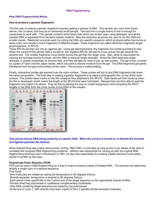
  • 1. DNA Fingerprinting How DNA Fingerprinting Works How to produce a genetic fingerprint: The first step to making a genetic fingerprint requires getting a sample of DNA. This sample can come from blood, semen, hair or saliva, and may be an extremely small sample. The root from a single strand of hair is enough for researchers to work with. This sample contains white blood cells which are broken open using detergent, and all the useable DNA is separated from the extra cellular material. Next the restriction enzymes are used to cut the DNA into smaller pieces. Restriction enzymes work by cutting the DNA at a specific sequence, which produces either blunt ends or sticky ends, and results in many fragments of different lengths. These fragments are called restriction fragments length polymorphisms, or RFLPs. These RFLPs are then put into an agarose gel. Using gel electrophoresis, the fragments are sorted according to size. When the current of the electric field is turned on, the negative RFLPs will start to move across the gel towards the positive end. The smaller fragments move farther across the gel than the larger ones. Also, alkali is responsible for causing the hydrogen bonds to break, and the DNA to become single-stranded. When the DNA becomes single- stranded, it causes nucleotides to become free, and they will later be used to pair up with probes. The gel is then covered by a piece of nylon and thin paper towels, which are used to absorb moisture from the gel. The DNA fragments get gently transferred from the gel to the surface of the nylon. This process is called blotting. Finally, radioactive probes get washed over the nylon surface. These probes will join to any DNA fragments that share the same composition. The final step to making a genetic fingerprint is to place a photographic film on top of the nylon surface. The probes leave marks on the film wherever they attached to the RFLPs. Dark bands will then show up when the film is developed, which marks the length of the RFLPs that were hybridized. Researchers are then able to read the fingerprint and match it to others. They do this by placing the xray on a light background, and comparing the RFLP lengths in the DNA from the crime scene, to the DNA of the suspect. This picture shows DNA being sorted by an electric field. When the current is turned on, it attracts the shortest and lightest particles the farthest. When fingerprinting was a fairly new process, during 1984-1990, it could take as long as four to six weeks of lab work to complete and compare DNA fingerprinting evidence. Jeffreys was responsible for coming up with the original DNA fingerprinting technique and in November of 1991, he also was responsible for creating a better test that could obtain results in as little as two days. Polymerase Chain Reaction (PCR) PCR can be used in DNA fingerprinting as a way to make numerous copies of isolated DNA. The process can selectively amplify a single copy of a desired sequence. First Cycle: -Dna molecules are melted by raising the temperature to 95 degrees Celcius. -Strands separate, temperature is lowered to 60 degrees Celcius. -Each primer binds specifically to the 3 prime end of the target sequence on the appropriate strands of DNA. -Primers direct taq polymerase to synthesize complementary nucleotides. -Only DNA containing target sequence are copied by taq polymerase. -At the end of cycle 1, both strands have been copied to form 2 partially double-stranded molecules.
  • 2. -These steps are repeated during the next cycles. After two cycles, four partially double-stranded molecules are produced, all containing the target sequence. -After many cycles, millions of copies of the DNA can be produced. -This is useful in DNA fingerprinting, as researchers can do many tests on the same DNA sample. What is DNA Fingerprinting? DNA fingerprinting is a process through which the unique genetic code of a person is identified and codified. DNA fingerprint is called so because just like the technology of fingerprint became very popular in the 1930s as means of crime detection, beginning in the 80s, DNA testing achieved a similar level of popularity in forensic science. Reasons Why It is Conducted Though fingerprints can be changed through surgical means or may be tampered with, the DNA code of every individual is completely unique and it therefore serves as a distinct marker of a person or evidence of his/her presence at a certain place. Even very small amounts of body fluids like sweat, mucus, blood and semen that the criminal inadvertently leaves behind on the scene of the crime can be later traced, and successfully reveal his/her identity conclusively, since DNA is present uniformly in each cell of an individual’s body. Thus, forensic DNA fingerprinting has become extremely popular as a method of crime detection. However, this method can also be used for medical purposes like detecting and predicting the chances of rare hereditary diseases that can be passed on from parents to their unborn children as well as in newborn babies. DNA fingerprinting is also extremely useful in research and development of cures for genetic disorders, as it helps locate and isolate the genetic patterns that cause certain diseases in particular families or racial groups, and thereby eventually shows the way to curing the condition. In addition, maintaining official records of the DNA fingerprints of each citizen in a state can help identify missing persons and casualties of accidents, as well as serve as a device of personal identification in important commercial transactions. DNA Fingerprinting DNA Fingerprinting is widely used in investigating and solving criminal crimes. Below are the steps involved for completing DNA fingerprinting. 1. Steps in DNA Fingerprinting A.-DNA is cut into fragments of specific sizes by certain enzymes called restriction enzymes. - Restriction enzymes are taken from bacteria - Each restriction enzymes recognizes and cuts DNA at a specific sequence - The DNA fragments can vary in number and size
  • 3. - Gel electrophoresis determine the numbers and sizes of DNA fragments procuduced by restriction enzyme treatment * If DNA is too small, the PCR is used to increase the quantity of DNA for analysis. The PCR makes many copies of the DNA in a small sample. Example; blood, saliva, hair, urine, or muscle. 2. The value of PCR (polymerase chain reaction) is that it makes many copies of the DNA in a small sample. 3. Restriction enzymes are taken from bacteria that use them to stop infection by viruses. 4. DNA probes are known sequences of DNA that will bind to their coomplementary DNA sequences wherever they are. It's used with the specific sequence that to the DNA fragement on a membrane with another sequence. 5'- TATATAGCTC-3' ---> 3'-ATATATCGAG-5' 5.ID criminals, ID remains, and genetic disorders are examples of uses of DNA fingerprinting Introduction Like the fingerprints that came into use by detectives and police labs during the 1930s, each person has a unique DNA fingerprint (Figure 1). Unlike a conventional fingerprint that occurs only on the fingertips and can be altered by surgery, a DNA fingerprint is the same for every cell, tissue, and organ of a person. It cannot be altered by any known treatment. Consequently, DNA fingerprinting is rapidly becoming the primary method for identifying and distinguishing among individual human beings.
  • 4. An additional benefit of DNA fingerprint technology is the diagnosis of inherited disorders in adults, children, and unborn babies. Even bloodstained clothing from Abraham Lincoln has been analyzed for evidence of a genetic disorder called Marfan's Syndrome. [Topics] The Structure of DNA The characteristics of all living organisms, including humans, are essentially determined by information contained within DNA that they inherit from their parents. The molecular structure of DNA can be imagined as a zipper with each tooth represented by one of four letters (A, C, G, or T) and with opposite teeth forming one of two pairs, either AT or GC (Figure 2). The information contained in DNA is the sequence of letters along the zipper. For example, the sequence ACGCT represents different information than the sequence AGTCC in the same way that the word "POST" has a different meaning from "STOP" or "POTS," even though they use the same letters. The traits of a human being are the result of information contained in the DNA code. Living organisms that look different or have different characteristics also have different DNA sequences. The more different the organisms, the more different are the DNA sequences. DNA fingerprinting is a very quick way to compare the DNA sequences of any two living organisms. [Topics] Making DNA Fingerprints DNA fingerprinting is a laboratory procedure that requires six steps (Figure 3):
  • 5. 1) Isolation of DNA. DNA must be recovered from the cells or tissues of the body. Only a small amount of tissue, like blood, hair, or skin, is needed. For example, the amount of DNA found at the root of one hair is usually sufficient. 2) Cutting, sizing, and sorting. Special enzymes called restriction enzymes are used to cut the DNA at specific places. For example, an enzyme called EcoR1, found in bacteria, will cut DNA only when the sequence GAATTC occurs. The DNA pieces are sorted according to size by a sieving technique called electrophoresis. The DNA pieces are passed through a gel made from seaweed agarose (a jelly-like product made from seaweed). This technique is the DNA equivalent of screening sand through progressively finer mesh screens to determine particle sizes. 3) Transfer of DNA to nylon. The distribution of DNA pieces is transferred to a nylon sheet by placing the sheet on the gel and soaking them overnight. 4-5) Probing. Adding radioactive or colored probes to the nylon sheet produces a pattern called the DNA fingerprint. Each probe typically sticks in only one or two specific places on the nylon sheet. 6) DNA fingerprint. The final DNA fingerprint is built by using several probes (5-10 or more) simultaneously. It resembles the bar codes used by grocery store scanners. [Topics] Uses of DNA Fingerprints
  • 6. DNA fingerprints are useful in several areas of society. They are used by professionals in human health and the justice system. Diagnosis of inherited disorders DNA fingerprinting is used to diagnose inherited disorders in both prenatal and newborn babies in hospitals around the world. These disorders may include cystic fibrosis, hemophilia, Huntington's disease, familial Alzheimer's, sickle cell anemia, thalassemia, and many others. Early detection of such disorders enables the medical staff to prepare themselves and the parents for proper treatment of the child. In some programs, genetic counselors use DNA fingerprint information to help prospective parents understand the risk of having an affected child. In other programs, prospective parents use DNA fingerprint information in their decisions concerning affected pregnancies. Developing cures for inherited disorders Research programs to locate inherited disorders on the chromosomes depend on the information contained in DNA fingerprints. By studying the DNA fingerprints of relatives who have a history of some particular disorder, or by comparing large groups of people with and without the disorder, it is possible to identify DNA patterns associated with the disease in question. This work is a necessary first step in designing an eventual genetic cure for these disorders. Forensic or criminal FBI and police labs around the U.S. have begun to use DNA fingerprints to link suspects to biological evidence-blood or semen stains, hair, or items of clothing-found at the scene of a crime. Since 1987, more than 150 cases have been decided with the assistance of DNA fingerprint evidence. Another important use of DNA fingerprints in the court system is to establish paternity in custody and child support litigation. In these applications, DNA fingerprints bring an unprecedented, nearly perfect accuracy to the determination. Southern blot From Wikipedia, the free encyclopedia Jump to: navigation, search A Southern blot is a method routinely used in molecular biology for detection of a specific DNA sequence in DNA samples. Southern blotting combines transfer of electrophoresis-separated DNA fragments to a filter membrane and subsequent fragment detection by probe hybridization. The method is named after its inventor, the British biologist Edwin Southern. [1] Other blotting methods (i.e., western blot, [2] northern blot, eastern blot, southwestern blot) that employ similar principles, but using RNA or protein, have later been named in reference to Edwin Southern's name. As the technique was eponymously named, Southern blot is capitalized as is conventional for proper nouns. The names for other blotting methods may follow this convention, by analogy. [3] Method 1. Restriction endonucleases are used to cut high-molecular-weight DNA strands into smaller fragments. 2. The DNA fragments are then electrophoresed on an agarose gel to separate them by size. 3. If some of the DNA fragments are larger than 15 kb, then prior to blotting, the gel may be treated with an acid, such as dilute HCl, which depurinates the DNA fragments, breaking the DNA into smaller pieces, thus allowing more efficient transfer from the gel to membrane. 4. If alkaline transfer methods are used, the DNA gel is placed into an alkaline solution (typically containing sodium hydroxide) to denature the double-stranded DNA. The denaturation in an alkaline environment may improve binding of the negatively charged thymine residues of DNA to a positively charged amino groups of membrane, separating it into single DNA strands for later hybridization to the probe (see below), and destroys any residual RNA that may still be present in the DNA. The choice of alkaline over neutral transfer methods, however, is often empirical and may result in equivalent results. [citation needed]
  • 7. 5. A sheet of nitrocellulose (or, alternatively, nylon) membrane is placed on top of (or below, depending on the direction of the transfer) the gel. Pressure is applied evenly to the gel (either using suction, or by placing a stack of paper towels and a weight on top of the membrane and gel), to ensure good and even contact between gel and membrane. If transferring by suction 20X SSC buffer is used to ensure a seal and prevent drying of the gel. Buffer transfer by capillary action from a region of high water potential to a region of low water potential (usually filter paper and paper tissues) is then used to move the DNA from the gel on to the membrane; ion exchange interactions bind the DNA to the membrane due to the negative charge of the DNA and positive charge of the membrane. 6. The membrane is then baked in a vacuum or regular oven at 80 °C for 2 hours (standard conditions; nitrocellulose or nylon membrane) or exposed to ultraviolet radiation (nylon membrane) to permanently attach the transferred DNA to the membrane. 7. The membrane is then exposed to a hybridization probe—a single DNA fragment with a specific sequence whose presence in the target DNA is to be determined. The probe DNA is labelled so that it can be detected, usually by incorporating radioactivity or tagging the molecule with a fluorescent or chromogenic dye. In some cases, the hybridization probe may be made from RNA, rather than DNA. To ensure the specificity of the binding of the probe to the sample DNA, most common hybridization methods use salmon or herring sperm DNA for blocking of the membrane surface and target DNA, deionized formamide, and detergents such as SDS to reduce non-specific binding of the probe. 8. After hybridization, excess probe is washed from the membrane (typically using SSC buffer), and the pattern of hybridization is visualized on X-ray film by autoradiography in the case of a radioactive or fluorescent probe, or by development of color on the membrane if a chromogenic detection method is used. Western blot From Wikipedia, the free encyclopedia Jump to: navigation, search Western blot using an antibody that recognizes proteins modified with lipoic acid. The western blot (sometimes called the protein immunoblot) is a widely accepted analytical technique used to detect specific proteins in the given sample of tissue homogenate or extract. It uses gel electrophoresis to separate native proteins by 3-D structure or denatured proteins by the length of the polypeptide. The proteins are then transferred to a membrane (typically nitrocellulose or PVDF), where they are stained with antibodies specific to the target protein. [1][2] There are now many reagent companies that specialize in providing antibodies (both monoclonal and polyclonal antibodies) against tens of thousands of different proteins. [3] Commercial antibodies can be expensive, although the unbound antibody can be reused between experiments. This method is used in the fields of molecular biology, biochemistry, immunogenetics and other molecular biology disciplines. Other related techniques include using antibodies to detect proteins in tissues and cells by immunostaining and enzyme- linked immunosorbent assay (ELISA).
  • 8. The method originated in the laboratory of Harry Towbin at the Friedrich Miescher Institute [1] . The name Western blot was given to the technique by W. Neal Burnette [4] and is a play on the name Southern blot, a technique for DNA detection developed earlier by Edwin Southern. Detection of RNA is termed northern blot and was developed by George Stark at Stanford [5] . Steps Tissue preparation Samples can be taken from whole tissue or from cell culture. Solid tissues are first broken down mechanically using a blender (for larger sample volumes), using a homogenizer (smaller volumes), or by sonication. Cells may also be broken open by one of the above mechanical methods. However, virus or environmental samples can be the source of protein and thus western blotting is not restricted to cellular studies only. Assorted detergents, salts, and buffers may be employed to encourage lysis of cells and to solubilize proteins. Protease and phosphatase inhibitors are often added to prevent the digestion of the sample by its own enzymes. Tissue preparation is often done at cold temperatures to avoid protein denaturing and degradation. A combination of biochemical and mechanical techniques – comprising various types of filtration and centrifugation – can be used to separate different cell compartments and organelles. Gel electrophoresis Main article: Gel electrophoresis The proteins of the sample are separated using gel electrophoresis. Separation of proteins may be by isoelectric point (pI), molecular weight, electric charge, or a combination of these factors. The nature of the separation depends on the treatment of the sample and the nature of the gel. This is a very useful way to identify a protein. By far the most common type of gel electrophoresis employs polyacrylamide gels and buffers loaded with sodium dodecyl sulfate (SDS). SDS-PAGE (SDS polyacrylamide gel electrophoresis) maintains polypeptides in a denatured state once they have been treated with strong reducing agents to remove secondary and tertiary structure (e.g. disulfide bonds [S-S] to sulfhydryl groups [SH and SH]) and thus allows separation of proteins by their molecular weight. Sampled proteins become covered in the negatively charged SDS and move to the positively charged electrode through the acrylamide mesh of the gel. Smaller proteins migrate faster through this mesh and the proteins are thus separated according to size (usually measured in kilodaltons, kDa). The concentration of acrylamide determines the resolution of the gel - the greater the acrylamide concentration the better the resolution of lower molecular weight proteins. The lower the acrylamide concentration the better the resolution of higher molecular weight proteins. Proteins travel only in one dimension along the gel for most blots. Samples are loaded into wells in the gel. One lane is usually reserved for a marker or ladder, a commercially available mixture of proteins having defined molecular weights, typically stained so as to form visible, coloured bands. When voltage is applied along the gel, proteins migrate through it at different speeds dependent on their size. These different rates of advancement (different electrophoretic mobilities) separate into bands within each lane.
  • 9. It is also possible to use a two-dimensional (2-D) gel which spreads the proteins from a single sample out in two dimensions. Proteins are separated according to isoelectric point (pH at which they have neutral net charge) in the first dimension, and according to their molecular weight in the second dimension. Transfer In order to make the proteins accessible to antibody detection they are moved from within the gel onto a membrane made of nitrocellulose or polyvinylidene difluoride (PVDF). The primary method for transferring the proteins is called electroblotting and uses an electric current to pull proteins from the gel into the PVDF or nitrocellulose membrane. The proteins move from within the gel onto the membrane while maintaining the organization they had within the gel. An older method of transfer involves placing a membrane on top of the gel, and a stack of filter papers on top of that. The entire stack is placed in a buffer solution which moves up the paper by capillary action, bringing the proteins with it. In practice this method is not used as it takes too much time; electroblotting is preferred. As a result of either "blotting" process, the proteins are exposed on a thin surface layer for detection (see below). Both varieties of membrane are chosen for their non-specific protein binding properties (i.e. binds all proteins equally well). Protein binding is based upon hydrophobic interactions, as well as charged interactions between the membrane and protein. Nitrocellulose membranes are cheaper than PVDF, but are far more fragile and do not stand up well to repeated probings. The uniformity and overall effectiveness of transfer of protein from the gel to the membrane can be checked by staining the membrane with Coomassie Brilliant Blue or Ponceau S dyes. Ponceau S is the more common of the two, due to its higher sensitivity and water solubility, the latter making it easier to subsequently destain and probe the membrane, as described below. [6]
  • 10. Blocking Since the membrane has been chosen for its ability to bind protein and as both antibodies and the target are proteins, steps must be taken to prevent the interactions between the membrane and the antibody used for detection of the target protein. Blocking of non-specific binding is achieved by placing the membrane in a dilute solution of protein - typically 3- 5% Bovine serum albumin (BSA) or non-fat dry milk (both are inexpensive) in Tris-Buffered Saline (TBS) or I-Block, with a minute percentage (0.1%) of detergent such as Tween 20 or Triton X-100. The protein in the dilute solution attaches to the membrane in all places where the target proteins have not attached. Thus, when the antibody is added, there is no room on the membrane for it to attach other than on the binding sites of the specific target protein. This reduces "noise" in the final product of the western blot, leading to clearer results, and eliminates false positives. Detection During the detection process the membrane is "probed" for the protein of interest with a modified antibody which is linked to a reporter enzyme; when exposed to an appropriate substrate this enzyme drives a colourimetric reaction and produces a color. For a variety of reasons, this traditionally takes place in a two-step process, although there are now one-step detection methods available for certain applications. Two steps Primary antibody The primary antibodies are generated when a host species or immune cell culture is exposed to protein of interest (or a part thereof). Normally, this is part of the immune response, whereas here they are harvested and used as sensitive and specific detection tools that bind the protein directly. After blocking, a dilute solution of primary antibody (generally between 0.5 and 5 micrograms/mL) is incubated with the membrane under gentle agitation. Typically, the solution is composed of buffered saline solution with a small percentage of detergent, and sometimes with powdered milk or BSA. The antibody solution and the membrane can be sealed and incubated together for anywhere from 30 minutes to overnight. It can also be incubated at different temperatures, with warmer temperatures being associated with more binding, both specific (to the target protein, the "signal") and non- specific ("noise"). Secondary antibody After rinsing the membrane to remove unbound primary antibody, the membrane is exposed to another antibody, directed at a species-specific portion of the primary antibody. Antibodies come from animal sources (or animal sourced hybridoma cultures); an anti-mouse secondary will bind to almost any mouse-sourced primary antibody, which allows some cost savings by allowing an entire lab to share a single source of mass-produced antibody, and provides far more consistent results. This is known as a secondary antibody, and due to its targeting properties, tends to be referred to as "anti- mouse," "anti-goat," etc. The secondary antibody is usually linked to biotin or to a reporter enzyme such as alkaline phosphatase or horseradish peroxidase. This means that several secondary antibodies will bind to one primary antibody and enhance the signal. Most commonly, a horseradish peroxidase-linked secondary is used to cleave a chemiluminescent agent, and the reaction product produces luminescence in proportion to the amount of protein. A sensitive sheet of photographic film is placed against the membrane, and exposure to the light from the reaction creates an image of the antibodies bound to the blot. A cheaper but less sensitive approach utilizes a 4-chloronaphthol stain with 1% hydrogen peroxide; reaction of peroxide radicals with 4-chloronaphthol produces a dark purple stain that can be photographed without using specialized photographic film.
  • 11. As with the ELISPOT and ELISA procedures, the enzyme can be provided with a substrate molecule that will be converted by the enzyme to a colored reaction product that will be visible on the membrane (see the figure below with blue bands). Another method of secondary antibody detection utilizes a near-infrared (NIR) fluorophore-linked antibody. Light produced from the excitation of a fluorescent dye is static, making fluorescent detection a more precise and accurate measure of the difference in signal produced by labeled antibodies bound to proteins on a western blot. Proteins can be accurately quantified because the signal generated by the different amounts of proteins on the membranes is measured in a static state, as compared to chemiluminescence, in which light is measured in a dynamic state. [7] A third alternative is to use a radioactive label rather than an enzyme coupled to the secondary antibody, such as labeling an antibody-binding protein like Staphylococcus Protein A or Streptavidin with a radioactive isotope of iodine. Since other methods are safer, quicker, and cheaper, this method is now rarely used; however, an advantage of this approach is the sensitivity of auto-radiography based imaging, which enables highly accurate protein quantification when combined with optical software (e.g. Optiquant). One step Historically, the probing process was performed in two steps because of the relative ease of producing primary and secondary antibodies in separate processes. This gives researchers and corporations huge advantages in terms of flexibility, and adds an amplification step to the detection process. Given the advent of high-throughput protein analysis and lower limits of detection, however, there has been interest in developing one-step probing systems that would allow the process to occur faster and with less consumables. This requires a probe antibody which both recognizes the protein of interest and contains a detectable label, probes which are often available for known protein tags. The primary probe is incubated with the membrane in a manner similar to that for the primary antibody in a two-step process, and then is ready for direct detection after a series of wash steps. Western blot using radioactive detection system Analysis After the unbound probes are washed away, the western blot is ready for detection of the probes that are labeled and bound to the protein of interest. In practical terms, not all westerns reveal protein only at one band in a membrane. Size approximations are taken by comparing the stained bands to that of the marker or ladder loaded during electrophoresis.
  • 12. The process is repeated for a structural protein, such as actin or tubulin, that should not change between samples. The amount of target protein is normalized to the structural protein to control between groups. This practice ensures correction for the amount of total protein on the membrane in case of errors or incomplete transfers. Colorimetric detection The colorimetric detection method depends on incubation of the western blot with a substrate that reacts with the reporter enzyme (such as peroxidase) that is bound to the secondary antibody. This converts the soluble dye into an insoluble form of a different color that precipitates next to the enzyme and thereby stains the membrane. Development of the blot is then stopped by washing away the soluble dye. Protein levels are evaluated through densitometry (how intense the stain is) or spectrophotometry. Chemiluminescent detection Chemiluminescent detection methods depend on incubation of the western blot with a substrate that will luminesce when exposed to the reporter on the secondary antibody. The light is then detected by photographic film, and more recently by CCD cameras which capture a digital image of the western blot. The image is analysed by densitometry, which evaluates the relative amount of protein staining and quantifies the results in terms of optical density. Newer software allows further data analysis such as molecular weight analysis if appropriate standards are used. Radioactive detection Radioactive labels do not require enzyme substrates, but rather allow the placement of medical X-ray film directly against the western blot which develops as it is exposed to the label and creates dark regions which correspond to the protein bands of interest (see image to the right). The importance of radioactive detections methods is declining due to its hazardous radiation [citation needed] , because it is very expensive, health and safety risks are high, and ECL (enhanced chemiluminescence) provides a useful alternative. Fluorescent detection The fluorescently labeled probe is excited by light and the emission of the excitation is then detected by a photosensor such as CCD camera equipped with appropriate emission filters which captures a digital image of the western blot and allows further data analysis such as molecular weight analysis and a quantitative western blot analysis. Fluorescence is considered to be one of the best methods for quantification, but is less sensitive than chemiluminescence. [8] Secondary probing One major difference between nitrocellulose and PVDF membranes relates to the ability of each to support "stripping" antibodies off and reusing the membrane for subsequent antibody probes. While there are well-established protocols available for stripping nitrocellulose membranes, the sturdier PVDF allows for easier stripping, and for more reuse before background noise limits experiments. Another difference is that, unlike nitrocellulose, PVDF must be soaked in 95% ethanol, isopropanol or methanol before use. PVDF membranes also tend to be thicker and more resistant to damage during use.
  • 13. 2-D gel electrophoresis Main article: Two-dimensional gel electrophoresis 2-dimensional SDS-PAGE uses the principles and techniques outlined above. 2-D SDS-PAGE, as the name suggests, involves the migration of polypeptides in 2 dimensions. For example, in the first dimension polypeptides are separated according to isoelectric point, while in the second dimension polypeptides are separated according to their molecular weight. The isoelectric point of a given protein is determined by the relative number of positively (e.g. lysine and arginine) and negatively (e.g. glutamate and aspartate) charged amino acids, with negatively charged amino acids contributing to a high isoelectric point and positively charged amino acids contributing to a low isoelectric point. Samples could also be separated first under nonreducing conditions using SDS-PAGE and under reducing conditions in the second dimension, which breaks apart disulfide bonds that hold subunits together. SDS-PAGE might also be coupled with urea-PAGE for a 2-dimensional gel. In principle, this method allows for the separation of all cellular proteins on a single large gel. A major advantage of this method is that it often distinguishes between different isoforms of a particular protein - e.g. a protein that has been phosphorylated (by addition of a negatively charged group). Proteins that have been separated can be cut out of the gel and then analysed by mass spectrometry, which identifies the protein. Please refer to reference articles for examples of the application of 2-D SDS PAGE.