
Empfohlen
Empfohlen
Weitere ähnliche Inhalte
Was ist angesagt?
Was ist angesagt? (20)
Andere mochten auch
Ähnlich wie Transmission electron microscope
Ähnlich wie Transmission electron microscope (20)
Kürzlich hochgeladen
Kürzlich hochgeladen (20)
Transmission electron microscope
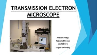
- 1. TRANSMISSION ELECTRON MICROSCOPE Presented by: Rajkamal Mohan (CHP15111) Tezpur University
- 2. CONTENTS Introduction and History Theoretical Background Instrumentation Sample Preparation Drawbacks of TEM
- 3. Introduction Microscopes are used to see objects that cannot be seen by naked eyes. The range can be between mm to nm. There are three main microscopic techniques: Optical Microscopy Scanning Probe Microscopy Electron Microscopy
- 4. Transmission Electron Microscopy (TEM) Transmission electron microscopy (TEM) is a microscopy technique where a beam of electrons is transmitted through a ultra thin specimen. An image is formed from the interaction of the electrons transmitted through the specimen; the image is magnified and focused onto an imaging device, such as a fluorescent screen, on a layer of photographic film, or to be detected by a sensor such as a CCD camera.
- 5. History 1897 1924 1929 1931 1934 J.J Thompson Louis deBroglie E. Ruska Knoll & Ruska Driest & Muller 1938 von Borries & Ruska Discovers electron Identifies wavelength for electron Ph.D thesis on magnetic lenses 1st electron microscope built Surpass resolution of the light microscope (LM) First practical electron microscope (EM) Seimens : 10 nm resolution J.J Thompson L. deBroglie E. Ruska M. Knoll
- 6. Theoretical Background Why we need Electron Microscope? Light microscopes are limited by the physics of light to 500x or 1000x magnification and a resolution of 0.2 micrometers. In the early 1930's there was a scientific desire to see the fine details of the interior structures of organic cells (nucleus, mitochondria...etc.). This required 10,000x plus magnification which was just not possible using Light Microscopes.
- 7. LM, resolving power ~0.25µm, maximum (useful) magnification is about 250µm/0.25µm = 1000X. Any magnification above this value represents empty magnification TEM at 60,000 volts has a resolving power of about 0.0025 nm. Maximum useful magnification of about 100 million times
- 8. Difference between light microscope and electron microscope FEATURES LIGHT MICROSCOPE ELECTRON MICROSCOPE Electromagnetic spectrum used Visible light 760nm (red) – 390nm Colours visible Electrons app. 4nm Monochrome Maximum resolving power approx. 200nm 0.2nm Maximum magnification x1000 – x1500 x500 000 Radiation source Tungsten or quartz halogen lamp High voltage (50kV) tungsten lamp Lenses Glass Magnets Interior Air-filled Vacuum Focusing screen Human eye (retina), photographic film fluorescent (TV) screen, photographic film
- 9. Instrumentation TEM characterizes samples simultaneously by diffraction and imaging techniques.
- 10. Instrumentation (Contd.) In a conventional transmission electron microscope, a thin specimen is irradiated with an electron beam of uniform current density. Electrons illuminate the specimen through a condenser lens system. Objective lens provides the formation of either image or diffraction pattern of the specimen. The electron intensity distribution is magnified with a lens system and viewed on a fluorescent screen. The image can be recorded by an image plate or digitally by a CCD camera.
- 11. Design of Transmission Electron Microscope A simplified ray diagram of a TEM consists of an electron source, condenser lens with aperture, specimen, objective lens with aperture, projector lens and fluorescent screen.
- 12. Electron Gun Electron beam is generated in the electron gun. Two basic types of guns are used: 1. Thermionic Gun: Based on two types of filaments: Tungsten(W) and Lanthanum Hexaboride(LaB6). 2. Field Emission Gun(FEG): Employs either a thermally assisted cold field emitter or Schottky emitter.
- 13. Condenser Lens Illuminates the specimen. Relatively weak lens. Longer focal length than objective or projector lens. May bring electron beam into focus directly upon specimen, above the specimen (over focusing) or below the specimen (under focusing).
- 14. Objective Lens Strong lens It has highly concentrated magnetic field and short focal length. Total magnification in the TEM is a combination of the magnification from the objective lens times the magnification of the intermediate lens times the magnification of the projector lens. Each of which is capable of approximately 100x. Mob X Mint X Mproj = Total Mag
- 15. Fluorescent Screen In TEM, screen coated with a material in the visible range, eg zinc sulphide, is installed beneath the projector lens in the path of the electron beam. Screen emits visible light when bombarded with electrons.
- 16. Vacuum System Electron can’t travel more than a few angstrom without colliding with gas molecules. Distance between photographic plate and electron gun is approximately 1 meter. Two types of vacuum pump are used 1. Rotary (mechanical) pump. 2. Diffusion pump (Oil or Mercury)
- 17. Some typical TEM models
- 18. TEM Sample Preparation The TEM sample should be thin, so special care must be taken while cutting a thin slice so that the specimen is not deformed during its preparation. Some common techniques are:- a) SPARK CUTTER: - In this, electric discharge between a wire and the specimen is used to cut the metal by removing small particles of metal from the surface of the specimen. b) FOCUSED ION BEAM (FIB):- A thin slice of the sample is cut by an ion beam on a scanning ion microscope. The main advantage of this method is that it allows selective thinning at desired locations by cutting trenches in the sample.
- 19. TEM Sample Preparation Some basic requirements Cleaning the surface of the specimen The proper cleaning of the surface of the sample is important because the surface can contain a variety of unwanted deposits, such as dust, silt, media components or other contaminants. The best way to clean the surface of specimen from contaminants is to carefully rinse them three times for 10 min in 0.1M cacodylic acid buffer (pH 7.3) at room temperature.
- 20. TEM Sample Preparation Fixatives Are chemicals that denature and precipitate cellular macromolecules. Primary fixation of the specimen Fixation can be achieved by perfusion and microinjection, immersion with vapours using various fixatives including aldehydes (glutaraldehyde), osmium tetroxide, tannic acid.
- 21. What information can TEM provide? RECTIONS ON THE BOTTOM SIDE ARE EXAMINED IN THIN OR FOIL SPECIMEN (TEM)
- 22. What information can TEM provide? Thickness of the specimen: The transmission of unscattered electrons is inversely proportional to the specimen thickness. Orientation, atomic arrangements and phases present: Given by incident electrons that are scattered by specimen atoms in an elastic fashion. These electrons follow Bragg's Law nλ=2dSinθ The incident electrons that are scattered by the same atomic spacing will be scattered by the same angle. These scattered electrons can be collated using magnetic lenses to form a pattern of spots; each spot corresponding to a specific atomic spacing (a plane). Diffraction pattern of a monocrystalline sample Selected area electron diffraction
- 23. What information can TEM provide? Inelastically scattered electrons can be utilized in two ways: Electron Energy Loss Spectroscopy (EELS) and Kikuchi Bands. Elemental composition and atomic bonding state: Determined by analyzing the energy with spectroscope attached under the electron microscope (Electron Energy Loss Spectroscopy). By selecting electrons with a specific loss energy, element distribution in specimen can be visualized. Kikuchi lines: Bands of alternating dark and bright lines related to the atomic spacing of the specimen. Appear in transmission electron diffraction patterns of relatively thick crystals due to Bragg reflection of inelastically scattered electrons. The Kikuchi lines pass straight through the transmitted and diffracted spots.
- 24. Limitations of TEM Sampling: Very small sample size. But fortunately we are dealing with nanostructures. Interpreting transmission images: TEM presents 2D images of 3D specimens. Electron Beam damage and Safety: TEM is a potential dangerous instrument that generates radiation level that is enough to kill human being. Specimen preparation: Your specimens have to be thin, very thin (has to be electron transparent) if you are going to get any information.
- 25. Examples of TEM images Matrix - β'-Ni2AlTi Precipitation in an Al-Cualloy Dislocations in superalloy
- 26. References [1] William R. Herguth, President, Guy Nadeau.Applications of Scanning Electron Microscopy and Energy Dispersive Spectroscopy (SEM/EDS) To Practical Tribology Problems. Senior Technical Associate Herguth Laboratories, Inc. [2] R.F. Egerton. Electron Energy-Loss Spectroscopy in the Electron Microscope. [3] M.Von Heimendahl, W.Bell, G.Thomas. Applications of Kikuchi line Analyses in Electron Microscopy. Journal of Applied Physics 35 (1964) 3614. [4] C. Richard Brundle, Charles A. Evans Jr, Shaun Wilson. Encyclopedia of materials characterization, Butterworth-Heinemann publications, 1992. [5] Joachim Mayer, Lucille A. Giannuzzi, Takeo Kamino, and Joseph Michael. TEM Sample Preparation and FIB-Induced Damage. Mrs Bulletin, volume 32, May 2007
- 27. THANK YOU