Pathophysiology review pt_i
•Als PPT, PDF herunterladen•
4 gefällt mir•1,964 views

Review of the pathophysiology part i Pathophysiology(病理生理学) Pathology uploaded by prabesh 杰诗

Empfohlen
Weitere ähnliche Inhalte
Was ist angesagt?
Was ist angesagt? (20)
Ähnlich wie Pathophysiology review pt_i
Ähnlich wie Pathophysiology review pt_i (20)
Mehr von Prabesh Raj Jamkatel
Mehr von Prabesh Raj Jamkatel (20)
Kürzlich hochgeladen
Kürzlich hochgeladen (20)
Pathophysiology review pt_i
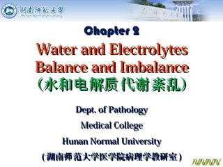
- 1. Dept. of PathologyDept. of Pathology Medical CollegeMedical College Hunan Normal UniversityHunan Normal University (( 湖南 范大学医学院病理学教研室师湖南 范大学医学院病理学教研室师 )) 1 Chapter 2Chapter 2 Water and ElectrolytesWater and Electrolytes Balance and ImbalanceBalance and Imbalance (水和 解 代 紊乱)电 质 谢(水和 解 代 紊乱)电 质 谢
- 2. 2 Water and ElectrolytesWater and Electrolytes Balance and ImbalanceBalance and Imbalance Physiological Basis of Water and SodiumPhysiological Basis of Water and Sodium MetabolismMetabolism Disorder of Other ElectrolytesDisorder of Other Electrolytes Regulation of Water and Sodium BalanceRegulation of Water and Sodium Balance Disorder of Water and Sodium MetabolismDisorder of Water and Sodium Metabolism
- 3. Distribution of Body Fluids Plasma 5% Interstitial 15% ICF 40% Extracellular fluid, ECF Intracellular fluid, ICF Transcellular fluid – secreted fluid (body cavities) (Third space) 1-2 % 3 ECF ICF
- 4. Factors Affecting Body Fluid Volume Fat, Sex, Age 4 Q: Do fat people have more or less body fluid?
- 5. Functions of Body Water Metabolism of biomoleculesMetabolism of biomolecules Body temperatureBody temperature LubricationLubrication Tissue constituent (boundTissue constituent (bound HH22O)O) 5
- 6. Intake (ml/day) Output (ml/day) Drinking 1000-1500 Food 700 Metabolism 300 Urine 1000-1500 (min: 500) Lungs 400 Skin 500 Stool 100 Total 2000-2500 (min: 1500) Total 2000-2500 Daily Balance of Water 6Q: Do infants require more or less water (per kg body weight)?
- 7. Distribution of Body Fluid ComponentsDistribution of Body Fluid Components Characteristics : a. Composition of electrolytes different between ICF and ECF b. Osmotic balance between ICF and ECF c. Electrically neutral in each compartment Blood Vessel Cell Membrane Proteins 7
- 8. Physiologic Functions of Electrolytes Maintenance of osmotic pressureMaintenance of osmotic pressure Generation of membrane potentialGeneration of membrane potential - Excitability of nerve and muscle- Excitability of nerve and muscle Participation in metabolism andParticipation in metabolism and functionfunction 8
- 9. Intake: 5 -10 g/d Absorption: Almost all by small intestine Excretion: Kidney (>97%), skin Sodium Balance ECF 50% ICF 10% Bone 40% ECF: Extracellular fluid ICF: Intracellular fluid Serum [NaSerum [Na++ ] 130~150 mmol/L] 130~150 mmol/L 9
- 10. 10 Water and ElectrolytesWater and Electrolytes Balance and ImbalanceBalance and Imbalance Physiological Basis of Water and SodiumPhysiological Basis of Water and Sodium MetabolismMetabolism Disorder of Other ElectrolytesDisorder of Other Electrolytes Regulation of Water and Sodium BalanceRegulation of Water and Sodium Balance Disorder of Water and Sodium MetabolismDisorder of Water and Sodium Metabolism
- 11. 11 Regulation of Body Fluids Two Levels: Neural - Thirst Hormones – Regulation through kidney Antidiuretic Hormone (ADH) Aldosterone (ADS) Atrial Natriuretic Peptide (ANP)
- 12. 12 Action of ADH: Role of Aquaporins PK = Protein Kinase PKa = Activated Protein Kinase Renal tubuleEpithelial cell
- 13. 13 Regulation of Body Fluids ADH Osmosis ↑ Distal tubules Reabsorption of H2O>Na+ Blood volume↓ Collecting ducts ADS Blood volume↓ Distal tubules Reabsorption of Na+ >H2O ↓Na+ /↑ K+ Collecting ducts Excretion of potassium Thirst Osmosis ↑ Thirst center Drinking water Blood volume↓ Regulator Stimulator Site of action Effect ANP Blood volume ↑ Distal tubules Excretion of sodium Collecting ducts Excretion of water
- 14. 14 Nephron ADH ADS ANP Regulation of Body Fluids by Hormones
- 15. 15 Water and Electrolytes BalanceWater and Electrolytes Balance and Imbalanceand Imbalance Physiological Basis of Water and SodiumPhysiological Basis of Water and Sodium MetabolismMetabolism Disorder of Other ElectrolytesDisorder of Other Electrolytes Regulation of Water and Sodium BalanceRegulation of Water and Sodium Balance Disorder of Water and Sodium MetabolismDisorder of Water and Sodium Metabolism
- 16. ECF↓ Hypovolemic ECF ↑ Hypervolemic ECF N Serum Na+ ↓ Hyponatremia Hypovolemic hyponatremia (Hypotonic dehydration) Hypervolemic hyponatremia (Water intoxication) Normovolemic hyponatremia Serum Na+ ↑ Hypernatremia Hypovolemic hypernatremia (Hypertonic dehydration) Hypervolemic hypernatremia (Salt intoxication) Normovolemic hypernatremia Serum Na+ N Hypovolemia (Isotonic dehydration) Hypervolemia (Edema) Classification of Water and Sodium Metabolic Disorders 16
- 17. Plasma ISF ICF Plasma ISF ICF Different Types of Water and Sodium Disorders Plasma ISF ICF ISF ICF Plasma Extracellula Intracellular 17
- 18. ECF↓ Hypovolemic ECF ↑ Hypervolemic ECF N Serum Na+ ↓ Hyponatremia Hypovolemic hyponatremia (Hypotonic) Hypervolemic hyponatremia (Water intoxication) Normovolemic hyponatremia Serum Na+ ↑ Hypernatremia Hypovolemic hypernatremia (Hypertonic) Hypervolemic hypernatremia (Salt intoxication) Normovolemic hypernatremia Serum Na+ N Hypovolemia (Isotonic) Hypervolemia (Edema) Classification of Water and Sodium Metabolic Disorders 22
- 19. 4. Pathogenesis of Edema I. Fluid interchange across the blood vessel - Abnormal distribution - Total amount of body fluid: N 23 Two types of balances disrupted II. Fluid interchange across the body - ↑ Retention of water and sodium - Total amount of body fluid: ↑
- 20. ① Capillary hydrostatic pressure (17 mmHg) ② Interstitial hydrostatic pressure (-6.5 mmHg) ③ Plasma colloidal osmotic pressure (28 mmHg) ④ Interstitial colloidal osmotic pressure (5 mmHg) The normal interchange of body fluid between plasma and interstitial fluid 24 (17 - (-6.5)) - (28 - 5) = 0.5 mmHg ① ② ③ ④
- 21. ① Increased capillary hydrostatic pressure ② Increased capillary permeability ③ Reduced plasma colloid osmotic pressure ④ Obstruction of lymph return 1) Imbalance of fluid interchange across the blood vessel Four Mechanisms: 25
- 22. 4. The most frequent clinical edema ① Cardiac edema: Right heart failure. This kind of edema usually shows up first in the legs and ankles. Why? Because good old gravity is pulling all that "loose" fluid straight down. So we call it “dependent edema”. 26
- 23. 27 ① Cardiac edema : Left heart failure – edema in the lungs (dyspnea).
- 24. 5. Alternations of metabolism and function Beneficial roles: (1) Diluting and neutralizing toxin(s) (2) Carrying antibodies and complements to edema region 28 Harmful roles: (1) Resulting in insufficient nutritional supply (2) Inducing dysfunctions of affected organs (3) May lead to death (edema of vital organs) ?
- 25. 29 Water and Electrolytes BalanceWater and Electrolytes Balance and Imbalanceand Imbalance Physiological Basis of Water and SodiumPhysiological Basis of Water and Sodium MetabolismMetabolism Disorder of Potassium MetabolismDisorder of Potassium Metabolism Regulation of Water and Sodium BalanceRegulation of Water and Sodium Balance Disorder of Water and Sodium MetabolismDisorder of Water and Sodium Metabolism
- 26. Potassium (K+ ): Distribution and Normal Functions ECF ICF (bone) 4.2 mmol/L (1.4%) 150 mmol/L (90%) K+ Distribution Serum [KSerum [K++ ] 3.5~5.5 mmol/L] 3.5~5.5 mmol/L 30
- 27. 31 Physiological Functions of K+ Cell metabolism Regulation of osmosis and pH Generation of resting potential
- 28. Generation of Resting Potential ← Resting Potential = K+ potential At resting: Plasma membrane permeability K + >> Na+ 32
- 29. 33 Serum K+ conc. < 3.5 mmol/L Disturbance ofDisturbance of Potassium MetabolismPotassium Metabolism Hypokalemia ( 低钾血症 ) Serum K+ conc. > 5.5 mmol/L Hyperkalemia ( 高钾血症 )
- 30. Changes of ECG in Hyperkalemia Delayed repolorization Flat T wave U wave Suppressed ST Speeded repolorization Peaked T wave Shortened Q-T 34
- 31. Dept. of PathologyDept. of Pathology Medical CollegeMedical College Hunan Normal UniversityHunan Normal University (( 湖南 范大学医学院病理学教研室师湖南 范大学医学院病理学教研室师 )) 35 Chapter 3Chapter 3 Acid-Base Balance andAcid-Base Balance and ImbalanceImbalance (酸 平衡紊乱)碱(酸 平衡紊乱)碱
- 32. 3636 Acid-Base Balance and ImbalanceAcid-Base Balance and Imbalance a.a. Acid-base homeostasisAcid-base homeostasis b.b. Parameters of acid-baseParameters of acid-base balancebalance c.c. Simple acid-base disturbanceSimple acid-base disturbance Metabolic acidosisMetabolic acidosis Respiratory acidosisRespiratory acidosis Metabolic alkalosisMetabolic alkalosis Respiratory alkalosisRespiratory alkalosis
- 33. Concepts of Acid and Base §2. Base: an acceptor of H+ . §1. Acid: a donor of hydrogen ions ( H+ ). 37
- 34. AcidsAcids Volatile acid ( 挥发 酸 ) H2CO3 Fixed acid ( 固定酸 ) Other acids 38
- 35. 2. Regulation of Acid-Base Balance2. Regulation of Acid-Base Balance Buffer Systems Blood Cells Bone Lungs Kidneys Three Levels 39
- 36. Buffer acid Buffer base Buffer systems in the bloodBuffer systems in the blood Ability 53 5 7 35 40
- 37. Cells Volatile acid (H2CO3) Fixed acids Lungs KidneysPlasma Production and Regulation of Acids and Bases Food Digestion Absorption Metabolism 41
- 38. 4242 Acid-Base Balance and ImbalanceAcid-Base Balance and Imbalance a.a. Acid-base homeostasisAcid-base homeostasis b.b. Parameters of acid-baseParameters of acid-base balancebalance c.c. Simple acid-base disturbanceSimple acid-base disturbance Metabolic acidosisMetabolic acidosis Respiratory acidosisRespiratory acidosis Metabolic alkalosisMetabolic alkalosis Respiratory alkalosisRespiratory alkalosis
- 39. 1. pH pH < 7.35: Acidosis pH > 7.45: Alkalosis §2. Normal value : 7.35 ~ 7.45 (average : 7.40) 43
- 40. Henderson-Hasselbalch EquationHenderson-Hasselbalch Equation pH = pKa + LogpH = pKa + Log [HCO[HCO33 -- ]] [H[H22COCO33]] = pKa + Log= pKa + Log 2424 1.21.2 = 6.1 + 1.3 = 7.4 44
- 41. 2. PaCO2 Partial pressure of carbon dioxide (CO2) in plasma (artery) Significance: respiratory parameter Normal Value: 33~46 mmHg (Average: 40 )PaCO2 ↑: Respiratory Acidosis Metabolic Alkalosis after compensation PaCO2 ↓: Respiratory Alkalosis Metabolic Acidosis after compensation 45
- 42. Normal Value: 22 ~ 27 mmol/L (Average: 24) [HCO3 - ] measured under “standard condition” 37~38°C Hb fully oxygenated PaCO2 @ 40 mmHg 37~38°C Hb fully oxygenated PaCO2 @ 40 mmHg 3. Standard Bicarbonate, SB SB ↑: Metabolic Alkalosis SB ↓: Metabolic Acidosis Not affected by respiration. Only reflecting metabolic factor. 46
- 43. Actual Bicarbonate, ABActual Bicarbonate, AB Reflecting: Both metabolic and respiratory factors HCO3 - measured under “actual condition”. Sealed off from air PaCO2 and O2 at original level Sealed off from air PaCO2 and O2 at original level Normal Value: the same as SB (24 mmol/L) 47
- 44. AB > SB, PaCO2 ↑ Respiratory acidosis (metabolic alkalosis after compensation) AB < SB, PaCO2 ↓ Respiratory alkalosis (metabolic acidosis after compensation) In physiological situation: AB = SB In pathological situation: AB ≠ SB AB vs. SB 48
- 45. 4.4. Buffer Base, BBBuffer Base, BB Meaning: BB ↑ - Metabolic alkalosis BB ↓ - Metabolic acidosis Normal: 45 ~ 52 mmol/L (Average: 48 ) The sum of all alkaline buffer substances in plasma (HCO3 - , HPO4 2- , Pr- , Hb- , HbO2 - ) 49
- 46. 5. Base Excess, BE The amount of a fixed acid or base that must be added to a blood sample to achieve a pH of 7.4 under standard condition. Normal value: -3.0 - +3.0 pH 7.35 7.4 7.45 BE -3.0 0 +3.0 Metabolic acidosis Normal Metabolic alkalosis §3. Meaning: 50
- 47. 6.6. Anion GapAnion Gap ,, AGAG The difference between undetermined anion (UA) and undetermined cation (UC) in the plasma (AG = UA - UC). AG↑ (AG>16): ↑ Fixed acids (metabolic acidosis) AG↓: little clinic meaning Undetermined AG = Na+ - (Cl- + HCO- 3) = 140 - (104 + 24) = 12 mmol/L (10 ~ 14 mmol/L) 51
- 48. 5252 Acid-Base Balance and ImbalanceAcid-Base Balance and Imbalance a.a. Acid-base homeostasisAcid-base homeostasis b.b. Parameters of acid-baseParameters of acid-base balancebalance c.c. Simple acid-base disturbanceSimple acid-base disturbance Metabolic acidosisMetabolic acidosis Respiratory acidosisRespiratory acidosis Metabolic alkalosisMetabolic alkalosis Respiratory alkalosisRespiratory alkalosis
- 49. pH Acidosis Respiratory [HCO3 - ]↓ H2CO3↑ Metabolic Alkalosis [HCO3 - ]↑ H2CO3 ↓ Metabolic Respiratory Types of Acid-Base DisturbanceTypes of Acid-Base Disturbance 53
- 50. Example 1. 1 diabetes patient : pH 7.32, HCO3 - 15 mmol/L, PaCO2 30 mmHg ; predict PaCO2 = 1.5×15 + 8±2 = 30.5±2 = 28.5 ~ 32.5 measured PaCO2 = 30, within 28.5 ~ 32.5; Therefore, simple MAc Equation : predict PaCO2 = 1.5×[HCO3 - ] + 8±2 Judgement : If measured PaCO2 within predicted PaCO2 , simple MAc If measured > predicted maximum, CO2 retention, MAc + RAc If measured < predicted minimum, CO2 too less, MAc + RAl Metabolic acidosis 54
- 51. 1.2 shock patient with pneumonia: pH 7.26 , HCO3 - 16 mmol/L , PaCO2 37 mmHg ; predicted PaCO2 = 1.5×16 + 8±2 = 32±2 = 30 ~ 34 measured PaCO2 =37, exceed predict maximum 34; Therefore, MAc + RAc Metabolic acidosis 55
- 52. 2.1 Pulmonary heart disease patient : pH 7.34, HCO3 - 31 , PaCO2 60 ; Predict HCO3 - = 24 + 0.4(60 - 40)±3 = 29 ~ 35 Measured HCO3 - = 31, within predicted, simple RAc Equation : predict HCO3 - = 24 + 0.4 PaCO△ 2 ±3 Example Judgement : If measured HCO3 - insofar as predict HCO3 - , simple RAc If measured > predict maximum, HCO3 - retention, RAc +MAl If measured < predict minimum, HCO3 - too less, RAc + MAc Respiratory acidosis 56
- 53. 2.2 Pulmonary heart disease patient given bicarbonate : pH 7.40 , HCO3 - 40 , PaCO2 67 ; Predict HCO3 - = 24 + 0.4(67 - 40)±3 = 31.8 ~ 37.8 Measured HCO3 - = 40, exceed predict maximum , RAc + MAl 2.3 Pulmonary heart disease patient : pH 7.22 , HCO3 - 20 , PaCO2 50 Predict HCO3 - = 24 + 0.4(50―40)±3 = 25 ~ 31 Measured HCO3 - =20, below predict minimum , RAc + MAc Respiratory acidosis 57
- 54. 3.1 Pyloric obstruction patient : pH 7.49 , HCO3 - 36 , PaCO2 48 ; Predict PaCO2 = 40 + 0.7(36-24)±5 = 43.4 ~ 53.4 Measured PaCO2 = 48, within predicted, simple MAl Equation : predict PaCO2 = 40 + 0.7 HCO△ 3 - ±5 Example Judgement : If measured PaCO2 insofar as predict PaCO2 , simple MAl If measured > predict maximum, CO2 retention, MAl + RAc If measured < predict minimum, CO2 too less, MAl + RAl Metabolic alkalosis 58
- 55. 3.2 Septic shock patient given excessive bicabonate and mechanical ventilation : pH 7.65 , HCO3 - 32 , PaCO2 30 ; Predict PaCO2 = 40 + 0.7(32-24)±5 = 40.6 ~ 50.6 Measured PaCO2 = 30, below predict minimum MAl + RAl 3.3 Pulmonary heart disease patient used the diuretics : pH 7.40 , HCO3 - 36 , PaCO2 60 ; Predict PaCO2 = 40 + 0.7(36-24)±5 = 43.4 ~ 53.4 Measured PaCO2 =60, exceed predict maximum MAl + RAc Equation : predict PaCO2 = 40 + 0.7 HCO△ 3 - ±5 Metabolic alkalosis 59
- 56. 4.1 Hysteria patient : pH 7.42 , HCO3 - 19 , PaCO2 29 ;Predict HCO3 - = 24 + 0.5(40 - 29)±2.5 = 16 ~ 21 Measured HCO3 - = 19, within predicted, simple RAl Equation : predict HCO3 - = 24 + 0.5 PaCO△ 2 ±2.5 Example Judgement : If measured HCO3 - insofar as predict HCO3 - , simple RAl If measured > predict maximum, HCO3 - retention, RAl + MAl If measured < predict minimum, HCO3 - too less, RAl + MAc Respiratory alkalosis 60
- 57. 4.2 ARDS patient with shock: pH 7.41 , HCO3 - 10.2, PaCO2 18 ; Predict HCO3 - = 24 + 0.5(18 - 40)±2.5 = 10.5 ~ 15.5 Measured HCO3 - = 10.2, below predict minimum, RAl + MAc Respiratory alkalosis 61
- 58. Changes of Blood Gas ParametersChanges of Blood Gas Parameters pHpH PaCOPaCO22 -- HHCOCO33 -- ABAB SBSB BBBB BEBE AcidosisAcidosis MetabolicMetabolic ↓↓ ↓↓ ↓↓↓↓ ↓↓ ↓↓ ↓↓ ↓↓ RespiratoryRespiratory ↓↓ ↑↑↑↑ ↑↑ ↑↑ ↑↑ ↑↑ ↑↑ AlkalosisAlkalosis MetabolicMetabolic ↑↑ ↑↑ ↑↑↑↑ ↑↑ ↑↑ ↑↑ ↑↑ RespiratoryRespiratory ↑↑ ↓↓↓↓ ↓↓ ↓↓ ↓↓ ↓↓ ↓↓ Metabolic: changes of pH and others at the same direction; Respiratory: changes of pH and other at the opposite direction. 62
- 59. Dept. of PathologyDept. of Pathology Medical CollegeMedical College Hunan Normal UniversityHunan Normal University (( 湖南 范大学医学院病理学教研室师湖南 范大学医学院病理学教研室师 )) 63 Chapter 4Chapter 4 FeverFever ( )发热( )发热
- 60. 6464 FeverFever a.a. IntroductionIntroduction b.b. Causes and MechanismCauses and Mechanism c.c. Stages and ManifestationsStages and Manifestations d.d. Alterations of Metabolism andAlterations of Metabolism and FunctionFunction e.e. Pathophysiological Basis ofPathophysiological Basis of Prevention and TreatmentPrevention and Treatment
- 61. heat loss peripheral thermo-sensors deep thermo- sensors set point blood vessel skeletal muscle heat production balance POAH sweat gland Regulation of Normal Body Temperature POAH: preoptic anterior hypothalamus (- body’s 65
- 62. 6666 FeverFever a.a. IntroductionIntroduction b.b. Causes and MechanismCauses and Mechanism c.c. Stages and ManifestationsStages and Manifestations d.d. Alterations of Metabolism andAlterations of Metabolism and FunctionFunction e.e. Pathophysiological Basis ofPathophysiological Basis of Prevention and TreatmentPrevention and Treatment
- 63. Pyrogenic activator Endogenous pyrogen (EP) EP producing cell Producing Releasing Process of Fever Development 67
- 64. Pyrogenic activators ( 发热激活物 ) are substances which can activate the EP-producing cells to produce and release endogenous pyrogen (EP). Concept Pyrogenic Activators 68
- 65. Pyrogenic activators Microbial pyrogens Bacteria Viruses Other microorganisms Non-microbial Pyrogenic substances Antigen-antibody complexes Component of complement cascade Steroids Anticancer drugs 69
- 66. Substances that areSubstances that are produced by EP-producingproduced by EP-producing cellscells under the action of pyrogenic activatorsunder the action of pyrogenic activators andand cause the increase in the thermoregulatory setcause the increase in the thermoregulatory set pointpoint in the hypothalamus.in the hypothalamus. Fever-inducing cytokines (large, hydrophilicFever-inducing cytokines (large, hydrophilic peptides).peptides). Endogenous Pyrogens (EPs) 70
- 67. Monocytes/Macrophages Endothelial cells Lymphocytes Tumor cells Endogenous Pyrogen (EP)- Producing Cells 71
- 68. Major Endogenous Pyrogens (EPs) Interleukin-1 (IL-1)Interleukin-1 (IL-1) Tumor necrosis factor (TNF)Tumor necrosis factor (TNF) Interferon (IFN)Interferon (IFN) Interleukin-6 (IL-6 )Interleukin-6 (IL-6 ) 72
- 69. POAH Thermoregulatory Center Positive regulatory center:Positive regulatory center: Located at preoptic anteriorLocated at preoptic anterior hypothalamus (POAH)hypothalamus (POAH) Warm-sensitive neuronsWarm-sensitive neurons Cold-sensitive neuronsCold-sensitive neurons Negative regulatory center:Negative regulatory center: Medial amydaloid nucleus (MANMedial amydaloid nucleus (MAN [[ 中杏仁核中杏仁核 ])]) Ventral septal area (VSAVentral septal area (VSA [[ 腹中膈腹中膈 ])]) Arcuate nucleus (ARCArcuate nucleus (ARC [[ 弓状核弓状核 ])]) 73
- 70. Routes for Endogenous Pyrogens toRoutes for Endogenous Pyrogens to Enter Thermoregulatory CenterEnter Thermoregulatory Center a.a. Passive transport via organum vasculosumPassive transport via organum vasculosum laminate terminal (OVLTlaminate terminal (OVLT [[ 小丘脑终板血管器小丘脑终板血管器 ], also called], also called supraoptic crestsupraoptic crest)) Most importantMost important a.a. Through stimulating vagus nerveThrough stimulating vagus nerve (( 迷走神经迷走神经 )) b.b. Active transport across the blood brain barrierActive transport across the blood brain barrier (BBB)(BBB) Important in pathological conditionsImportant in pathological conditions EPs can not directly act on thermoregulatory center because of BBB. 74
- 71. Central Mediators of FeverCentral Mediators of Fever - The positive regulatory mediators- The positive regulatory mediators Prostaglandin E2 (PGE2)Prostaglandin E2 (PGE2) Corticotrophin-releasing hormone (CRH)Corticotrophin-releasing hormone (CRH) Cyclic adenosine monophosphate (cAMP)Cyclic adenosine monophosphate (cAMP) Nitric oxide (NO)Nitric oxide (NO) NaNa++ /Ca/Ca2+2+ ratioratio 75
- 72. Prostaglandin E2 (PGE2) PGE2 can induce fever when injected into cerebralPGE2 can induce fever when injected into cerebral ventricles.ventricles. Bacterial endotoxin and EP can stimulate theBacterial endotoxin and EP can stimulate the hypothalamus to produce PGE2.hypothalamus to produce PGE2. Cyclooxygenase inhibitor can inhibit the production ofCyclooxygenase inhibitor can inhibit the production of PGE2.PGE2. PGE2PGE2 ↑↑ in cerebrospinal fluid during fever.in cerebrospinal fluid during fever. 76
- 74. Febrile CeilingFebrile Ceiling (Fever Limit)(Fever Limit) Upper limit of the febrile response.Upper limit of the febrile response. Human core body temperature almost neverHuman core body temperature almost never rises above 41 -42 during fever.℃ ℃rises above 41 -42 during fever.℃ ℃ - This phenomenon is called- This phenomenon is called febrile ceilingfebrile ceiling.. Regulated by negative fever mediators.Regulated by negative fever mediators. 78
- 75. Negative Central Regulatory MediatorsNegative Central Regulatory Mediators •Arginine vasopressin (AVP)Arginine vasopressin (AVP) - ADH- ADH •Lipocortin-1 (LC-1)Lipocortin-1 (LC-1) •αα-Melanocyte stimulating hormone-Melanocyte stimulating hormone ((αα-MSH)-MSH) 79
- 76. Pyrogenic activator Endogenous pyrogen (EP) EP producing cell Producing Releasing Pathogenesis of Fever 80
- 77. 8181 FeverFever a.a. IntroductionIntroduction b.b. Causes and MechanismCauses and Mechanism c.c. Stages and ManifestationsStages and Manifestations d.d. Alterations of Metabolism andAlterations of Metabolism and FunctionFunction e.e. Pathophysiological Basis ofPathophysiological Basis of Prevention and TreatmentPrevention and Treatment
- 78. Three stages of feverThree stages of fever I: Fervescence stageI: Fervescence stage II: Persistent febrile stageII: Persistent febrile stage III: Defervescence stageIII: Defervescence stage Stages and Manifestations of Fever I II III 82
- 79. 8383 FeverFever a.a. IntroductionIntroduction b.b. Causes and MechanismCauses and Mechanism c.c. Stages and ManifestationsStages and Manifestations d.d. Alterations of Metabolism andAlterations of Metabolism and FunctionFunction e.e. Pathophysiological Basis ofPathophysiological Basis of Prevention and TreatmentPrevention and Treatment
- 80. Metabolic Changes During FeverMetabolic Changes During Fever Basal metabolic rate increases by 13% with 1℃Basal metabolic rate increases by 13% with 1℃ elevation in body temperature.elevation in body temperature. Glycolysis → Lactate ↑Glycolysis → Lactate ↑ Adipose tissue utilization → Ketone ↑, Weight lossAdipose tissue utilization → Ketone ↑, Weight loss Glycogen degradation → Blood sugar ↑Glycogen degradation → Blood sugar ↑ Vitamin consumption ↑Vitamin consumption ↑ 84
- 81. Systematic ChangesSystematic Changes •Nervous systemNervous system •Cardiovascular systemCardiovascular system •Respiratory systemRespiratory system •Digestive systemDigestive system •Immune systemImmune system 85
- 82. Beneficial Effects of FeverBeneficial Effects of Fever - Self defense- Self defense Fever often increases the anti-infectionFever often increases the anti-infection capacity of the body.capacity of the body. The anti-tumor activity is also augmented duringThe anti-tumor activity is also augmented during fever.fever. EP can induce the acute phase response.EP can induce the acute phase response. 86
- 83. Dept. of PathologyDept. of Pathology Medical CollegeMedical College Hunan Normal UniversityHunan Normal University (( 湖南 范大学医学院病理学教研室师湖南 范大学医学院病理学教研室师 )) 87 Chapter 5Chapter 5 StressStress ( 激)应( 激)应
- 84. 8888 StressStress a.a. IntroductionIntroduction b.b. Stress ResponsesStress Responses c.c. Alterations of Metabolism andAlterations of Metabolism and FunctionFunction d.d. Stress and DiseasesStress and Diseases
- 85. What Is Stress? A series ofA series of non-specific systemic adaptation responsesnon-specific systemic adaptation responses ofof the body to anythe body to any strong stimulusstrong stimulus.. A state of tension that can lead to disharmony orA state of tension that can lead to disharmony or disruption of the homeostasis of the body.disruption of the homeostasis of the body. 89
- 86. A stimulus or agent that induces stress.A stimulus or agent that induces stress. StressorsStressors Psychological or socio-culturalPsychological or socio-cultural Intrinsic enrionment of the bodyIntrinsic enrionment of the body Extrinsic factors (Physical, chemical, biological)Extrinsic factors (Physical, chemical, biological) Threat to self-esteem ( 自尊心 ), human relationships, accident, etc. Cold, heat, toxins, drugs, bacteria, etc. Homeostasis, disease, cancer, etc. StressorStressor 90
- 87. EustressEustress (( 良性 激应 )) Preparing for Holidays Preparing for a job interview, presentation, etc. DistressDistress (( 劣性 激应 )) Traffic accidentTraffic accident BurnBurn TumorTumor Dual Effects of Stress 91
- 88. 9292 StressStress a.a. IntroductionIntroduction b.b. Stress ResponsesStress Responses c.c. Alterations of Metabolism andAlterations of Metabolism and FunctionFunction d.d. Stress and DiseasesStress and Diseases
- 89. Stress ResponsesStress Responses Cellular ResponsesCellular Responses Heat Shock ProteinsHeat Shock Proteins Acute Phase ProteinsAcute Phase Proteins NeuroendocrineNeuroendocrine ResponsesResponses Locus ceruleus-Locus ceruleus- norepinephrine (LC-NE)norepinephrine (LC-NE) Hypothalamus-Hypothalamus- pituitary-adrenal cortexpituitary-adrenal cortex (HPA)(HPA) 93
- 90. The LC-NE System Stressor LC-NELC-NE Central effects Peripheral effectsExcitement, alert, nervousness, anxiety Changes of organ systems NE Sympathetic nerveSympathetic nerve 94
- 91. The HPA SystemThe HPA System Stressor Central effects CRH↑ Depression, anxiety, anorexia Peripheral effects GCs↑ ACTH↑ HPA: Hypothalamus-pituitary-adrenal cortex CRH: Corticotropin-releasing hormone ACTH: Adrenocorticotropic hormone GCs: Glucocorticoids (Hypothalamus ) (Pituitary gland) (Adrenal cortex) 95
- 92. Cellular Responses to StressCellular Responses to Stress In response to sustained stressors, cells arouse a series ofIn response to sustained stressors, cells arouse a series of intracellular signal transduction and activation of certain genesintracellular signal transduction and activation of certain genes and synthesize some protective proteins, including:and synthesize some protective proteins, including: Heat shock proteins (HSPs)Heat shock proteins (HSPs) Acute phase proteins (APPs)Acute phase proteins (APPs) 96
- 93. HSP Family HSP110 Small molecule HSP (HSP27, HSP10, etc.) HSP90 HSP70 HSP60 HSP40 Ubiquitin Class Intracellular location Cytoplasm/Nucleus Cytoplasm/ER Cytoplasm/Nucleus/ER/Mito Cytoplasm/Mito Cytoplasm/ER Cytoplasm/ER/Nucleus Cytoplasm/Nucleus 97
- 94. Degradation Folding/Modification Protein (Functional) Translation Poly- peptide mRNA Transcription DNA 5’ 3’ Denat ure HSP HSPs: “Molecular Chaperones” HSPs help protein:HSPs help protein: FoldingFolding RenaturationRenaturation TranslocationTranslocation DegradationDegradation 98
- 95. 【【 Acute phase proteins, APPsAcute phase proteins, APPs 】】 A class of proteins whose plasma concentrationsA class of proteins whose plasma concentrations increase (increase (positive acute phase proteinspositive acute phase proteins) during the) during the acute phase response.acute phase response. APPs are secretory proteins.APPs are secretory proteins. 【【 AAcute phase responsecute phase response 】】 A quick non-specific defensive response elicited inA quick non-specific defensive response elicited in response to stress or inflammation –response to stress or inflammation – secretion ofsecretion of proteins to plasmaproteins to plasma.. Acute Phase Proteins 99
- 96. Name Mol. Wt. (kDa) Peak Time (h) Main Functions Group I: > 1,000-fold increase C-reactive protein 105 6-10 Complement activation Serum amyloid A 160 6-10 Cholesterol clearance Group II: > 2-4-fold increase α1-acid glycoprotein 40 24 Promote fibroblast growth α1- antichymotrypsin 68 10 Inhibit cathepsin G Haptoglobin 100 24 Inhibit cathepsins B, H, L Fibrinogen 340 24 Coagulation, tissue repair Group III: <2-fold increase Ceruloplasmin 151 48-72 Inhibit free radicals Complement C3 180 48-72 Chemotaxis, mast cell degranulation Common Acute Phase Proteins 100
- 97. 101101 StressStress a.a. IntroductionIntroduction b.b. Stress ResponsesStress Responses c.c. Alterations of Metabolism andAlterations of Metabolism and FunctionFunction d.d. Stress and DiseasesStress and Diseases
- 98. Blood Cardiovascular Respiratory ↑ Viscosity ( 粘滞度 ) ↑Contractibility ↑Heart rate ↑BP (→ Hypertension) Blood redistribution Dilation of bronchi Nervous Excitement, anxiety, anger Digestive Anorexia, gluttony (↑ appetite) Stress ulcer Effects of CAs on Organ Systems 102
- 99. HormoneHormone EffectEffect ACTH Glucagon Thyroid hormone Parathyroid hormone Calcitonin Renin Erythropoietin Insulin ↑ ↑ ↑ ↑ ↑ ↑ ↑ ↓ Effects of CAs on Hormone Secretion - a positive regulatory mechanism 103
- 100. ↑↑ Metabolic rateMetabolic rate ↑↑ Breakdown of fatty acids and proteinsBreakdown of fatty acids and proteins →→ Weight loss, weakness,Weight loss, weakness, ↓↓immunityimmunity ↓↓ Synthesis of biomoleculesSynthesis of biomolecules ↑↑ Blood sugarBlood sugar →→ HyperglycemiaHyperglycemia Effects of CAs on MetabolismEffects of CAs on Metabolism 104
- 101. Glucagon ↑ Insulin ↓ CAs ↑ α cells of pancreas β cells of pancreas Stressor Sugar ↑ Stress Hyperglycemia 105
- 102. GCs: Promoting Adaptation Anti-insulin – ↑ blood sugar Enhance the effect of CAs → ↑ BP Enhance the metabolic rate of the body Stabilize the lysosome membrane 106
- 103. GCs: Adverse Effects ↓Immune response ↓ Growth and development ↓ Sex glands ↓ Protein and collagen synthesis 107
- 104. 108108 StressStress a.a. IntroductionIntroduction b.b. Stress ResponsesStress Responses c.c. Alterations of Metabolism andAlterations of Metabolism and FunctionFunction d.d. Stress and DiseasesStress and Diseases
- 105. Section 4 Stress and Diseases Stress disease Stress-related diseases Stress ulcer Hypertension Coronary heart disease Atherosclerosis Irritable bowel syndrome Depression 109
- 106. Protective Mechanism of Gastric Mucosa 110
- 107. H+ H+ H+ H+ H+ H+ H+ H+ H+ H+ H+ H+ H+ Ischemia HCO3 - Mucus Blood flow ↓ Stress H+ Diffusion Lumen Ischemia of mucosaIschemia of mucosa – basic mechanism– basic mechanism Diffusion of HDiffusion of H++ from lumen to mucosafrom lumen to mucosa 111 Mechanisms of Stress Ulcer Muco sa
- 108. Dept. of PathologyDept. of Pathology Medical CollegeMedical College Hunan Normal UniversityHunan Normal University (( 湖南 范大学医学院病理学教研室师湖南 范大学医学院病理学教研室师 )) 112 Chapter 6Chapter 6 HypoxiaHypoxia (缺 )氧(缺 )氧
- 109. 113113 HypoxiaHypoxia a.a. IntroductionIntroduction b.b. Parameters of HypoxiaParameters of Hypoxia c.c. Classification, Etiology, andClassification, Etiology, and MechanismMechanism d.d. Alterations of Metabolism andAlterations of Metabolism and Function in the BodyFunction in the Body e.e. Pathophysiological Basis ofPathophysiological Basis of TreatmentTreatment
- 110. Normal Process of Oxygen Acquiring and Utilization Air Lungs Ventilation Blood Tissue utilizationDiffusion External respiration Internal respirationAir transportation Perfusion Oxygen supply Oxygen usage 114
- 111. 115115 HypoxiaHypoxia a.a. IntroductionIntroduction b.b. Parameters of HypoxiaParameters of Hypoxia c.c. Classification, Etiology, andClassification, Etiology, and MechanismMechanism d.d. Alterations of Metabolism andAlterations of Metabolism and Function in the BodyFunction in the Body e.e. Pathophysiological Basis ofPathophysiological Basis of TreatmentTreatment
- 112. Parameters for Evaluation of Hypoxia PO2: Partial pressure of O2 C-O2max: O2 binding capacity C-O2: Blood O2 content SO2: O2 saturation Da-vO : Difference in arterio-venous O 116
- 113. 20 40 60 80 100 0 20 40 60 80 100 PO2(%) PO2 (mmHg) Oxygen Dissociation Curve 2,3-DPG ↑ [H+]↑ (pH ↓) CO2 ↑ Temp ↑ 2,3-DPG↓ [H+] ↓ (pH↑) CO2↓ Temp ↓ Hb-O2 affinity? Hb-O2 affinity? 117
- 114. The binding of 2,3-DPG prevents binding of O2. Effect of 2,3-DPG on O2 Binding Glycerate 2,3-Diphosphoglycerate 118
- 115. 119119 HypoxiaHypoxia a.a. IntroductionIntroduction b.b. Parameters of HypoxiaParameters of Hypoxia c.c. Classification, Etiology, andClassification, Etiology, and MechanismMechanism d.d. Alterations of Metabolism andAlterations of Metabolism and Function in the BodyFunction in the Body e.e. Pathophysiological Basis ofPathophysiological Basis of TreatmentTreatment
- 116. Types of Hypoxia Air Lungs ① Hypotonic Blood Tissue utilization ② Hemic ③ Circulatory ④ Histogenous 120
- 117. 3.1 Hypotonic Hypoxia Hypotonic hypoxia is characterized by the decrease of PaO2 (< 60 mmHg). Also called Hypoxic Hypoxia. 121
- 118. Etiology and Mechanism Decreased O2 level of inspired air Hypoventilation Diffusion abnormality Venous-to-arterial shunt (tetralogy of Fallot) ↓O2 Diffusion abnormality Venous-to- arterial shunt Hypo- ventilation 122
- 119. 3.2 Hemic Hypoxia Refers to decreasedRefers to decreased quantity of Hb in the bloodquantity of Hb in the blood or altered affinity of Hb foror altered affinity of Hb for oxygen.oxygen. Also calledAlso called HematogenousHematogenous oror IsotonicIsotonic Hypoxia.Hypoxia. 123
- 120. Etiology and Mechanism Quantity of Hb changed (Anemia)Quantity of Hb changed (Anemia) Quality of Hb changedQuality of Hb changed → ↓→ ↓ ability of Hb to bind Oability of Hb to bind O22 Carbon monoxide (CO) poisoningCarbon monoxide (CO) poisoning form Carboxyhemoglobin (HbCO)form Carboxyhemoglobin (HbCO) FeFe3+3+ poisoningpoisoning form Methemoglobin (HbFeform Methemoglobin (HbFe3+3+ )) 124
- 121. 3.3 Circulatory Hypoxia Circulatory hypoxia refers to inadequateCirculatory hypoxia refers to inadequate blood flow leading to inadequateblood flow leading to inadequate oxygenation of the tissues.oxygenation of the tissues. Also called Hypokinetic Hypoxia.Also called Hypokinetic Hypoxia. 125
- 122. Etiology and mechanism Systemic circulation obstacle Shock Local circulation obstacle Left heart failure Thrombosis Arterial stenosis (narrowing) Tissue congestion, tissue ischemia 126
- 123. 3.4 Histogenous hypoxia Even though the amount of oxygenEven though the amount of oxygen delivered to tissue is adequate, the tissuedelivered to tissue is adequate, the tissue cells can not make use of the oxygencells can not make use of the oxygen supplied to them.supplied to them. Also called Dysoxidative Hypoxia.Also called Dysoxidative Hypoxia. 127
- 124. Mitochondrial injuryMitochondrial injury Cyanide poisoningCyanide poisoning ArsenideArsenide RadiationRadiation Bacterial toxinsBacterial toxins Oxygen free radicalOxygen free radical inhibit the function of the mitochondriainhibit the function of the mitochondria Deficiency of B group vitamins (BDeficiency of B group vitamins (B22 or PP)or PP) Coenzymes required for oxidative phosphorylation.Coenzymes required for oxidative phosphorylation. Causes of Histogenous Hypoxia 128
- 125. Characteristic Changes of Different Types of Hypoxia 129
- 126. Type PaO2 C-O2max Ca-O2 SaO2 Da-vO2 Hypotonic ↓ N ↓ ↓ ↓ Hemic N ↓ ↓ N ↓ Circulatory N N N N ↑ Histogenous N N N N ↓ Changes of Blood Oxygen Parameters in Different Types of Hypoxia 130
- 127. 131131 HypoxiaHypoxia a.a. IntroductionIntroduction b.b. Parameters of HypoxiaParameters of Hypoxia c.c. Classification, Etiology, andClassification, Etiology, and MechanismMechanism d.d. Alterations of Metabolism andAlterations of Metabolism and Function in the BodyFunction in the Body e.e. Pathophysiological Basis ofPathophysiological Basis of TreatmentTreatment
- 128. Section 4. Alterations of Metabolism and Function Respiratory system Circulatory system Hematologic system Central nervous system Tissues and cells 132
- 129. Central respiratory failureCentral respiratory failure - Periodic breathing- Periodic breathing Cheyne-Stoke respirationCheyne-Stoke respiration Biot’s breathingBiot’s breathing High altitude pulmonary edema (HAPE)High altitude pulmonary edema (HAPE) Clinical Manifestations Biot’s breathing Cheyne-StokeCheyne-Stoke 4.1 Respiratory system 133
- 130. 4.2 Circulatory system Increased cardiac output (CO) and heart rate (HR)Increased cardiac output (CO) and heart rate (HR) Redistribution of blood flowRedistribution of blood flow Dilation of heart and brain vesselsDilation of heart and brain vessels Hypoxic Pulmonary Vasoconstriction (Hypoxic Pulmonary Vasoconstriction (HPV)HPV) Capillary proliferationCapillary proliferation Hypoxia → HIF (hypoxia-inducible factor) →Hypoxia → HIF (hypoxia-inducible factor) → VEGF → Capillary growthVEGF → Capillary growth 134
- 131. 4.3 Hematologic System Increase in RBCs and HbIncrease in RBCs and Hb Hypoxia → HIF → EPOHypoxia → HIF → EPO ↑↑ 2,3-DPG2,3-DPG (produced from glycolysis)(produced from glycolysis) →→ ODC shift (left or right?)ODC shift (left or right?) • goodgood for Ofor O22 release in the tissue;release in the tissue; • badbad for Ofor O22 binding in the lungsbinding in the lungs 135
- 132. 4.4 Central nervous system Acute hypoxiaAcute hypoxia HeadacheHeadache Poor memoryPoor memory Inability to make judgmentInability to make judgment DepressionDepression Chronic hypoxiaChronic hypoxia Unable to concentrateUnable to concentrate FatigueFatigue DrowsinessDrowsiness Cerebral edema and neuron injury →Cerebral edema and neuron injury → worsen hypoxia → deathworsen hypoxia → death 136
- 133. ↑↑ Ability to use of oxygenAbility to use of oxygen (All types except histogenous hypoxia)(All types except histogenous hypoxia) ↑↑ Number and density of mitochondriaNumber and density of mitochondria ↑↑ Activity of mitochondrial enzymesActivity of mitochondrial enzymes ↑↑ GlycolysisGlycolysis ↑↑ Capillary densityCapillary density ↓↓ Metabolic stateMetabolic state ↑↑ Myoglobin (OMyoglobin (O22 reservoir)reservoir) 4.5 Tissues and Cells 137
- 134. Dept. of PathologyDept. of Pathology Medical CollegeMedical College Hunan Normal UniversityHunan Normal University (( 湖南 范大学医学院病理学教研室师湖南 范大学医学院病理学教研室师 )) 138 Chapter 7Chapter 7 ShockShock (休克)(休克)
- 135. 139139 ShockShock a.a. IntroductionIntroduction b.b. Pathogenesis of ShockPathogenesis of Shock c.c. Alterations of Metabolism andAlterations of Metabolism and FunctionFunction d.d. Pathophysiological Basis ofPathophysiological Basis of TreatmentTreatment
- 136. Etiological factorsEtiological factors Microcirculation failureMicrocirculation failure Shock Cell injury and organ dysfunctionsCell injury and organ dysfunctions Effective circulatory volumeEffective circulatory volume ↓↓ 140
- 137. ↓↓Blood volumeBlood volume Heart failureHeart failure ↑↑Blood vesselBlood vessel capacitycapacity HypovolemicHypovolemic CardiogeniCardiogeni cc VasogenicVasogenic EffectiveEffective circulatorycirculatory volume↓volume↓ ShockShock General Categories of ShockGeneral Categories of Shock 141
- 138. 142142 ShockShock a.a. IntroductionIntroduction b.b. Pathogenesis of ShockPathogenesis of Shock c.c. Alterations of Metabolism andAlterations of Metabolism and FunctionFunction d.d. Pathophysiological Basis ofPathophysiological Basis of TreatmentTreatment
- 139. 1.1. Sympathetic activationSympathetic activation rather thanrather than sympathetic failure during shocksympathetic failure during shock 2. Essential issue of shock2. Essential issue of shock -- Not:Not: BP↓BP↓ -- But:But: Blood flow↓ → Tissue perfusion↓Blood flow↓ → Tissue perfusion↓ Microcirculation TheoryMicrocirculation Theory 143
- 140. Pathogenesis of Shock Microcirculation Changes Cellular Mechanisms Humoral Mechanisms
- 141. Microcirculation Changes Three Stages of Microcirculation Changes Stage I: Ischemic hypoxia stageStage I: Ischemic hypoxia stage Stage II: Stagnant hypoxia stageStage II: Stagnant hypoxia stage Stage III: Refractory stageStage III: Refractory stage
- 142. Normal Ischemic hypoxia stage Ischemic Hypoxia Stage Microcirculatory changes Arteriole +++ Sphincter ++++ Venule + 146
- 143. Inflow ↓ ↓ & outflow ↓ Characteristics of MC perfusion Arteriole +++ Sphincter ++++ Venule + Ischemic Hypoxia Stage Inflow < outflow ↓ Opening of true capillaries 147
- 144. 148 AutoAuto BloodBlood Transfusion During Stage ITransfusion During Stage I The “first defense line” in shockThe “first defense line” in shock
- 145. Capillary hydrostatic pressure ↓Capillary hydrostatic pressure ↓ Absorption of fluid into blood vessels ↑Absorption of fluid into blood vessels ↑ AutoAuto FluidFluid TransfusionTransfusion AutoAuto FluidFluid Transfusion During Stage ITransfusion During Stage I ↓↓ Opening of true capillariesOpening of true capillaries The “second defense line” in shockThe “second defense line” in shock 149
- 146. Three Stages of Microcirculation Changes Stage I: Ischemic hypoxia stageStage I: Ischemic hypoxia stage Stage II: Stagnant hypoxia stageStage II: Stagnant hypoxia stage Stage III: Refractory stageStage III: Refractory stage Microcirculation Changes
- 147. Change of microcirculation Normal Stagnant hypoxia stage Stagnant Hypoxia Stage Arteriole + Sphincter - Venule +++ 151
- 148. 152 ConstrictionConstriction of Arteriolesof Arterioles ConstrictionConstriction of Venulesof Venules DilationDilation ofof ArteriolesArterioles ConstrictionConstriction of Venulesof Venules Stage IIStage IIStage IStage I Cell Adhesion Molecules Stagnant Hypoxia Stage
- 149. Inflow ↑ & outflow ↓ Characteristics of MC perfusion Venule +++ ↑ CAMs Inflow > outflow Stagnant Hypoxia Stage ↑ Opening of capillary Arteriole + Sphincter - 153
- 150. Three Stages of Microcirculation Changes Stage I: Ischemic hypoxia stageStage I: Ischemic hypoxia stage Stage II: Stagnant hypoxia stageStage II: Stagnant hypoxia stage Stage III: Refractory stageStage III: Refractory stage Microcirculation Changes
- 151. Microcirculatory Changes - paralyzed and collapsed Normal Stage III Refractory Stage 155
- 152. Mechanism for DIC Development 1) Increased blood viscosity 2) Coagulation system activated 3) Platelet aggregation and adhesion 156
- 153. Refractory Mechanisms 1) DIC1) DIC 2) Failure of vital organs2) Failure of vital organs 3) No-reflow phenomenon3) No-reflow phenomenon Refractory Stage 157
- 154. No-Reflow Phenomenon The failure of blood to reperfuse an ischemic area after theThe failure of blood to reperfuse an ischemic area after the physical obstruction has been removed.physical obstruction has been removed. Microcirculatory perfusion is not improved obviously;Microcirculatory perfusion is not improved obviously; Blood flow in capillary is not recovered.Blood flow in capillary is not recovered. 158
- 155. Initial Cause Effective Circulatory Volume ↓ MC Ischemia MC Stasis MC Failure Cell damage Organ failure Compensatory Decompensatory Refractory MODS Stage I Stage II Stage III Shock Pathogenesis - Summary
- 156. Pathogenesis of Shock Microcirculation Changes Cellular Mechanisms Humoral Mechanisms
- 157. Cell DamageCell Damage Cell Membrane DamageCell Membrane Damage NaNa++ /Ca/Ca2+2+ pump dysfunctionpump dysfunction NaNa++ /Ca/Ca2+2+ inflowinflow ,, KK++ outflowoutflow Cellular edemaCellular edema Lysosomal Damage Swelling and vacuole formation Lysosomal enzyme release Cell autolysis Mitochondrial DamageMitochondrial Damage Acidosis→Respiratory enzymes ↓Acidosis→Respiratory enzymes ↓ Hypoxia→ATP↓Hypoxia→ATP↓ 161
- 158. Pathogenesis of Shock Microcirculation Changes Cellular Mechanisms Humoral Mechanisms
- 159. Humoral Mechanisms Vasoactive Substances Inappropriate Inflammatory Response
- 160. Vasoactive SubstancesVasoactive Substances VasoconstrictorsVasoconstrictors VasodilatorsVasodilators 164 Endothelin Angiotensin ADH (Vasopressin) ANP CAs Histamine 5-Hydroxytryptamine NO PGE2 PGI2 TXA2 Bradykinin β-endorphin
- 161. Humoral Mechanisms Vasoactive Substances Inappropriate Inflammatory Response - Systemic Inflammatory Response Syndrome
- 162. An inflammatory state affecting the whole body,An inflammatory state affecting the whole body, frequently a response of the immune system to anfrequently a response of the immune system to an infectious or noninfectious insult.infectious or noninfectious insult. Key IssuesKey Issues ▲▲ Disseminated activation of inflammatory cellsDisseminated activation of inflammatory cells ▲▲ Inflammatory mediator spilloverInflammatory mediator spillover Systemic Inflammatory Response SyndromeSystemic Inflammatory Response Syndrome (SIRS)(SIRS) 166
- 163. Development of SIRS Causative Factor Inflammatory Cells Inflammatory mediators Cascade 167
- 164. Pro-inflammatory mediatorsPro-inflammatory mediators TNFαTNFα 、、 IL-1IL-1 、、 IL-2IL-2 、、 IL-6IL-6 、、 IL-8IL-8 、、 IFNIFN 、、 LTsLTs 、、 PAFPAF 、、 TXATXA22 Anti-inflammatory mediatorsAnti-inflammatory mediators IL-4IL-4 、、 IL-10IL-10 、、 IL-13IL-13 、、 PGEPGE22 、、 PGIPGI22 Inflammatory MediatorsInflammatory Mediators 168
- 165. 169169 ShockShock a.a. IntroductionIntroduction b.b. Pathogenesis of ShockPathogenesis of Shock c.c. Alterations of Metabolism andAlterations of Metabolism and FunctionFunction d.d. Pathophysiological Basis ofPathophysiological Basis of TreatmentTreatment
- 166. Alterations of Metabolism and Function Metabolic Disorders Water, Electrolytes and Acid-Base Disturbance Multiple Organ Dysfunction Syndrome
- 167. Organ Dysfunction During Shock 0 10 20 30 40 50 60 70 80 90 100 Lung Liver Kidney GI Heart Incidence 171
- 168. Multiple Organ Dysfunction SyndromeMultiple Organ Dysfunction Syndrome (MODS)(MODS) Pathogenesis of MODS:Pathogenesis of MODS: IschemiaIschemia HypoxiaHypoxia AcidosisAcidosis Uncontrolled inflammatory responseUncontrolled inflammatory response - SIRS- SIRS 172
- 169. 173173 ShockShock a.a. IntroductionIntroduction b.b. Pathogenesis of ShockPathogenesis of Shock c.c. Alterations of Metabolism andAlterations of Metabolism and FunctionFunction d.d. Pathophysiological Basis ofPathophysiological Basis of TreatmentTreatment
- 170. 2) Prevent cell damage and protect cell function 3) Block the effect of inflammatory mediators 4) Prevent onset of DIC and MOSF 1) Improve MC Prevention & treatment according to Pathogenesis 174
- 171. Treatment principles Treat microcirculatory stasis Stagnant Hypoxia Stage ②. Volume replacement “Infusion as much as required” ①. Acidosis correction ③. Vasoactive drugs (Vasodilators vs. Vasoconstrictors) 175
- 172. Dept. of PathologyDept. of Pathology Medical CollegeMedical College Hunan Normal UniversityHunan Normal University (( 湖南 范大学医学院病理学教研室师湖南 范大学医学院病理学教研室师 )) 176 Chapter 8Chapter 8 Disturbance of HemostasisDisturbance of Hemostasis (凝血与抗凝血平衡紊乱)(凝血与抗凝血平衡紊乱)
- 173. 177177 Disturbance of HemostasisDisturbance of Hemostasis a.a. Coagulation and anticoagulationCoagulation and anticoagulation homeostasishomeostasis b.b. Disseminated intravascularDisseminated intravascular coagulation (DIC)coagulation (DIC)
- 174. a.a. Coagulation SystemCoagulation System b.b. Anticoagulation SystemAnticoagulation System c.c. Fibrinolytic SystemFibrinolytic System The Three Hemostasis Systems
- 175. The ”Classic” Coagulation System XII XIIa XI XIa IX IXa II IIa I Ia (Fibrin) Phospholipid, Ca++ , VIII Phospholipid, Ca++ , V 179 Prothrombin activator formation Thrombin formation Fibrin formation X Xa Intrinsic Fibrin net XIIIa VIIa VII Tissue factor (III) Ca++ T F Extrinsic X II: Prothrombin I: Fibrinogen
- 176. 1.1. FromFrom body fluid (plasma)body fluid (plasma) (1)(1) Antithrombin (AT- )Ⅲ ⅢAntithrombin (AT- )Ⅲ Ⅲ (2)(2) Thrombomodulin (TM) - protein C systemThrombomodulin (TM) - protein C system (3)(3) Tissue factor pathway inhibitor (TFPI)Tissue factor pathway inhibitor (TFPI) (4) Heparin(4) Heparin 2. From Cells2. From Cells (1) Vascular endothelial cells (VEC)(1) Vascular endothelial cells (VEC) (2) Monocyte-macrophage system(2) Monocyte-macrophage system (3) Liver cells(3) Liver cells Anticoagulation System 180
- 177. Protein S FVIIIa FVa Endothelial cell The Effect of Protein C Thrombomodulin Protein C Activated protein C ThrombinEPCR 181 EPCR: Endothelial ↑ Release of tPA, uPA
- 178. Fibrinolytic Pathway Plasminogen Plasmin Fibrin/Fibrinogen Fibrin/Fibrinogen degradation products (FDP) 182 Plasminogen Activator Inhibitor (PAI) Antiplasmin Plasminogen Activator Tissue-type (tPA) Urokinase-type (uPA) Thrombin, F a, F aⅫ Ⅺ Kallikrei n
- 179. 183183 Disturbance of HemostasisDisturbance of Hemostasis a.a. Coagulation and anticoagulationCoagulation and anticoagulation homeostasishomeostasis b.b. Disseminated intravascularDisseminated intravascular coagulation (DIC)coagulation (DIC)
- 180. Disseminated intravascular coagulation (DIC) a.a. ConceptConcept b.b. CausesCauses c.c. PathogenesisPathogenesis d.d. Precipitating factorsPrecipitating factors e.e. Clinic manifestationsClinic manifestations f.f. Pathophysiological basis of prevention andPathophysiological basis of prevention and treatment of DIC (3P, DD)treatment of DIC (3P, DD)
- 181. 185 SYSTEMIC ACTIVATION OF COAGULATION Intravascula r deposition of fibrin Depletion of platelets and coagulation factors Thrombosis of blood vessels Bleeding Organ failure DEATHDEATH Hypercoagulable state Hypocoagulable state
- 182. Disseminated intravascular coagulation (DIC) a.a. ConceptConcept b.b. CausesCauses c.c. PathogenesisPathogenesis d.d. Precipitating factorsPrecipitating factors e.e. Clinic manifestationsClinic manifestations f.f. Pathophysiological basis of prevention andPathophysiological basis of prevention and treatment of DIC (3P, DD)treatment of DIC (3P, DD)
- 183. Causes of DIC • Infectious diseasesInfectious diseases • MalignancyMalignancy • TraumaTrauma • Obstetrical emergencyObstetrical emergency • OthersOthers 187
- 184. Disseminated intravascular coagulation (DIC) a.a. ConceptConcept b.b. CausesCauses c.c. PathogenesisPathogenesis d.d. Precipitating factorsPrecipitating factors e.e. Clinic manifestationsClinic manifestations f.f. Pathophysiological basis of prevention andPathophysiological basis of prevention and treatment of DIC (3P, DD)treatment of DIC (3P, DD)
- 185. Stage ofStage of hypercoagulabilithypercoagulabilit yy Stage ofStage of hypocoagulabilityhypocoagulability Stage ofStage of secondary fibrinolysissecondary fibrinolysis 189
- 186. Disseminated intravascular coagulation (DIC) a.a. ConceptConcept b.b. CausesCauses c.c. PathogenesisPathogenesis d.d. Precipitating factorsPrecipitating factors e.e. Clinic manifestationsClinic manifestations f.f. Pathophysiological basis of prevention andPathophysiological basis of prevention and treatment of DIC (3P, DD)treatment of DIC (3P, DD)
- 187. Precipitating FactorsPrecipitating Factors • Impairment of clearance mechanismImpairment of clearance mechanism - Mononuclear phagocyte system dysfunction- Mononuclear phagocyte system dysfunction • Liver DiseaseLiver Disease - Synthesis of coagulation factors- Synthesis of coagulation factors ↓↓ • Hypercoagulable state of bloodHypercoagulable state of blood - Pregnancy- Pregnancy • Microcirculation dysfunctionMicrocirculation dysfunction - Acidosis, plasma viscosity- Acidosis, plasma viscosity ↑↑, platelet aggregation, platelet aggregation 191
- 188. Disseminated intravascular coagulation (DIC) a.a. ConceptConcept b.b. CausesCauses c.c. PathogenesisPathogenesis d.d. Precipitating factorsPrecipitating factors e.e. Clinic manifestationsClinic manifestations f.f. Pathophysiological basis of prevention andPathophysiological basis of prevention and treatment of DIC (3P, DD)treatment of DIC (3P, DD)
- 189. Clinical Manifestations • BleedingBleeding • Circulatory disturbance - shockCirculatory disturbance - shock • Multiple organ dysfunction syndromeMultiple organ dysfunction syndrome • Microangiopathic hemolytic anemiaMicroangiopathic hemolytic anemia 193
- 191. Anemia During DIC 195 Microangiopathic hemolytic anemia (MAHA) Formation of Schistocytes
- 192. Schistocyte Formation force Fibrin strands RBC RBC RBC fragments 196
- 193. Disseminated intravascular coagulation (DIC) a.a. ConceptConcept b.b. CausesCauses c.c. PathogenesisPathogenesis d.d. Precipitating factorsPrecipitating factors e.e. Clinic manifestationsClinic manifestations f.f. Pathophysiological basis of prevention andPathophysiological basis of prevention and treatment of DIC (3P, DD)treatment of DIC (3P, DD)
- 194. • Tests for fibrinolysis •Fibrinogen degradation products (FDP) •D-dimer •Plasma protamine paracoagulation test (3P) 198 Laboratory Tests for DIC
- 195. Fbg: Fibrinogen FM: Fibrin monomer Fbn: Fibrin Pln: Plasmin 3P Test: Plasma Protamine Paracoagulation Test Purpose: Examining the existence of FDPPurpose: Examining the existence of FDP protamine Dissociating FM Fibrin multimer (Clot) Fbg FM Fbn Ⅱa XⅢa FDP Pln Pln FM—X (Solublecomplex) (A, B, C X,Y) 199
- 196. Fbg FM Fbn Ⅱa XⅢa Pln Primary fibrinolysis A,B,C,X,Y + D-Monomer A,B,C,X,Y + D-Dimer Secondary Pln fibrinolysis D-Dimer Test Purpose: Examining the existence of secondary fibrinolysisPurpose: Examining the existence of secondary fibrinolysis 200
- 197. Dept. of PathologyDept. of Pathology Medical CollegeMedical College Hunan Normal UniversityHunan Normal University (( 湖南 范大学医学院病理学教研室师湖南 范大学医学院病理学教研室师 )) 201 Chapter 9Chapter 9 Ischemia-Reperfusion InjuryIschemia-Reperfusion Injury (缺血(缺血 -- 再灌注 )损伤再灌注 )损伤
- 198. 202202 Ischemia-Reperfusion InjuryIschemia-Reperfusion Injury a.a. OverviewOverview b.b. EtiologyEtiology c.c. PathogenesisPathogenesis d.d. Alterations of Metabolism andAlterations of Metabolism and FunctionFunction e.e. Pathophysiological Basis ofPathophysiological Basis of Prevention and TreatmentPrevention and Treatment
- 199. Ischemia Concept Injury More injury Reperfusion “A paradox” After prolonged ischemia, reestablishment of blood flow (reperfusion) does not relieve ischemic injury; On the contrary, it aggravates the tissue injury.
- 200. 204204 Ischemia-Reperfusion InjuryIschemia-Reperfusion Injury a.a. OverviewOverview b.b. EtiologyEtiology c.c. PathogenesisPathogenesis d.d. Alterations of Metabolism andAlterations of Metabolism and FunctionFunction e.e. Pathophysiological Basis ofPathophysiological Basis of Prevention and TreatmentPrevention and Treatment
- 201. Coronary Artery Bypass Graft (CABG) Percutaneous Transluminal Coronary Angioplasty (PTCA) Shock resuscitation (fluid infusion) Organ transplantation Thrombolytic therapy Etiology Ischemia followed by reperfusion
- 202. Factors Influencing IR Injury a.a. Duration of ischemiaDuration of ischemia b.b. Collateral circulationCollateral circulation c.c. Dependency on oxygen supplyDependency on oxygen supply d.d. Condition of reperfusionCondition of reperfusion 206
- 203. Effect of Duration of Ischemia on IRI Ischemia time (min) Reperfusion time (min) Ventricular tachycardia (%) Ventricula r fibrillation (%) Mortality (%) 2 10 0 0 0 5 10 47.6 47.6 25.8 10 10 30.0 40.0 10.0 15 10 9.0 0 0 207
- 204. 208208 Ischemia-Reperfusion InjuryIschemia-Reperfusion Injury a.a. OverviewOverview b.b. EtiologyEtiology c.c. PathogenesisPathogenesis d.d. Alterations of Metabolism andAlterations of Metabolism and FunctionFunction e.e. Pathophysiological Basis ofPathophysiological Basis of Prevention and TreatmentPrevention and Treatment
- 205. Pathogenesis of IR Injury a.a. Role of OFR/ROSRole of OFR/ROS b.b. Calcium overloadCalcium overload c.c. Activation of neutrophilsActivation of neutrophils
- 206. Free Radicals Oxygen Free Radicals Reactive Oxygen Species Non-Free Radicals (Oxygen- containing) Non-Oxygen Free Radicals O2 . OH . LO . 1 O2 H2O2 OONO- L . Cl . CH3 . The Relationship Between Free radicals and Reactive Oxygen Species
- 207. Mechanism of OFR Increase During IR InjuryMechanism of OFR Increase During IR Injury a.a. Increased OFR productionIncreased OFR production b.b. Decreased OFR clearanceDecreased OFR clearance Role of ORF/ROS Injurious Effects of OFRInjurious Effects of OFR
- 208. 212 Generation of OFR a.a. Xanthine oxidase pathwayXanthine oxidase pathway b.b. Neutrophils pathwayNeutrophils pathway c.c. Mitochondria pathwayMitochondria pathway
- 209. Xanthine Oxidase Pathway Xanthine dehydrogenase Xanthine oxidase (XO) ATP↓ Ischemia [Ca2++ ]i↑ ATP ADP AMP HypoxanthineXanthineO2 . _ H2O2 + + O2 Reperfusion Uric acidO2 . _ +H2O2 + XOO2 OH. Ca2++ -dependent proteinase Adenosine
- 210. 214 Generation of OFR a.a. Xanthine oxidase pathwayXanthine oxidase pathway b.b. Neutrophils pathwayNeutrophils pathway c.c. Mitochondria pathwayMitochondria pathway
- 211. OFROFR Respiratory burstRespiratory burst (呼吸爆(呼吸爆 )发)发 Activation of neutrophilsActivation of neutrophils IschemiaIschemia Neutrophils Pathway Oxygen consumptionOxygen consumption↑↑ ReperfusionReperfusion OO22 ↑↑ Chemoattractants (CChemoattractants (C33,, LTBLTB44)) Kill pathogenKill pathogen Damage tissueDamage tissue
- 212. 216 Generation of OFR a.a. Xanthine oxidase pathwayXanthine oxidase pathway b.b. Neutrophils pathwayNeutrophils pathway c.c. Mitochondria pathwayMitochondria pathway
- 213. Generation of Endogenous OFR O2( 98% ) 4e _ +4H + 2H2O + ATP Cytochrome oxydase e _ O2 e _ +2H + H2O2 OH. e _ +H + H2O e _ +H + H2O ( 1-2% ) _ SOD O2 . FeFe 33 ++
- 214. Ischemia Mitochondria Pathway Ca2+ entering mito ATP↓ OO22 .. __ ↑↑e _ Reperfusion O2 OO22 + O2( 98% ) 4e _ +4H + 2H2O Cytochrome oxydase e _ O2 e _ +2H + H2O2 OH. e _ +H + H2O e _ +H + H2O_ O2 . × (2%)
- 215. Mechanism of OFR Increase During IR InjuryMechanism of OFR Increase During IR Injury a.a. Increased OFR productionIncreased OFR production b.b. Decreased OFR clearanceDecreased OFR clearance Role of ORF/ROS Injurious Effects of OFRInjurious Effects of OFR
- 216. 220 Clearance of OFR a.a. Enzymatic clearanceEnzymatic clearance SOD (Superoxide dismutase)SOD (Superoxide dismutase) CAT (Catalase)CAT (Catalase) b.b. Non-enzymatic clearanceNon-enzymatic clearance
- 217. Enzymatic Clearance of OFR e _ O2 e _ +2H + H2O2 e _ +H + ( 1-2% ) _ O2 . H2O + O2 SOD CAT Mn-SOD CuZn-SOD Amyotrophic lateral sclerosis (ALS) Mutation Stephen Hawking
- 218. 222 Clearance of OFR a.a. Enzymatic clearanceEnzymatic clearance b.b. Non-enzymatic clearanceNon-enzymatic clearance
- 219. Non-enzymatic OFR scavengersNon-enzymatic OFR scavengers Vitamins (Vit C, Vit E)Vitamins (Vit C, Vit E) CeruloplasminCeruloplasmin Dimethyl sulfoxide (DMSO)Dimethyl sulfoxide (DMSO) AllopurinolAllopurinol Glutathione (GSH)Glutathione (GSH) H2O2 + 2GSH 2H2O+ GSSG
- 220. Mechanism of OFR Increase During IR InjuryMechanism of OFR Increase During IR Injury a.a. Increased OFR productionIncreased OFR production b.b. Decreased OFR clearanceDecreased OFR clearance Role of ORF/ROS Injurious Effects of OFRInjurious Effects of OFR
- 221. Injurious Effects of OFR
- 222. Pathogenesis of IR Injury a.a. Role of ORF/ROSRole of ORF/ROS b.b. Calcium overloadCalcium overload c.c. Activation of neutrophilsActivation of neutrophils
- 223. Mechanisms of Calcium Overload NaNa++ -Ca-Ca2+2+ exchanger dysfunctionexchanger dysfunction Damage in cell membraneDamage in cell membrane Damage in organelle (Mito or SR) membraneDamage in organelle (Mito or SR) membrane Na+ -Ca2+ Exchanger Ca2+ Pump Ca2+ [Ca2+ ]e : 10-3 M [Ca2+ ]i : 10-7 M Ca2+ channel Mito SR Ca2+ Na + Ca 2+ Ca 2+ SR: Sarcoplasmic reticulum
- 224. Pathogenesis of IR Injury a.a. Role of ORF/ROSRole of ORF/ROS b.b. Calcium overloadCalcium overload c.c. Activation of neutrophilsActivation of neutrophils
- 225. IR injuryIR injury Activation of Neutrophils Chemokines (LTs, PAF, Kinin) Adhesion molecules ↑↑ (integrin, ICAM-1)(integrin, ICAM-1) Activation of Neutrophils Granzymes (elastase, collagenase) OFR Cytokines
- 226. 230230 Ischemia-Reperfusion InjuryIschemia-Reperfusion Injury a.a. OverviewOverview b.b. EtiologyEtiology c.c. PathogenesisPathogenesis d.d. Alterations of Metabolism andAlterations of Metabolism and FunctionFunction e.e. Pathophysiological Basis ofPathophysiological Basis of Prevention and TreatmentPrevention and Treatment
- 227. OO22 ••-- HH22OO22 HOClHOCl •• OOHH GutGut Heart &Heart & vesselsvessels Lungs &Lungs & airwaysairways Brain &Brain & nervesnerves 231 IR Injury to Important OrgansIR Injury to Important Organs
- 228. ArrhythmiaArrhythmia Ventricular fibrillationVentricular fibrillation Ventricular TachycardiaVentricular Tachycardia Myocardial dysfunctionMyocardial dysfunction COCO ↓↓ Myocardial stunningMyocardial stunning Reversible reduction of the function of heartReversible reduction of the function of heart contraction after reperfusion.contraction after reperfusion. Restored after a few hours or days.Restored after a few hours or days. Myocardial IR Injury
- 229. 233233 Ischemia-Reperfusion InjuryIschemia-Reperfusion Injury a.a. OverviewOverview b.b. EtiologyEtiology c.c. PathogenesisPathogenesis d.d. Alterations of Metabolism andAlterations of Metabolism and FunctionFunction e.e. Pathophysiological Basis ofPathophysiological Basis of Prevention and TreatmentPrevention and Treatment
- 230. Reduce ischemia Control reperfusion conditions Scavenge OFR Relieve Ca2+ overload Improve metabolism - Energy supplementation - Cell protectors Prevention and Treatment of IR Injury
- 231. Lower pressureLower pressure Lower flow speedLower flow speed Lower temperatureLower temperature Lower pHLower pH Lower CaLower Ca2+2+ and Naand Na++ ↓↓ OFR and edemaOFR and edema ↓↓ CaCa2+2+ overloadoverload ↓↓ metabolism →↓ energymetabolism →↓ energy consumptionconsumption Control Reperfusion ConditionsControl Reperfusion Conditions
Hinweis der Redaktion
- http://v.qihuang99.com/player/1777.html?1777-0-1
- Cavities: gastrointestinal cavity; abdominal (peritoneal) cavity.
- The percentage of plasma is constant among different people groups. Fat tissue: 30% water Skeletal muscle: 70-80% water
- Lots of reactions take place in water. 1 g water evaporates, takes away 2405 J of energy. Water in plasma (ratio: 84%) is mobile. Water in tissue: mostly collagen-bound. Water in the heart (ratio: 79%) is mostly bound (proteins, polysaccharides) (not mobile).
- Carbohydrates =&gt; CO2 and water. Miminum requuirement of water: 1500 ml/day. Miniumum urine volume per day: 500 ml (从尿排代谢废物35g/日)(最大浓度6~8g%)。 24小时尿量少于400毫升或者每小时尿量少于17毫升为少尿(oliguria)。24小时尿量少于100毫升叫做无尿(anuria)或者闭尿。
- Potential: K and Ca K+: Glycogen synthesis
- 5-10 g/d: 100~200 mmol/d Skin excretion &lt;3%. Xiangya Part I stops here. (多吃多排;少吃少排;不吃不排)
- Aldosterone (ADS) is a 盐皮质激素.
- Aquaporins (also known as water channels), AQPs. A group of proteins, AQP0-9. PKa phosphorylates AQP2. Aquaporin 2 translocates to the cell membrane of the collecting ducts principal cells. Water channels increase membrane permeability to water and promote very rapid movement of water through the cell membrane.
- In parentheses are called in the clinic.
- From left to right: Hypotonic dehydration (Hypovolemic hyponatremia); Hypertonic dehydration (Hypovolemic hypernatremia); Water intoxication (Hypervolemic hyponatremia)
- Water in ISF goes to ICF and also to plasma (because of protein).
- Isotonic hypovolemia occurs the most frequently in surgery department.
- mmHg: millimeter of mercury. Lymphatic vessel takes back the 0.5 mmHg. Lymphatics have a high compensatory capacity.
- 全身水肿时,体重能敏感地反映细胞外液容量的变化。因而动态检测体重的增减,是观察水肿消长的最有价值的指标,它比观察皮肤凹陷体征更敏感。
- Why insufficient nutritional supply – because edema increases the distance to the tissue.
- K+ disturbance may lead to cardiac arrhythmias, paralysis.
- Metabolism: K+ associated with enzymes used for glycogen and protein synthesis. Hypokalemia will inhibit the secretion of insulin.
- Polarity is formed by the difference of K+ concentration between inside and outside. Under resting conditions, cell membrane is only permeable to K+. (resting potential is called equilibrium potential for K+ (K+平衡电位 ). 当神经细胞处于静息状态时,k+通道开放(Na+通道关闭),这时k+会从浓度高的膜内向浓度低的膜外运动,使膜外带正电,膜内带负电。膜外正电的产生阻止了膜内k+的继续外流,使膜电位不再发生变化,此时膜电位称为静息电位。 Tetrodotoxin is a lethal toxin found in pufferfish that inhibits the voltage-sensitive sodium channel, halting action potentials.
- Disorders are big problems in clinic. http://v.qihuang99.com/player/1777.html?1777-0-2
- Normally, acid substances are much more than alkaline ones, when people take regular diet.
- All fixed acids can be buffered by these buffer systems.
- CO2 can also bind to HB in the plasma.
- pH is the negative logarithm of H+ concentration. pH is measured in the artery blood.
- PaCO2 is mainly regulated by respiration (elimination). PaCO2 is the best respiratory parameter. The respiratory control of CO2 is so efficient that CO2 retention does not develop even if CO2 production is largely increased (when respiratory function is normal).
- Hb fully oxygenated – meaning 100% oxygen saturation. SB increases also in respiratory acidosis after compensation of the kidneys. SB decreases also in respiratory alkalosis after compensation of the kidneys.
- AB = [HCO3-]
- Not necessary to compare between AB and SB, since PaCO2 is the best respiratory parameter. AB,SB均↓→代酸 AB,SB均↑→代碱
- Reflects metabolic situation.
- For pH higher than 7.4, an acid must be added – the BE value is positive.
- Na+, Cl-, HCO3- are determined ions. Undetermined anions include: negatively charged proteins, phosphate, sulfate, lactate, ketone bodies, etc.
- Pyloric obstruction leads to vomiting.
- 在原发呼吸障碍时,pH值和PaCO2改变方向相反;在原发代谢障碍时,pH值和PaCO2改变方向相同。
- http://v.qihuang99.com/player/1777.html?1777-0-2
- Deep thermosensors are in the blood. POAH: preoptic anterior hypothalamus (视前区-下丘脑前部)
- Pyrogenic activator (called exogenous pyrogen initially) Endogenous pyrogen (EP)
- Other microorganisms include fungus, Spirochetes, parasites. Pyretic (similar to pyrogenic)
- Originally called granulocytic/leukocytic pyrogens.
- Initially people thought neutrophils are EP-producing cells (actually they are, but they are much weaker than monocytes.) These are almost all immune cells.
- Essence: small molecule proteins.
- Active transport by cytokine-specific carriers across the blood brain barrier (BBB) The organum vasculosum of the lamina terminalis (OVLT) (or supraoptic crest) is one of the circumventricular organs of the brain. OVLT as a circumventricular organ has special atypical blood-brain barrier. Cerebellum 英 [ˌserə&apos;beləm] 美 [ˌserə&apos;beləm] n.【医】小脑
- Arachidonic acid other than PGE2 may be involved as central mediators.
- Generation of arachidonic acid metabolites and their roles in inflammation Robbins and Cotran Pathologic Basis of Disease 7th edition
- Pyrogenic activator (called exogenous pyrogen initially) Endogenous pyrogen (EP)
- Dynamic curve of set point Curve of normal body temperature
- Protein degradation, negative nitrogen balance.
- acute phase response: seen in acute inflammation.
- http://v.qihuang99.com/player/1777.html?1777-0-2
- Injection of atropin, formaldehyde, morphine, etc.
- CRH transgenic mice have a lower learning and perception ability CRH: Corticotropin-releasing hormone ACTH: Adrenocorticotropic hormone (ACTH), also known as corticotropin GCs: Glucocorticoids (Normal:25~37 mg/d, Stress:100 mg/d)
- 大家在学细胞生物学时都学过,机体的遗传信息储存在基因组DNA中,以基因组DNA为模版可以复制出mRNA,以 mRNA为模板通过翻译生成多肽,在正常状态下,这些核糖体上新合成的多肽链还不是成熟的蛋白质,他们要经过正确的折叠形成一定的空间构型,并且移位到正确的位置才能成为一个有功能的成熟蛋白质。而这个过程就需要热休克蛋白的辅助。 此外,在应激时各种应激原导致蛋白质受损、变性,使之成为伸展的或错误折叠的多肽链,他们的疏水区域又重新暴露在外,因此容易形成蛋白质聚集物,对细胞造成严重损伤。这时HSP又可以防止蛋白质变性、聚集并且帮助变性蛋白质解聚和复性。如果蛋白质受损过于严重,HSP还可以帮助受损蛋白降解,防止其对细胞的损伤。
- Ceruloplasmin: 血浆铜蓝蛋白 Negative APPs: albumin, proalbumin, transferrin.
- Xiangya only talks about the first 4. these effects are mainly caused by CAs. Blood: also↑ Fatty acids and ketones Viscosity increase: platelets number and aggregation and WBCs increase. Redistribution: Heart vessel dilates, brain vessel not changed, skin vessels contracts. Gluttony: eating too much. CA: also leads to decreased immunity and disordered endocrine.
- Growth hormone is also increased by CAs A positive regulatory loop. 一个放大机制。
- These effects are mainly caused by CAs.
- Prolonged hyperglycemia will cause glycosuria (even diabetes)
- GCs also suppress inflammation (as part of immunity). Stress make people grow slow. CRH → ↓growth hormone GCs → ↓ response of tissues to IGF-1 GCs → ↓thyroid hormone axis
- Directly caused by stress: stress ulcer. Related: Hypertension, coronary heart disease, asclerosis.
- Mucus layer (mucus and HCO3-) (secreted by gastric epithelium), about 500 uM, covering and protecting mucosa.
- Decreased blood flow can’t carry away H+. Decreased regeneration capacity also contributes to stress ulcer.
- http://v.qihuang99.com/player/1777.html?1777-0-2
- Upper region: PO2 &gt; 60 mmHg Midregion: PO2 60-40 mmHg, SO2 90-75% Lower region: PO2 &lt; 40 mmHg
- Glycerate or 2,3-DPG (2,3-Diphosphoglycerate, 2,3-Bisphosphoglycerate, 2,3-Diphosphoglyceric acid) are intermediates in glycolysis.
- ①②③ relates to oxygen supply; ④ relates to oxygen usage.
- If lower than 30 mmHg, the patient will die.
- Hypoventilation (tumor) Impaired ventilation-perfusion imbalance (alveolar interstitial inflammation)
- In this type of hypoxia, there can be an adequate amount of oxygen available in the arterial blood, but the problem is the lack of hemoglobin a protein in our RBCs or the inability to carry oxygen Anemia is having a low blood count, the oxygen is unable to reach the body because the number of blood cells is low in the body. Carbon monoxide poisoning is very commonly seen especially because carbon monoxide is undetectable unless you have alarms in your house. Also related to smoking. Methemoglobinemia is a condition in which iron atoms oxidize into a ferric state which eliminate the ability for hemoglobin to carry oxygen. Can be seen in patients receiving iNO. Red blood cells are normally circular shaped and the oxygen can bind with the cell Sickle cell anemia is a genetic condition in which the patient produces hemoglobin that is shaped like sickle or banana-shaped. Results in the inability to carry oxygen which lead to hypoxia and pain crises
- Carbon monoxide has an affinity for hemoglobin that is 210x greater than oxygen. CO in blood at 0.1%, SO2 will decrease by 50%, leading to death. Carbon monoxide poisoning is very commonly seen especially because carbon monoxide is undetectable unless you have alarms in your house. Also related to smoking. ODC shift to the left decreases the amount of oxygen released.
- Histotoxic hypoxia in which quantity of oxygen reaching the cells is normal, but the cells are unable to use the oxygen effectively, due to disabled oxidative phosphorylation enzymes. Cyanide toxicity is one example.
- Arsenide: arsenic trioxide Vit PP: Nicotinamide (the constituent of NAD AND NADP) Q: What’s the most important function of mitochondria?
- Low PaO2 stimulates the chemoreceptor in carotid and aortic body, which causes increase of ventilation. Biot’s breathing: no breathing between normal breathing.
- ODC will right shift. EPO is released by kidney
- Drowsiness: sleepy
- http://v.qihuang99.com/player/1777.html?1777-0-20
- Effective circulatory volume↓ will cause effective perfusion ↓
- absorption of fluid from interstitial space
- Nowadays, more people think that CAMs are the main mechanisms. Tousoulis, D et al. Heart 2006;92:441-444
- Left: pre-resistance Right: post-resistance
- 3rd mechanism for DIC is TXA2-PGI2 disturbance.
- Cell membrane damage also causes lipid peroxidation induced by oxygen free radicals. Membrane potential will also change.
- Isoforms[edit] There are three isoforms (identified as ET-1, -2, -3) with varying regions of expression and binding to at least four known endothelin receptors, ETA, ETB1, ETB2 and ETC.[4] ADH是下丘脑视上核释放。
- Although the definition of SIRS refers to it as an &quot;inflammatory&quot; response, it actually has pro- and anti-inflammatory components.
- 一般说来,炎症介质是在局部产生的,在血浆中测不到。 IFN是由活化的T细胞和NK细胞释放的。
- Transfusion first than treating fracture. BP-increasing drugs result in higher mortality in shock patients. Use vasodilators (not vasoconstrictors – but for anaphylactic shock, use vasoconstrictors). 抗炎:抗NF-B核移位 — 皮质激素、抗TNF抗体、抗PGs和LTs — 皮质醇(“氢化可的松”)、非类固醇抗炎药(消炎痛)、抗OFR — 别嘌呤醇 改善细胞功能:ATP制剂、糖皮质激素应用,可稳定细胞膜、溶酶体膜,抗炎、抗过敏(长期使用引起电介质紊乱,感染扩散,一般疗程要短。)
- Volume replacement: 2-3 time volumes of lost blood. Parameters to evaluate whether infusion is enough: 1) urine volume; 2) CVP; 3) PAWP; 4) HCT (35-45%). Vasodilators: 654-2; atropin.
- Disorders are big problems in clinic. http://v.qihuang99.com/player/1777.html?1777-0-2
- Three stages and three complexes. Ca++ also called factor IV.
- AT-III is serine protease inhibitor produced by the liver.
- Protein S (also known as S-Protein) is a vitamin K-dependent plasma glycoprotein synthesized in the endothelium. The best characterized function of Protein S is its role in the anti coagulation pathway, where it functions as a cofactor to Protein C in the inactivation of Factors Va and VIIIa. Thrombomodulin functions as a cofactor in the thrombin-induced activation of protein C in the anticoagulant pathway by forming a 1:1 stoichiometric complex with thrombin.
- Kallikrein: 激肽释放酶
- shock, and multiple organ dysfunction and microangiopathic hemolytic anemia can occur.
- The mechanisms that activate or &quot;trigger&quot; DIC act on processes that are involved in normal hemostasis; namely, the processes of platelet adhesion and aggregation and the contact-activated (intrinsic) and tissue-activated (extrinsic) pathways of coagulation. 1. Activation of the extrinsic coagulation system by tissue factor expressed on cell surfaces: trauma, cancer, obstetric events, ascites (shunt) etc; 2. Activation of intrinsic coagulation system by causing injury to endothelial cells: hypotension, endotoxin, heat stroke, vasculitis, aneurysm, hemangioma etc; 3. Activation of coagulation factors, e.g., factor Ⅹ by cancers, factor Ⅱ by snake venoms; All of these lead to thrombin generation that in the presence of failure of the control mechanisms results in intravascular coagulation. This in turn, can lead to thrombosis and consumption of platelets, fibrinogen, and other coagulation factors. Bleeding can be caused by depletion of these essential hemostatic components; by the anticoagulant effects of fibrinogen/fibrin degradation products; and by further depletion of fibrinogen, factor Ⅴ, and factor Ⅷ by plasmin if generated in excess by the secondary fibrinolysis or if uninhibited due to diminished antiplasmin levels.
- Mononuclear phagocyte system clears activated coagulation factors.
- 裂体碎片
- 3. Tests for fibrinolysis (1) Fibrinogen degradation products (FDP): There is usually an increased level of FDP. (2) D-dimer: Assay of plasma D-dimer is very useful for evaluation of patients with DIC. Increased levels indicate that cross-linked fibrin generated by thrombin has been digested by plasmin. (3) Plasma protamine paracoagulation test (3P) (4) Euglobulin lysis time
- http://v.qihuang99.com/player/1777.html?1777-0-16
- During ischemia, ROS generation is reduced. However, during reperfusion, ROS production is greatly increased (one of the major mechanisms of IR injury).
- CABG: 冠状动脉旁路搭桥术 PTCA: 经皮冠状动脉扩张术
- Abundance in collateral circulation (侧枝循环), not easy for IR injury.
- The sodium-calcium exchanger (often denoted Na+/Ca2+ exchanger, NCX, or exchange protein) is an antiporter membrane protein that removes calcium from cells. It uses the energy that is stored in the electrochemical gradient of sodium (Na+) by allowing Na+ to flow down its gradient across the plasma membrane in exchange for the countertransport of calcium ions (Ca2+). The NCX removes a single calcium ion in exchange for the import of three sodium ions.[1] The exchanger exists in many different cell types and animal species.[2] The NCX is considered one of the most important cellular mechanisms for removing Ca2+.[2] Should be intracellular calcium.
- LBT4 increase is because of breakdown of membrane lipid leading to increase of AA.
- O2-: superoxide anion; OH-: hydroxyl radical. O2. is the basis for production of other OFR; OH. is the most active and potent endogenous OFR. Catalase (CAT) makes H2O2 to become H2O and O2.
- Mn (Manganese) (锰) [‘mæŋɡəniːz] (not to confuse with Mg (Magnesium)(镁) [mæɡ’niːziəm] ). Cu (Copper) [&apos;kɑːpər] Hawking (physicist and cosmologist from U of Cambridge) suffers from a rare early-onset, slow-progressing form of amyotrophic lateral sclerosis (ALS,肌萎缩性脊髓侧索硬化征), also known as motor neuron disease or Lou Gehrig&apos;s disease, that has gradually paralysed him over the decades.[20] He now communicates using a single cheek muscle attached to a speech-generating device. Hawking married twice and has three children.全身关节不能活动,不能说话。
- Allopurinol inhibit the transformation from xanthine dehydrogenase to xanthine oxidase. Reaction of GSH to GSSG requires GSH-Px (glutathione peroxidase).
- Lipids, proteins, and DNA can be oxidized.
- Calcium overload (a pathological process) does not equal to calcium increase. Two types of calcium channel: VOCC: Voltage-operated Ca2+ channel; ROCC: Receptor-operated Ca2+ channel. Mito contains 500-fold more calcium than cytosol. Intracellular IP3 increases, calcium is released from ER to the cytoplasm. Na-Ca exchanger is not a pump, does not need energy. It depends on Na-Ca concentration. (exchange of Na vs. Ca at 3:1). This [Ca2+]e:10-3 ~ 10-2M; [Ca2+]i:10-8 ~10-7M
- Stunning (心肌顿抑) is a protective mechanism, leading to decreased oxygen consumption.
- Cell protectors: metallothionein, taurate (牛磺酸)
- Lower pH to decrease the difference between extracellular and intracellular [H+], reducing Na-Ca exchange. Lower Na will reduce the activation of Na-Ca exchanger.