

Empfohlen
Empfohlen
Weitere ähnliche Inhalte
Was ist angesagt?
Was ist angesagt? (20)
Andere mochten auch
Andere mochten auch (16)
Ähnlich wie LAB
Ähnlich wie LAB (20)
Kürzlich hochgeladen
Kürzlich hochgeladen (20)
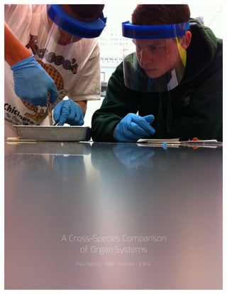
LAB
- 2. Purpose There are many differences between the organ systems of different animal species. In this lab, the bodies of a frog and a fetal pig were dissected to have their organs observed and compared with each other and the human body. The lab also allows the observer to learn about the internal organs in general with hands-on experience. I hypothesized that the frog and the pig would have relatively similar organ structures with the exception of the intestines and presence of the umbilical cord. Materials • Safety Goggles or Mask, Surgical Gloves, and a Lab Apron • Forceps • Preserved Frog or Fetal Pig • 6 to 10 Dissecting Pins • Dissecting Tray and Paper Towels • Plastic Storage Bag and Twist Tie • Scissors • Dissecting Needle or Probe • Camera • Metric Ruler • A Lot of String
- 3. Procedure: Frog Dissection 1. Put on safety goggles or mask, gloves, and a lab apron. 2. Place a frog on the dissection tray. Examine the fingers on its forelegs to determine its sex. If it has thick pads on its thumbs, it is male. Male frogs also tend to be smaller than females. 3. Identify the mouth, external nares, tympani, eyes, and nictitating membranes on the head. 4. Turn the frog on its back and pin down the legs. Cut the hinges of the mouth and open it wide. Use a probe to find and identify the vomerine teeth, maxillary teeth, internal nares, tongue, openings to the Eustachian tubes, esophagus, pharynx, and the glottis. 5. Look for the opening to the frog’s cloaca between the hind legs. Use the forceps to lift the skin and use scissors to cut along the center of the body from the cloaca to the lip. Turn back the skin, cut toward the side of each leg, and pin the skin flat. The cloaca is the structure that receives eggs, sperm, urine, and wastes from the body. 6. Lift and cut through the muscles and breast bone to open up the body cavity. If the frog is female, the abdominal cavity may be filled with dark- colored eggs. If this is the case with your frog, remove the eggs from the frog. 7. Use the provided diagrams to identify the organs of the digestive system. Take photos of the organs and label them.
- 4. Photos: Frog Dissection 1 2
- 5. 3 4
- 6. 5 6
- 7. 7 9 8
- 8. 10 11
- 9. 1. Fat Bodies Spaghetti shaped structures that have a bright orange or yellow color, if you have a particularly fat frog, these fat bodies may need to be removed to see the other structures. Usually they are located just on the inside of the abdominal wall. 2. Peritoneum A spider web like membrane that covers many of the organs, you may have to carefully pick it off to get a clear view. 3. Liver The largest structure of the body cavity. This brown colored organ is composed of three parts, or lobes. The liver consists of a right lobe, left anterior lobe, and left posterior lobe. The liver is not primarily an organ of digestion; it does secrete a digestive juice called bile. Bile is needed for the proper digestion of fats. 4. Heart At the top of the liver, the heart is a triangular structure. The left and right atrium can be found at the top of the heart. A single ventricle located at the bottom of the heart. The large vessel extending out from the heart is the conus arteriosis. 5. Lungs Locate the lungs by looking underneath and behind the heart and liver. They are two spongy organs.
- 10. 6. Gall Bladder Lift the lobes of the liver. There will be a small green sac under the liver. This is the gall bladder, which stores bile. 7. Stomach Curving from underneath the liver is the stomach. The stomach is the first major site of chemical digestion. Frogs swallow their meals whole. Follow the stomach to where it turns into the small intestine. The pyloric sphincter valve regulates the exit of digested food from the stomach to the small intestine. 8. Small Intestine Leading from the stomach. The first straight portion of the small intestine is called the duodenum. The curled portion is the ileum. A membrane called the mesentery holds the ileum together. Note the blood vessels running through the mesentery, they will carry absorbed nutrients away from the intestine. Absorption of digested nutrients occurs in the small intestine. 9. Large Intestine As you follow the small intestine down, it will widen into the large intestine. The large intestine is also known as the cloaca in the frog. The cloaca is the last stop before wastes, sperm, or urine exit the frog's body. 10. Spleen Return to the folds of the mesentery. This dark red spherical object serves as a holding area for blood.
- 11. 11. Esophagus Return to the stomach and follow it upward, where it gets smaller is the beginning of the esophagus. The esophagus is the tube that leads from the frog’s mouth to the stomach. Open the frog’s mouth and find the esophagus, poke your probe into it and see where it leads. Procedure: Fetal Pig Dissection Part 1: External Anatomy 1. Obtain a fetal pig and rise off the excess preservative by holding it under a running faucet. Lay the pig on its side in the dissecting pan and locate the dorsal, ventral, and lateral surfaces. Also locate the anterior and posterior ends. 2. Measure the pig’s length from snout to base of tail to determine its approximate age. The pig we received measured 35cm, longer than the average newborn pig. We then assumed that the pig was going to be born soon after it was killed and preserved. 3. Examine the pig’s head. Locate the eyelids and the external ears or pinnae. Find the external nostrils. 4. Study the pig’s appendages and examine the pig’s toes. Count and record the number of toes and type of hoof the pig has. The pig had 4 toes. 5. Locate the umbilical cord. With scissors, cut across the cord about 1cm from the body. Examine the 3 openings in the umbilical cord.
- 12. 6. Lift the pig’s tail to find the anus. Study the ventral surface of the pig and note the tiny bumps called mammary papillary. In females, these structures connect to the mammary glands. 7. Determine the sex of the pig by looking for the urogenital opening. On females, this opening is located near the anus. On males, the opening is located near the umbilical cord. 8. If the pig is female, you should also note that the urogenital papilla is present near the genital opening. Males do not have urogenital papilla. 9. Observe the eyes of the pig. Carefully remove the eyelid so that you can view the eye underneath. The eyes were well developed and the pigs were most likely born with their eyes shut. 10. Carefully lay the pig on one side in the dissecting pan and cut away the skin from the side of the face and upper neck to expose the masseter muscle that works the jaw, lymph nodes, and salivary glands. 11. With scissors, make a 3cm incision in each corner of the pig’s mouth. Spread the jaws open and examine the tongue. Observe the palate on the roof of the mouth. I could feel my own hard and soft palates with my tongue. 12. Note the taste buds, or sensory papillae, on the side of the tongue. Locate the esophagus at the back of the mouth. Feel the edge of the mouth for teeth. Locate the canine and incisor teeth. The fetal pig had teeth. Humans are not born with teeth.
- 13. 13. Locate the epiglottis, a cone-shaped structure at the back of the mouth. A flap of skin helps to close this opening when a pig swallows. Find the opening to the esophagus. 14. Clean up the materials. Tie the legs of the pig together using the string (front two and back two), and label the pig with your name(s) using a piece of tape that is attached to on of the legs. Part 2: The Incision 1. Place the fetal pig ventral side up in the dissecting tray. 2. Tie a string securely around a front limb. Run the string under the tray, pull it tight, and tie it to the other front limb. Repeat this procedure with the hind limbs to hold the legs apart so you can examine internal structures. 3. Study the diagram below. The dashed lines numbered 1-5 show the first set of incisions that you will make. To find the exact location for the incision marked 2, press along the thorax with your fingers to find the lower edge of the ribs. This is where you will make incision 2. 4. With scissors, make the incisions in order, beginning with 1. Be sure to keep the tips of your scissors pointed upward because a deep cut will destroy the organs below. Also, remember to cut away from yourself_ 5. After you have made your incisions through the body wall, you will see the peritoneum, a thin layer of tissue that lines the body cavity. Cut through the peritoneum along the incision lines.
- 14. 6. Spread the flaps of the body wall apart. Cut the umbilical vein, which extends through the liver. Once the vein is cut, carefully pull the flap of skin, including the end of the umbilical cord between the hind legs. You are now able to see the organs of the abdominal cavity. 7. If time remains continue with part B, the digestive tract. Otherwise, clean up and return your materials and pig as you did on day 1. Part B: Digestive System 1. Locate the diaphragm, a sheet of muscle that separates the abdominal cavity from the thoracic cavity. Find the most obvious structure in the abdominal cavity, the brownish-colored liver. There were 4 lobes. 2. Find the tube-like esophagus, which joins the mouth and the stomach. Food moves down the esophagus by muscular contractions after being softened by saliva in the mouth. Follow the esophagus and locate the soft, sac-like stomach beneath the liver. 3. Using scissors, cut along the outer curve of the stomach. Open the stomach and note the texture of its inner walls. These ridges inside the stomach are called rugae and increase the area for the release of digestive enzymes. The stomach may not be empty because fetal pigs swallow amniotic fluid. 4. The pig has a digestive system, which is classified as monogastric or nonruminant. Humans also have this type of digestive system. They have one stomach (mono=one, gastric=stomach). Locate the entrance to the
- 15. stomach or esophageal area, the cardiac region, which is largest, and the pyloric region where the stomach narrows to join to the small intestine. 5. At the end of the stomach, there is a sphincter, or ring-shaped muscle to control food leaving the stomach and entering the duodenum. Locate the cardiac sphincter at the junction of the stomach and esophagus, and the pyloric sphincter at the junction of the stomach and small intestine. Fetal pigs receive their nourishment from their mother through the umbilical cord. 6. Identify the first part of the small intestine, the U-shaped duodenum, which connects to the lower end of the stomach. Pancreatic juice, made by the pancreas, and bile, made by the liver and stored in the gall bladder, are add to food here to continue digestion. 7. Study the rest of the small intestine. Notice that it is a coiled, narrow tube, held together by tissue called mesentery. The soupy, partly digested food that enters the small intestine from the stomach is called chyme. 8. Carefully cut through the mesentery and uncoil the small intestine. Note and record its length in centimeters. The mid-section is called the jejunum, while the last section is called the ileum. The small intestine measured 250cm long. 9. With scissors, remove a 3-cm piece of the lower small intestine. Cut it open and rinse it out.
- 16. 10. Observe the inner surface of the small intestine. Run your finger along it and note its texture. Using a magnifying glass, examine the villi, the tiny projections that line the small intestine and increase the surface area for absorption. 11. Follow the small intestine until it reaches the wider, looped large intestine. Cut the mesentery and unwind the large intestine (CAREFUL!) or colon. Measure and record its length. The large intestine measured 20cm long. 12. At the junction of the large and small intestine, locate a blind pouch called the caecum. The caecum has no known function in the pig. Notice that the large intestine leads into the rectum, a tube that runs posteriorly along the dorsal body wall. The rectum carries wastes to the opening called the anus where they are eliminated. 13. Locate the thin, white pancreas beneath the stomach and duodenum. Pancreatic juice flows through pancreatic ducts to the duodenum. 14. Between the lobes of the liver, find the small, greenish-brown gall bladder. Locate the hepatic duct, which carries bile from the liver to the gall bladder. 15. Find the spleen, a long, reddish-brown organ wrapped around the stomach. The spleen filters out old red blood cells and produces new ones for the fetus. 16. On the diagram (crude drawing), label the pig's body organs.
- 17. 17. Clean up your materials and work area. Wrap the pig in damp paper towels and put it in a plastic bag. Obtain a piece of masking tape and label your bag with your names. Return your lab equipment and pig to the supply cart and then thoroughly wash your hands with soap. Photos: Fetal Pig Dissection 1. Liver and Gall Bladder (above the heart)
- 18. 8. Spleen (next to the liver) 3. Heart (between lungs and liver) 4. Stomach (Connected to Esophagus and small intestines by sphincters)
- 19. 5. Small Intestines (connect stomach and large intestine) 6. Large Intestines (Connect small intestine and rectum) 7. Lungs (lie over the heart) 8
- 20. Conclusion This lab test made it clear that while the digestive systems in mammals and amphibians share many of the same traits, there are differences that may affect the eating habits of each branch. In this conclusion, the features of the human organs will also be compared to the structures of the frog and pig. My hypothesis was in fact correct, as the pig and frog did have different intestine lengths, and the frog was missing an umbilical cord (a little obvious). The two major systems that were looked at closely are the circulatory system, which works to deliver supplies throughout an organism’s body, and the digestive system, which digests food to extract nutrients and excrete waste. The esophagus of the frog was much shorter than that of the fetal pig. Pigs have a much longer esophagus proportional to their body, similar to humans. A longer esophagus will give the animal’s salivary enzymes a longer period of time to digest food. The frog had a shorter esophagus, most likely because it contained more enzymes in the stomach to digest the food further along in the digestive process. The pig contained fewer enzymes, and because they are quite similar to humans, one can hypothesize that humans also have fewer enzymes than a frog. There are some ideas that may help to further explain this. A frog tends to swallow its food whole (such as insects), while a pig takes more time to grind its food down with its teeth. A frog would need more enzymes in its stomach to make up for the lack of physical digestion in its mouth. Animals with more advanced diets need longer digestive systems, because they don’t have enough
- 21. enzymes to break the food down quickly enough. The process to extract the nutrients and minerals is also much more advanced. A longer esophagus will contribute to a longer digestive system, while a short one contributes to a digestive system that does not require a very long digestive tract. The structures of the stomachs of the pig and frog were very different from each other. The frog’s stomach was much more of a channel as part of the digestive tract, while the pig’s stomach’s structure was much more sack-like. Frogs don’t need to contain their food in their stomach for very long, because their multitude of enzymes will digest it more rapidly. Pigs’ stomachs need to contain the food for a longer period of time to allow for the enzymes to digest it successfully. Many other mammals, including humans have the same stomach structures. A pig’s pancreas produces digestive enzymes for the small intestines and insulin, that controls the blood sugar level. This is similar to human beings. Too much or to little insulin can lead to diabetes. A frog’s pancreas also produces digestive enzymes. The frog’s pancreas is relatively larger than the pig’s when compared to the size of the small intestines. This may be because it has to produce more enzymes as described in the paragraph above. The frog had a single slit between its legs called the cloaca. All waste and reproductive products are excreted through this opening. The pig has two openings, however: one for feces (the anus) and another for urinary and reproductive functions. The male genitals are located below the umbilical cord, and the female reproductive organs are located next to the anus of the pig.
- 22. Once again, humans share more similarities with pigs, but the structures are still very different. Human females have a separate urinary tract, and the reproductive systems of males are located well beneath the naval cavity. The lungs of the pig were very large in relationship to those of the frog. The pig requires more oxygen (hence the larger lungs) to support its larger, more intricate circulatory system. Frogs have smaller lungs, because they use their skin for gas exchange, as well. Humans’ lungs are similar to pigs’ lungs, because their circulatory systems are very similar. The mammalian heart is much more advanced than the frog’s. It has a total of four chambers, wheras the frog’s has only three chambers. A mammal’s heart separates oxygenated blood from deoxygenated blood to accomplish higher efficiency when delivering oxygen to a larger body. A frog’s heart partially mixes oxygenated blood with deoxygenated blood, because it is less important for the blood to be completely oxygenated, as it takes a much shorter time for the blood to be reoxygenated in the frog’s smaller body. Comparatively, the sizes of the livers of the pig and the frog were very similar. The functions of the liver are the very same: to produce bile, store glycogen, and to detoxify poisons. Because of the similarities between the livers of frogs and pigs, it is very likely that the human liver has similar functions and a similar relative size. The pig and frog gall bladders look very different from each other. The pig’s gall bladder hugs the underside of the liver and is colored the same brown tint, and the frog’s gall bladder is small and green, attached to the back surface
- 23. of the liver. The function of the gall bladder is to store bile that has been produced by the liver and to deliver the bile to the small intestines. There is not a clear reason why there is so much color variation between the two gall bladders, so it would be difficult to hypothesize what a human’s gall bladder might look like (the fact was later researched to find that the human gall bladder is usually green). There was most likely some discoloration of the pig’s gall bladder, as well, according to external sources. During our lab, we could not locate the gall bladder in our pig. We listed this as an experimental error. The small intestine of the fetal pig was, comparatively, much longer than that of the frog. It measured to be 250cm long, while the frog’s small intestines measured only 5cm long. This relates back to the findings surrounding the relationship between the total length of the digestive tract and the diet of the animals (discussed in paragraphs 2 and 3 of the conclusion). If one considers the length of the pig (because humans are also mammals), he or she can calculate his or her intestine size. It was calculated that, a person measuring 170cm tall would have small intestines measuring approximately 1214cm long, or enough to make 81 hot dogs. The function of the large intestine is to store waste before it is excreted from the body. The large intestine of the pig was discovered to be much shorter than the small intestine, measuring at only 20cm long. Interestingly though, the frog’s large intestine matched the length of its small intestine at 5cm long. Because the large intestine has no digestive function, the sizes of the large
- 24. intestines in the pig and frog are comparable. The same rule can be applied to humans. By performing these dissections, one is able to conclude that there are clear differences between humans, pigs, and frogs. Most of them are due to the fact that frogs are amphibians and have followed a very different evolutionary path from mammals such as humans and pigs, but many similarities can be drawn between the branches. Many of the organs are very similar to each other and provide similar functions in three very different bodies in a facinating display of the deeply connected biological world we live in. One also knows, as a result of this lab, how the different digestive systems have adapted to the diet and surrounding climate of the host animals. This turned out to be a very successful lab, with a very minimal amount of experimental errors. It was a good demonstration of what occurs in the natural world all the time and was able to open up one’s perspective to observe living creatures in a more intellectual fashion. If I could change one thing, however, I would have the option to dissect an animal that did not have all of its blood drained from its body (which tends to make it look lifeless and uninviting). Sources Urinary and Reproductive Systems http://goo.gl/A1ATd A Frog’s Lungs and How They Work http://goo.gl/UUVU6 Normal Heart Rate of Frogs http://goo.gl/fhy0R Circulatory System of a Pig http://goo.gl/LbIS1 Primary Functions of the Liver http://goo.gl/98WDt