The Most Attractive Pune Call Girls Budhwar Peth 8250192130 Will You Miss Thi...
Characterization of materials lec2
1. 07/04/2014
1
Lec 29
XPS/AES Instrumentation
In order to achieve the necessary UHV and clean the surface of chamber
and sample, the vacuum chamber must be baked at an elevated
temperature (250–350°C) and pumped. This baking process allows the gas
molecules adsorbed onto the chamber walls to be pumped out. An
additional requirement for the chamber is magnetic shielding, because the
trajectory of signal electrons is strongly affected by any magnetic field,
even the Earth’s magnetic field.
Source Guns (X-ray Gun)
An electron spectrometer system contains an X-ray gun for XPS analysis. The
working principles of the X-ray gun are similar to the X-ray tube used for
X-ray diffractometry. X-ray photons are generated by high-energy
electrons striking a metal anode, commonly Al or Mg for XPS spectrometry.
The X-ray gun produces a characteristic X-ray line to excite atoms of the
surface to be analyzed. XPS uses both non-monochromatic and
monochromatic X-ray sources.
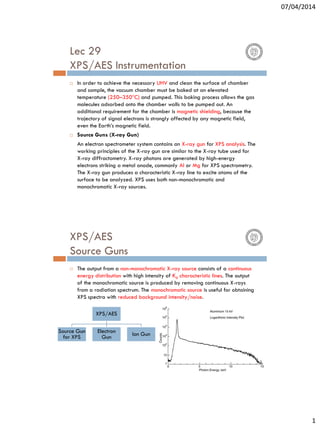
XPS/AES
Source Guns
The output from a non-monochromatic X-ray source consists of a continuous
energy distribution with high intensity of Kα characteristic lines. The output
of the monochromatic source is produced by removing continuous X-rays
from a radiation spectrum. The monochromatic source is useful for obtaining
XPS spectra with reduced background intensity/noise.
XPS/AES
Source Gun
for XPS
Electron
Gun Ion Gun
2. 07/04/2014
2
Source Gun
Energies of the characteristic X-ray lines used in XPS are lower than those
used in X-ray diffractometry. For example, the energies of AlKα and MgKα
are 1.48 and 1.25 keV, respectively. But energies of CuKα and MoKα, which
are commonly used in X-ray diffractometry are 8.04 keV and 17.44 keV,
respectively.
The reason to choose lower energy X-rays (soft characteristic X-rays) is their
narrow line width i.e. their range of energy. XPS requires a line width <1.0
eV to ensure good energy resolution. Both AlKα and MgKα exhibit line
widths <1.0 eV and have sufficient energies for photoelectron excitation.
Different from the X-ray tube used in an X-ray diffractometer, a single X-
ray gun in XPS has both Al and Mg anodes as shown in next Figure. Such a
gun structure makes an easy switching between MgKα and AlKα radiation.
Source Gun
3. 07/04/2014
3
Electron Gun
The electron guns used in AES analysis is similar to those used in electron
microscopy. LaB6 and FE guns are commonly used in electron spectrometers.
The LaB6 gun can provide an electron beam of high brightness with spatial
resolution of 200 nm after being focused by an electromagnetic lens.
FE guns are increasingly used in modern instruments. These guns provide
superior brightness and higher spatial resolution than LaB6. In addition, their
emitting surface remains clean during operation without adsorption of gas
molecules.
Ion Gun
The functions of an ion gun are twofold. First, it provides a high energy ion
flux to clean sample surfaces before examination. This is necessary because
the signal electrons come from the surface atom layers of a sample. Sample
surfaces are commonly contaminated with adsorbed hydrocarbons, water
vapor and oxides that need to be removed before surface analysis.
Ion Gun
The second function of the ion gun is to sputter out sample atoms layer by
layer so that an elemental depth profile can be revealed. The ion gun
produces an argon ion beam by either electron impact or gaseous
discharge. This beam has an energy level of 0.5-5.0 keV, and can be
focused to a diameter down to several tens of micrometers. The ion beam
can scan a surface area as large as 10×10 sq mm.
4. 07/04/2014
4
XPS Instrumentation
Electron Energy Analysers
An electron energy analyzer is required to obtain XPS and Auger spectra.
The most commonly used analyzer is called the hemispherical sector
analyzer (HSA). The analyzer is composed of two concentric hemispheres
with radii R1 and R2. Negative potentials V1 and V2 are applied to the
inner and outer hemispheres, respectively.
The applied potential generates a median equipotential surface with radius
of Ro. The potential along the median surface (Vo) is called the pass energy
of the HSA. A slit at one end of the HAS allows electrons from the sample to
enter, and a slit at the other end lets electrons pass through to an electron
detector. The HSA only allows the electrons with energy E=eVo, which are
injected tangentially to the median surface, to pass through its channel and
reach the detector.
5. 07/04/2014
5
Instrumentation
Characteristics of Electron Spectra
XPS Spectra
An XPS spectrum can have three types of peaks on a background: photo-
emission from core electron levels, photo-emission from valence levels and
Auger emission excited by X-rays.
The core-level photoelectron peaks are the primary peaks for elemental
analysis. The valence-level peaks are those at low binding energy (0–20 eV)
and are primarily useful in studies of the electronic structure of materials. The
Auger peaks, arising from X-rays excited Auger process, are also useful for
chemical analysis.
Next Figure shows an XPS spectrum of a clean silver surface in which the core-
level peaks are marked as 3s, 3p and 3d. The valence-level peak is marked as
4d and the Auger peaks are marked as MNN.
XPS spectra have a step-like background, increasing with binding energy. They
result from inelastic scattering of photoelectrons in a solid. It should be noted
that the X-ray radiation is commonly not exactly monochromatic. The
photoelectron emission can also be excited by continuous X-rays, which are
commonly associated with characteristic X-ray radiation. Such photoelectron
emission generates a background in the low binding energy region of spectrum.
6. 07/04/2014
6
XPS of Ag excited by MgKα
XPS Spectra
Shake-up satellites are the extra peaks which result from interaction
between a photoelectron and a valence electron. A photoelectron can
excite (shake-up) a valence electron to a higher energy level and thereby
lose a few electron volts of kinetic energy. This will create a satellite peak
associated with a core-level peak of photoelectrons as shown in Figure.
Shake-up satellites are useful for chemical analysis. For example, a shake-
up satellite is associated with the Cu2p of CuO, but it is absent around the
Cu 2p peak of Cu2O.
For certain transition and rare earth metals with unpaired electrons in 3d
and 4f shells, the shake-up satellites produce strong peaks.
Multiplet splitting of a core-level peak may occur in a compound that has
unpaired electrons in its valence level. For example, the core-level peak of
Ni2p3/2 of NiO shows multiplet splitting.
Multiplet splitting is also useful in chemical analysis. For example, Ni(OH)2
can be distinguished from NiO because the 2p3/2 of Ni(OH)2 does not
have multiplet splitting.
7. 07/04/2014
7
XPS Spectra
XPS Spectra
Plasmon loss generates another type of satellite peaks; these peaks do not
provide useful information but they complicate a spectrum. Plasmon loss
refers to the energy loss of a photoelectron because it excites collective
vibrations in conduction electrons in a metal.
The vibrations require a characteristic amount of energy. Such characteristic
amounts of photoelectron energy loss in photoelectrons will generate
satellite peaks as shown in Figure. Plasmon loss peaks may occur in XPS
spectra of clean metal surfaces and also in Auger spectra.