This presentation explains Physiology of blood, Variations in blood cells-Oral manifestations and Clinical importance, Blood groups and Transfusion of blood
2. BLOOD
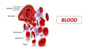
• Blood is a connective tissue in fluid form.
• ‘Fluid of life’: Carries oxygen from lungs to all parts of the body and
carbon dioxide from all parts of the body to the lungs.
• ‘Fluid of growth’: Carries nutritive substances from the digestive system
and hormones from endocrine gland to all the tissues.
• ‘Fluid of health’: Protects the body against the diseases and gets rid of
the waste products and unwanted substances by transporting them to
the excretory organs like kidneys.
3. PHYSICAL CHARACTERISTICS
• Color: Red in color.
Arterial blood: Scarlet red- More oxygen
Venous blood: Purple red- More carbon dioxide.
• Volume: Normal adult -5 L. In a newborn baby, the volume is 450 ml. It
increases during growth and reaches 5 L at the time of puberty. In females, it
is slightly less and is about 4.5 L.
In a normal young healthy adult : 8% of the body weight in a normal healthy
individual weighing about 70 kg.
• pH: Blood is slightly alkaline and its pH in normal conditions is 7.4.
4. • Specific gravity:
Total blood : 1.052 to 1.061
Blood cells : 1.092 to 1.101
Plasma : 1.022 to 1.026
• Viscosity: Blood is five times more viscous than water. It is mainly
due to red blood cells and plasma proteins.
5. • Nutritive
• Respiratory
• Excretory
• Transport of hormones and enzymes
• Regulation of water balance: Water in blood is freely interchangeable with interstitial
fluid.
• Regulation of acid-base balance: Plasma proteins, Hb acts as buffer
• Regulation of body temperature
• Storage: Water, proteins, glucose, Na, K – Used during starvation or electrolyte loss.
• Defensive : Neutrophils , Monocytes: Phagocytosis
Lymphocytes: Immunity
Eosinophils: Detoxification, disintegration and removal of foreign bodies.
BLOOD FUNCTIONS
6. • Blood contains blood cells(45%) which are called formed elements and the liquid portion known
as plasma(55%).
BLOOD COMPOSITION
If blood is collected in a hematocrit tube along with a
suitable anticoagulant and centrifuged for 30 minutes at a
speed of 3000 revolutions per minute (rpm), the red blood
cells settle down at the bottom having a clear plasma at the
top. Volume of red blood cells expressed in percentage is
called the hematocrit value or packed cell volume (PCV).
Serum is the clear straw-colored fluid that oozes from blood clot. When the
blood is shed or collected in a container, it clots. In this process, the
fibrinogen is converted into fibrin and the blood cells are trapped in this
fibrin forming the blood clot. After about 45 minutes, serum oozes out of the
blood clot. Volume of the serum is almost the same as that of plasma (55%).
It is different from plasma only by the absence of fibrinogen, i.e. serum
contains all the other constituents of plasma except fibrinogen. Fibrinogen is
absent in serum because it is converted into fibrin during blood clotting.
Thus, SERUM = PLASMA – FIBRINOGEN
7.
8. • Normal values:
Total proteins : 7.3 g/dL (6.4 to 8.3 g/dL)
Serum albumin: 4.7 g/dL
Serum globulin : 2.3 g/dl
Fibrinogen : 0.3 g/dL
• ALBUMIN/GLOBULIN RATIO: Normal A/G ratio is 2 : 1.
• MOLECULAR WEIGHT:
Albumin : 69,000
Globulin : 1,56,000
Fibrinogen : 4,00,000.Thus, the molecular weight of fibrinogen is greater than that of other
two proteins.
• Plasma proteins are responsible for the oncotic or osmotic pressure in the blood. Osmotic
pressure exerted by proteins in the plasma is called colloidal osmotic (oncotic) pressure.
Normally, it is about 25 mm Hg. Albumin plays a major role in exerting oncotic pressure.
PLASMA PROTEINS
9. • Specific gravity of the plasma proteins is 1.026.
• Acceptance of hydrogen ions is called buffer action. The plasma proteins have 1/6 of total
buffering action of the blood.
ORIGIN OF PLASMA PROTEINS:
• In embryonic stage, the plasma proteins are synthesized by the
mesenchyme cells. The albumin is synthesized first and other proteins are
synthesized later.
• In adults, the plasma proteins are synthesized mainly from
reticuloendothelial cells of liver. The plasma proteins are synthesized also
from spleen, bone marrow, disintegrating blood cells and general tissue
cells. Gamma globulin is synthesized from B lymphocytes.
10. FUNCTIONS OF PLASMA PROTEINS
1. Coagulation of blood – Fibrinogen to fibrin
2. Defense mechanism of blood – Immunoglobulins
3. Transport mechanism – α Albumin, β globulin transport hormones, gases, enzymes, etc.
4. Maintenance of osmotic pressure in blood
5. Acid-base balance
6. Provides viscosity to blood
7. Provides suspension stability of RBC
8. Reserve proteins
9. Role in ESR- Globulin, Fibrinogen accelerate the formation of rouleaux formation
10.Role in production of Trephone substances necessary for nourishment of tissue cells in culture.
11. • Non-nucleated formed elements in the blood.
• Also known as erythrocytes (Erythros = red)- due to the presence of the colouring
pigment called hemoglobin.
• Play a vital role in transport of respiratory gases.
• RBCs are larger in number compared to the other two blood cells.
RED BLOOD CELLS
12. MORPHOLOGY OF RBC
i. NORMAL SHAPE
Normally, the RBCs are disk shaped and biconcave (dumbbell
shaped). Central portion is thinner and periphery is thicker. The
biconcave contour of RBCs has some mechanical and functional
advantages.
1. Biconcave shape helps in equal and rapid diffusion of oxygen
and other substances into the interior of the cell.
2. Large surface area is provided for absorption or removal of
different substances.
3. Minimal tension is offered on the membrane when the
volume of cell alters.
4. Because of biconcave shape, while passing through minute
capillaries, RBCs squeeze through the capillaries very easily
without getting damaged.
MORPHOLOGY OF RBC’s
13. ii. NORMAL SIZE:
• Diameter : 7.2 µ (6.9 to 7.4 µ)
• Thickness : At the periphery it is thicker with 2.2 µ and at the center it is thinner with 1
µ.This difference in thickness is because of the biconcave shape.
• Surface area : 120 sq µ.
• Volume : 85 to 90 cu µ.
14. iii. NORMAL STRUCTURE:
NON-NUCLEATED
• Absence of nucleus in human RBC - DNA is also absent.
• Other organelles such as mitochondria and Golgi apparatus also are absent in RBC.
• Because of absence of mitochondria, the energy is produced from glycolytic process.
15. • RBC has a special type of cytoskeleton, which is made up of Actin and
Spectrin.
• Both the proteins are anchored to transmembrane proteins by means of
another protein called ankyrin.
• Absence of Spectrin results in hereditary spherocytosis. In this condition,
the cell is deformed, losses its biconcave shape and becomes globular
(spherocytic). The spherocyte is very fragile and easily ruptured
(hemolyzed) in hypotonic solutions.
16. • ROULEAUX FORMATION:
When blood is taken out of the blood vessel, the
RBCs pile up one above another like the pile of
coins. This property of the RBCs is called
rouleaux (pleural = rouleau) formation .It is
accelerated by plasma proteins globulin and
fibrinogen.
• SPECIFIC GRAVITY of RBC is 1.092 to 1.101.
PROPERTIES OF RBC’s
17. • PACKED CELL VOLUME (PCV):
Is the proportion of blood occupied by RBCs expressed in percentage. It is also called
hematocrit value.
It is 45% of the blood and the plasma volume is 55%.
• SUSPENSION STABILITY:
During circulation, the RBCs remain suspended uniformly in the blood. This property of the
RBCs is called the suspension stability.
18. • Average lifespan of RBC is about 120 days.
• After the lifetime the senile (old) RBCs are destroyed in reticuloendothelial system.
DETERMINATION OF LIFESPAN OF RED BLOOD CELLS
• Lifespan of the RBC is determined by radioisotope method.
• RBCs are tagged with radioactive substances like radioactive iron or radioactive
chromium.
• Life of RBC is determined by studying the rate of loss of radioactive cells from
circulation.
LIFE SPAN OF RBC’s
19. • When the cells become older (120 days), the cell membrane becomes more fragile.
• Diameter of the capillaries is less or equal to that of RBC. Younger RBCs can pass
through the capillaries easily. However, because of the fragile nature, the older cells
are destroyed while trying to squeeze through the capillaries.
• The destruction occurs mainly in the capillaries of red pulp of spleen because the
diameter of splenic capillaries is very small. So, the spleen is called ‘Graveyard of
RBCs’. Destroyed RBCs are fragmented and hemoglobin is released from the
fragmented parts. Hemoglobin is immediately phagocytized by macrophages of the
body, particularly the macrophages present in liver (Kupffer cells), spleen and bone
marrow.
FATE OF RBC’s
20. HEMOGLOBIN IRON + GLOBIN+ PORPHYRIN
• Iron combines with the protein called apoferritin to form
ferritin, which is stored in the body and reused later.
• Globin enters the protein depot for later use.
• Porphyrin is degraded into bilirubin, which is excreted by liver
through bile.
• Daily 10% RBCs, which are senile, are destroyed in normal
young healthy adults. It causes release of about 0.6 g/dL of
hemoglobin into the plasma. From this 0.9 to 1.5 mg/dL
bilirubin is formed.
21. Erythropoiesis is the process of the origin, development and maturation of erythrocytes. Hemopoiesis
or hematopoiesis is the process of origin, development and maturation of all the blood cells.
SITE OF ERYTHROPOIESIS
IN FETAL LIFE: Occurs in three stages:
ERYTHROPOIESIS
Mesoblastic Stage First two months of
intrauterine life
Mesenchyme of yolk
sac
Hepatic Stage From third month of
intrauterine life
Liver – Main organ
Spleen, Lymphoid
organs also involved.
Myeloid Stage last three months of
intrauterine life
Red bone marrow and
liver.
22. IN NEWBORN BABIES, CHILDREN AND ADULTS:
RBCs are produced only from the red bone marrow.
Upto 20 years of age Red bone marrow of all
bones (long bones and all
the flat bones).
After 20 years of age Membranous bones like
vertebra, sternum, ribs,
scapula, iliac bones and skull
bones and from the ends of
long bones. After 20 years of
age, the shaft of the long
bones becomes yellow bone
marrow because of fat
deposition and looses the
erythropoietic function.
Though bone marrow is the site
of production of all blood cells,
comparatively 75% of the bone
marrow is involved in the
production of leukocytes and
only 25% is involved in the
production of erythrocytes. But
still, the leukocytes are less in
number than the erythrocytes,
the ratio being 1:500. This is
mainly because of the lifespan of
these cells. Lifespan of
erythrocytes is 120 days whereas
the lifespan of leukocytes is very
short ranging from one to ten
days. So the leukocytes need
larger production than
erythrocytes to maintain the
required number
23. STEM CELLS
Uncommitted pluripotent hemopoietic stem cell
Committed pluripotent hemopoietic stem cell
Lymphoid stem cell
Colony forming unit-
GM
Colony forming unit-
M
Colony forming blastocyte
Colony forming unit-E
Granulocytes Megakaryocyte
L NE B E M P
25. STAGES OF ERYTHROPOIESIS IMPORTANT EVENTS
PROERYTHROBLAST SYNTHESIS OF HEMOGLOBIN STARTS
EARLY NORMOBLAST NUCLEOLI DISAPPEAR
INTERMEDIATE NORMOBLAST HEMOGLOBIN STARTS APPEARING
LATE NORMOBLAST NUCLEUS DISAPPEAR
RETICULOCYTE RETICULUM IS FORMED.
CELL ENTERS CAPILLARY FROM SITE OF
PRODUCTION
MATURED ERYTHROCYTE RETICULUM DISAPPEARS
CELL ATTAINS BICONCAVITY
26. FACTORS NECESSARY FOR ERYTHROPOIESIS
• GENERAL FACTORS
Erythropoietin
Thyroxine
Hemopoietic growth factors
Vitamins
• MATURATION FACTORS
Vitamin B12 (Cyanocobalamin)
Intrinsic factor of castle
Folic acid
27. • Hemoglobin (Hb) is the iron containing coloring matter of red blood cell (RBC).
• It is a chromoprotein forming 95% of dry weight of RBC and 30% to 34% of wet weight.
• Function of hemoglobin is to carry the respiratory gases- Oxygen and Carbon dioxide.
• It also acts as a buffer.
• Molecular weight of hemoglobin is 68,000.
Average hemoglobin (Hb) content in blood is 14 to 16 g/dL. However, the value varies
depending upon the age and sex of the individual.
Age: At birth : 25 g/dL
After 3rd month : 20 g/Dl
After 1 year : 17 g/dL
From puberty onwards : 14 to 16 g/dL
At the time of birth, hemoglobin content is very high because of increased number of
RBCs.
Sex: In adult males : 15 g/dL, In adult females : 14.5 g/dL
HEMOGLOBIN
28. FUNCTIONS OF Hb:
1.TRANSPORT OF OXYGEN:
When oxygen binds with hemoglobin, a physical process called
oxygenation occurs, resulting in the formation of oxyhemoglobin. The iron
remains in ferrous state in this compound. Oxyhemoglobin is an unstable
compound and the combination is reversible, i.e. when more oxygen is
available, it combines with hemoglobin and whenever oxygen is required,
hemoglobin can release oxygen readily. When oxygen is released from
oxyhemoglobin, it is called reduced hemoglobin or ferrohemoglobin.
29. 2. TRANSPORT OF CARBON DIOXIDE:
When carbon dioxide binds with hemoglobin, carbhemoglobin is formed. It is
also an unstable compound and the combination is reversible, i.e. the carbon
dioxide can be released from this compound. The affinity of hemoglobin for
carbon dioxide is 20 times more than that for oxygen.
3. BUFFER ACTION:
Hemoglobin acts as a buffer and plays an important role in acid base balance
30. STRUCTURE OF HEMOGLOBIN:
• Conjugated protein.
• It consists of a protein combined with an iron
containing pigment.
• Protein part: Globin , Iron containing pigment: Heme.
• Heme also forms a part of the structure of myoglobin
(oxygen binding pigment in muscles) and neuroglobin
(oxygen binding pigment in brain).
• IRON:
Normally, it is present in ferrous (Fe2+) form. It is in
unstable or loose form. In some abnormal conditions,
the iron is converted into ferric (Fe3+) state, which is a
stable form.
31. • PORPHYRIN:
The pigment part of heme is called porphyrin. It is formed by four pyrrole rings
(tetrapyrrole) called, I, II, III and IV. The pyrrole rings are attached to one another by
methane (CH4) bridges. The iron is attached to ‘N’ of each pyrrole ring and ‘N’ of globin
molecule.
• GLOBIN:
Globin contains four polypeptide chains. Among the four polypeptide chains, two are β
chains and two are α-chains.
POLYPEPTIDE CHAIN MOLECULAR WEIGHT AMINO ACIDS
α-chain 15,126 141
β-chain 15,866 146
32. • Hemoglobin is of two types:
1. Adult hemoglobin – HbA
2. Fetal hemoglobin – HbF
Replacement of fetal hemoglobin by adult hemoglobin starts
immediately after birth. It is completed at about 10th to 12th week after
birth. Both the types of hemoglobin differ from each other structurally
and functionally.
STRUCTURAL DIFFERENCE:
In adult hemoglobin, the globin contains two α-chains and two β-chains.
In fetal hemoglobin, there are two α chains and two γ-chains instead of
β-chains.
FUNCTIONAL DIFFERENCE:
Functionally, fetal hemoglobin has more affinity for oxygen than that of
adult hemoglobin. And, the oxygen hemoglobin dissociation curve of
fetal blood is shifted to left.
34. • Erythrocyte sedimentation rate (ESR) is the rate at which the erythrocytes settle
down. Normally, the red blood cells (RBCs) remain suspended uniformly in circulation.
This is called suspension stability of RBCs.
If blood is mixed with an anticoagulant and allowed to stand on a vertical
tube, the red cells settle down due to gravity with a supernatant layer of clear plasma.
• ESR is also called sedimentation rate, sed rate or Biernacki reaction. It was first
demonstrated by Edmund Biernacki in 1897.
DETERMINATION OF ESR
There are two methods to determine ESR.
1. Westergren’s method
2. Wintrobe’s method
ERYTHROCYTE SEDIMENTATION RATE
35. 1. WESTERGREN’S METHOD
Westergren Tube is 300 mm long and opened on both
ends. It is marked 0 to 200 mm from above downwards.
Westergren tube is used only for determining ESR. 1.6 mL
of blood is mixed with 0.4 mL of 3.8% sodium citrate
(anticoagulant) and loaded in the Westergren tube. The
ratio of blood and anticoagulant is 4:1. The tube is fitted
to the stand vertically and left undisturbed. The reading is
taken at the end of 1 hour.
36. • WINTROBE’S METHOD
Wintrobe tube is a short tube opened on only one end. It is 110
mm long with 3 mm bore. Wintrobe tube is used for determining
ESR and PCV. It is marked on both sides. On one side the marking is
from 0 to 100 (for ESR) and on other side from 100 to 0 (for PCV).
About 1 mL of blood is mixed with anticoagulant,
ethylenediaminetetraacetic acid (EDTA). The blood is loaded in the
tube up to ‘0’ mark and the tube is placed on the Wintrobe stand.
And, the reading is taken after 1 hour.
37. Westergren Method Wintrobe Method
In males : 3 to 7 mm in 1 hour
In females : 5 to 9 mm in 1 hour
Infants : 0 to 2 mm in 1 hour
In males : 0 to 9 mm in 1 hour
In females : 0 to 15 mm in 1 hour
Infants : 0 to 5 mm in 1 hour
38. Packed cell volume (PCV) is the proportion of blood
occupied by RBCs, expressed in percentage. It is the volume
of RBCs packed at the bottom of a hematocrit tube when
the blood is centrifuged. It is also called HEMATOCRIT
VALUE OR ERYTHROCYTE VOLUME FRACTION (EVF).
METHOD OF DETERMINATION:
Blood is mixed with the anticoagulant
ethylenediaminetetraacetic acid (EDTA) or heparin and filled
in hematocrit or Wintrobe tube (110 mm long and 3 mm
bore) up to 100 mark. The tube with the blood is
centrifuged at a speed of 3000 revolutions per minute (rpm)
for 30 minutes. RBCs packed at the bottom form the packed
cell volume and the plasma remains above this. In between
the RBCs and the plasma, there is a white buffy coat, which
is formed by white blood cells and the platelets
PACKED CELL VOLUME
39. Determination of PCV helps in:
1. Diagnosis and treatment of anemia
2. Diagnosis and treatment of Polycythemia
3. Determination of extent of dehydration and recovery from dehydration after
treatment
4. Decision of blood transfusion.
NORMAL PCV: In males = 40% to 45%
In females = 38% to 42%
40. PHYSIOLOGICAL VARIATIONS OF ESR PATHOLOGICAL VARIATIONS OF ESR
1. Age: ESR is less in children and infants because of
more number of RBCs.
2. Sex: It is more in females than in males because of
less number of RBCs.
3. Menstruation: The ESR increases during
menstruation because of loss of blood and RBCs
4. Pregnancy: From 3rd month to parturition, ESR
increases up to 35 mm in 1 hour because of
hemodilution.
ESR increases in diseases such as the following
conditions:
1. Tuberculosis
2. All types of anemia except sickle cell anemia
3. Malignant tumours
4. Rheumatoid arthritis
5. Rheumatic fever
6. Liver diseases.
ESR decreases in the following conditions:
1. Allergic conditions
2. Sickle cell anemia
3. Peptone shock
4. Polycythemia
5. Severe leukocytosis
VARIATIONS IN ESR,PCV
41. PCV increases in:
1. Polycythemia
2. Dehydration
3. Dengue shock syndrome: Dengue fever (tropical disease caused by flavivirus
transmitted by mosquito Aedes aegypti) of grade III or IV severity.
PCV decreases in:
1.Anemia
2. Cirrhosis of liver
3. Pregnancy
4. Hemorrhage due to ectopic pregnancy (pregnancy due to implantation of fertilized
ovum in tissues other than uterine wall), which is characterized by vaginal bleeding.
42. 1.MEAN CORPUSCULAR VOLUME (MCV) :
MCV is the average volume of a single RBC and it is expressed in cubic
microns (cu µ). Normal MCV is 90 cu µ (78 to 90 cu µ).
When MCV is normal, the RBC is called normocyte. When MCV increases,
the cell is known as a macrocyte and when it decreases, the cell is called
microcyte.
• In Pernicious anemia and Megaloblastic anemia, the RBCs are macrocytic
in nature.
• In Iron deficiency anemia the RBCs are microcytic.
BLOOD INDICES
43. 2. MEAN CORPUSCULAR HEMOGLOBIN (MCH):
MCH is the quantity or amount of hemoglobin present in one RBC. It is expressed in micro-
microgram or picogram (pg). Normal value of MCH is 30 pg (27 to 32 pg).
3. MEAN CORPUSCULAR HEMOGLOBIN CONCENTRATION (MCHC):
• MCHC is the concentration of hemoglobin in one RBC. It is the amount of hemoglobin
expressed in relation to the volume of one RBC. So, the unit of expression is percentage.
• This is the most important absolute value in the diagnosis of anemia. Normal value of
MCHC is 30% (30% to 38%).
• When MCHC is normal, the RBC is Normochromic.
• When the MCHC decreases, the RBC is known Hypochromic.
• In Pernicious anemia and Megaloblastic anemia, RBCs are macrocytic and normochromic
or hypochromic.
• In Iron deficiency anemia, RBCs are microcytic and hypochromic.
44. 4. COLOR INDEX (CI):
• Color index is the ratio between the percentage of hemoglobin and the
percentage of RBCs in the blood.
• Actually, it is the average hemoglobin content in one cell of a patient compared
to the average hemoglobin content in one cell of a normal person.
• Normal color index is 1.0 (0.8 to 1.2). It was widely used in olden days.
• However, it is useful in determining the type of anemia. It increases in macrocytic
(pernicious) anemia and megaloblastic anemia. It is reduced in iron deficiency
anemia. And, it is normal in normocytic normochromic anemia.
45. • Anemias are a group of diseases
characterised by a decrease in
haemoglobin, RBC’s or PCV resulting
in decreased oxygen carrying
capacity of blood due to
a. Blood loss
b. Increased destruction of RBC’s
(Hemolysis)
c. Decreased production of RBC’s.
ANEMIA
46. A.BASED ON MORPHOLOGY
1. MACROCYTIC ANEMIA:
• Where cells are larger than normal
• MCV > 100
MEGALOBLASTIC ANEMIA:
• Cells are larger than normal due to impaired DNA Synthesis
• Can be caused by inadequate dietary intake, decreased absorption or inadequate
utilization.
• Vitamin B12 deficiency- Decreased absorption of B12 in the gut due to deficiency of
intrinsic factor which is caused by alcohol dependence, pernicious anemia etc
• Folate deficiency – Due to hyperutilization during pregnancy, hemolytic anemia,
myelofibrosis, malignancy, chronic inflammatory disorders or growth spurt.
• Drugs can also cause anemia by reducing absorption of folate(ex. Phenytoin) or by
interfering with corresponding metabolic pathways ( ex. Methotrexate)
47. 2. MICROCYTIC ANEMIA
• RBC’s are lesser in size than normal
• MCV<80fL
• MCHC <32g/Dl
IRON DEFICIENCY ANEMIA:
• Develops when body stores of iron drop too low to support normal RBC production.
• Inadequate dietary iron, impaired iron absorption, bleeding, loss of body iron in urine may be the
cause.
SICKLE- CELL ANEMIA:
• RBC’s become sickle shaped
• Presence of abnormal haemoglobin- HbS
• Genetic causes: Abnormal genes which are inherited from parents
• Present from birth
48. • Most infants don’t present symptoms until they
are 5-6 years of age.
• Causes painful swelling of hands and feet called
dactylitis, fatigue, jaundice in children.
• Some complications: Acute pain ( sickle cell or
vaso- occlusive crisis), acute chest syndrome,
stroke, pulmonary hypertension, liver
complications etc
49. 3.NORMOCYTIC ANEMIA:
• Common- Older age
• Decrease in Hb and Hematocrit but not MCV, MCH or
MCHC
• Anemias of chronic disease is a hypo proliferative anemia
with chronic infectious or inflammatory processes or
tissue injury.
• Pathogenesis shortened RBC survival, impaired bone
marrow response and disturbance of iron metabolism.
Causes:
Recent blood loss
Hemolysis
Bone marrow failure
Anemias of chronic diseases
Renal failure
Endocrine disorders
Myelodysplastic anemia
50. B.BASED ON ETIOLOGY
1.Deficiency
• Iron
• Vitamin B12
• Folic acid
• Pyridoxine
2. Central- caused by impaired bone marrow function
• Anemia of chronic disease
• Anemia of elderly ( Normocytic)
• Malignant bone marrow disorders
• Myelodysplastic syndrome, Leukemia, Aplastic anemia, Multiple myeloma
3. Peripheral
• Bleeding ( Hemorrhage)
• Hemolysis( Hemolytic anemia)
51. APLASTIC ANEMIA
• Syndrome of bone marrow failure characterised by
Peripheral pancytopenic and marrow hypoplasia.
• Although often normocytic, mild macrocytosis can also be
observed in association with erythropoiesis and elevated
fetal Hb levels.
• Primarily due to bone marrow failure
• On morphological evaluation, hemopoietic elements in
the bone marrow are less than 25% and they are largely
replaced with flat cells.
• Symptoms:
Fatigue, Dyspnea, Paleness, tachycardia, frequent infections
• Mostly genetic cause
52.
53. 3. BASED ON PATHOPHYSIOLOGY
A. Excessive blood loss
• Recent hemorrhage
• Trauma
• Peptic ulcer
• Gastritis
• Haemorrhoids
B. Chronic hemorrhage
• Vaginal bleeding
• Peptic ulcer
• Intestinal parasites
• Aspirin and other NSAID’s
c. Excessive RBC Destruction
• RBC antibodies, Heriditary
• Drugs, Disorders of Hb synthesis
• Physical trauma to RBC’s
D. Inadequate production of mature RBC’s
• Deficiency of nutrients ( B12, Folic acid, iron,
protein)
E. Conditions with infiltration of bone marrow
• Lymphoma
• Leukemia
• Myelofibrosis
• Carcinoma
F. Endocrine abnormalities
• Hypothyroidism
• Adrenal insufficiency
• Pituitary insufficiency
G. Chronic renal disease
F. Chronic inflammatory disease:
• Granulomatous diseases
• Collagen vascular diseases
H. Hepatic diseases
55. IRON DEFICIENCY ANEMIA
• Iron deficiency anemia is a chronic, microcytic, hypochromic type of anemia, which occurs either
due to inadequate absorption or excessive loss of iron from the body.
• It is the most common type of anemia and the erythrocytes besides being hypochromic,
microcytic are also severely decreased in number.
CAUSES:
• Inadequate absorption of iron in the body
• Excessive loss of blood
• Increased demand for RBC
• Decreased intake of iron in the body
56. CLINICAL MANIFESTATIONS
Fatigue, easy tiring, light
headedness and lack of energy.
Palpitations, dizziness and
sensitivity to cold.
Lemon-tinted pallor of the skin and
generalised weakness.
Koilonychia ( Spoon-shaped finger
nails) is an important feature and
the patients often exhibit
increased brittleness and cracking
of the finger nails.
Splitting of hair is also commonly
seen.
Pica
ORAL MANIFESTATIONS
Pallor of oral mucosa and gingiva with atrophy and
loss of keratinization( Atrophic mucositis).
Atrophic glossitis with patchy or diffuse loss or
flattening of tongue papillae and glossodynia.
Tongue appears smooth, bald and red with a glazed
appearance, it may be tendered and have burning
sensation ( glossopyrosis).
Dysphagia, recurrent apthous ulcers and candidiasis
of oral mucosa.
Abnormal bleeding from ulcers, faulty wound
healing and angular chelitis ( caused by C. albicans
and it presents reddening, cracking, fissuring and
discomfort in commissural region) are common.
A manifestation of Iron deficiency anemia:
Plummer- Vinson syndrome – Dysphagia,
Oesophageal webs, Glossitis, Iron deficiency
anemia.
57. PERNICIOUS ANEMIA/ ADDISON’S ANEMIA
• Refers to anemia characterised by impaired RBC maturation secondary to insufficient Vitamin B12
due to defective intrinsic factor required for its absorption through intestinal wall.
• It is due to atrophy of the gastric mucosa because of autoimmune destruction of parietal cells.
The gastric atrophy results in decreased production of intrinsic factor and poor absorption of
vitamin B12, which is the maturation factor for RBC.
• RBCs are larger and immature with almost normal or slightly low hemoglobin level. Synthesis of
hemoglobin is almost normal in this type of anemia. So, cells are macrocytic and
normochromic/hypochromic.
• Before knowing the cause of this anemia, it was very difficult to treat the patients and the
disease was considered to be fatal. So, it was called pernicious anemia.
• Pernicious anemia is common in old age and it is more common in females than in males. It is
associated with other autoimmune diseases like disorders of thyroid gland, Addison’s disease, etc.
58. CLINICAL MANIFESTATIONS
Generalised weakness,
fatigue, palpitations,
nausea, vomiting, dyspnea,
headache, weight loss due
to decreased oxygen
carrying capacity of blood.
Smooth, dry, yellow skin.
Neurological
manifestations: Tingling
sensation, parasthesia and
numbness of extremities
due to peripheral nerve
degeneration.
Degeneration of myelin
sheath.
Ataxia ( Muscular
incoordination).
ORAL MANIFESTATIONS
Glossitis, glossodynia, loss of taste
sensation, glossopyrosis.
“Beefy red” colour tongue with
patchy ulcerations on the dorsum and
lateral borders.
Sometimes, atrophy and
inflammation of filiform papillae
produces “ bald” appearance of the
tongue and the condition is called
“ Hunter’s glossitis or Moeller’s
glossitis”.
Burning sensation in other mucosal
sites.
Focal areas of atrophy, erythema or
hyperpigmentation may be seen in
oral cavity.
59. MEGALOBLASTIC ANEMIA
• Megaloblastic anemia is due to the deficiency of another maturation factor called folic acid. Here,
the RBCs are not matured.
• The DNA synthesis is also defective, so the nucleus remains immature. The RBCs are
megaloblastic and hypochromic.
• Features of pernicious anemia appear in megaloblastic anemia also. However, neurological
disorders may not develop.
Glossitis:
Filiform
papillae disappear first,
but in advanced cases
fungiform papillae are lost
and the tongue becomes
smooth and “fiery red” in
colour.
60. SICKLE CELL ANEMIA
• Sickle cell anemia is an inherited blood disorder,
characterized by sickle shaped red blood cells. It is also
called hemoglobin SS disease or sickle cell disease.
• Sickle cell anemia is due to the abnormal hemoglobin
called hemoglobin S (sickle cell hemoglobin). In this, α
chains are normal and β chains are abnormal. The
molecules of hemoglobin S polymerize into long chains and
precipitate inside the cells. Because of this, the RBCs attain
sickle (crescent) shape and become more fragile leading to
hemolysis.
• Sickle cell anemia occurs when a person inherits two
abnormal genes (one from each parent).
61. CLINICAL MANIFESTATIONS
Delayed physical growth and
development.
Malaise, weakness, jaundice.
Pallor and loss of appetite.
Loss of consciousness in severe
cases- Sickle cell crisis ( Increased
risk of blocking of capillaries results
in ischemic damage)
Children do not develop symptoms
until late in first year.
Extreme susceptibility to
infections, renal failure and CNS
disturbances are common.
ORAL MANIFESTATIONS
Oral mucosal pallor can be seen and
sometimes oral mucosa may be yellowish in
colour due to hemolytic jaundice.
Asymptomatic pulpal necrosis, anesthesia
and parasthesia of the mandibular nerve.
Decreased bony density with coarse
trabecular pattern between root apex of
tooth and the inferior border of mandible.
Extraction of tooth may lead to
development of osteomyelitis especially in
mandible.
Thrombosis of blood vessels and infraction
in jaw bone often produce “ painful crisis”.
RADIOLOGIC FEATURE: a. Skull radiographs-
“Hair on end” appearance due to multiple
small spicules across the calvarium.
b. IOPA: “Step- ladder” like trabeculae
between posterior teeth
62. THALASSEMIA
• Thalassemia is an inherited disorder, characterized by abnormal hemoglobin. It is also known as
COOLEY’S ANEMIA OR MEDITERRANEAN ANEMIA.
• Thalassemia is of two types: i. α thalassemia ii. β thalassemia.
• The β thalassemia is very common among these two- “Classic or Major thalassemia”
• In normal hemoglobin, number of α and β polypeptide chains is equal. In thalassemia, the
production of these chains become imbalanced because of defective synthesis of globin genes.
This causes the precipitation of the polypeptide chains in the immature RBCs, leading to
disturbance in erythropoiesis. The precipitation also occurs in mature red cells, resulting in
hemolysis.
• α-Thalassemia: Occurs in fetal life or infancy. In this α chains are less, absent or abnormal. In
adults, β chains are in excess and in children, γ chains are in excess. This leads to defective
erythropoiesis and hemolysis. The infants may be stillborn or may die immediately after birth.
• β-Thalassemia : In βthalassemia, β chains are less in number, absent or abnormal with an excess
of αchains. The α chains precipitate causing defective erythropoiesis and hemolysis.
63. CLINICAL MANIFESTATIONS
Mongoloid facies with
prominent forehead,
depressed nasal bridge,
prominent cheek bones
and protrusion of maxilla.
Severe anemia and
frequent jaundice with
yellowish pallor of the skin,
hepatosplenomegaly, fever,
weakness, lethargy.
ORAL MANIFESTATIONS
Bimaxillary protrusion with painless
enlargement of jaw bones.
Spacing or flaring of maxillary anterior teeth.
Pallor of oral mucosa
Xerostomia due to salivary gland dysfunction
as a result of iron overload.
Severe malocclusion- enlargement of jaws
with open bite.
Prominent malar bones
Delayed pneumatisation of maxillary sinus
Retracted upper lips
Discolouration of teeth due to iron overload.
RADIOGRAPHIC FINDINGS:
a. Skull: “Hair on end” or “ Crew-cut”
appearance
b. Jaw bones: Salt and pepper appearance
c. Ribs: “Rib within a rib” due to increased
radiodensity within or overlapping the
medullary spaces of the ribs.
64. APLASTIC ANEMIA
• Aplastic anemia is a rare life threatening haemorrhagic disease
characterised by general lack of bone marrow activity, that results in
decreased formation of RBC, WBC and platelet cells.
• Decreased production of RBC- Anemia
WBC- Leucopenia
Platelets- Thrombocytopenia
• In aplastic anemia, the bone marrow shows lack of maturation
of hemopoietic stem cells.
PANCYTOPENIA
65.
66. CLINICAL MANIFESTATIONS
Weakness,
lightheadedness, dyspnea
and fatigue due to slight
physical exertion.
Marked pallor of skin and
petechiae.
Frequent episodes of
epistaxis and bruises.
Numbness and tingling of
extremities
Generalised edema of the
body and tachycardia
Fever and severe infections
due to neutropenia.
Severe and fatal
hemorrhages.
ORAL MANIFESTATIONS
Petechiae, Ecchymoses, Purpuric
spots and frank hematoma
formation in the oral cavity.
Spontaneous gingival bleeding,
occasional uncontrolled
hemorrhages and epistaxis
frequently due to platelet deficiency.
Extreme pallor of oral mucosa
Multiple areas of ulcerations in oral
mucosa, gingiva and pharynx.
Gingival hyperplasia
Fulminating conditions like
bacteremia or septicaemia may
develop from simple oral infections.
67. TREATMENT:
1.Iron deficiency anemia:
• Oral iron therapy- ferrous iron salts which are not enteric coated and not slow or sustained release is
recommended at a daily dosage of 200mg in two or 3 divided doses.
• Dietary iron: Best- Meat, fish, poultry
• Parenteral iron: Required where there is malabsorption from diet, intolerance of oral iron.
2. Vitamin B12 Deficiency:
• Oral cobalamin: 1-2 mg daily for 1-2 weeks followed by 1 mg daily.
• Parenteral therapy given if neurological symptoms are present- cyanocobalamin 1000 micrograms
daily.
3.Folate deficiency anemia:
Oral folate 1 mg daily for 4 months. If malabsorption daily dose increased to 5 mg.
4. Anemia of chronic disease:
• Treatment must focus on correcting reversible causes.
• Epoetin alfa is a human erythropoietin produced in cell culture during recombinant DNA technology
which stimulates erythropoiesis.
68. • In renal failure: 50-100 units/kg thrice weekly
• If no increase in Hb after 6-8 weeks of administration then increase to 150 units/ kg thrice weekly
or in patients with AIDS to 300 units/kg weekly.
5. Hemolytic anemia:
• Administration of folic acid supplements
• Corticosteroids such as Prednisone prevents phagocytosis of antibody covered RBC’s and thus
useful in autoimmune hemolytic anemia.
6. Thalassemia:
• Folic acid supplements and iron chelation therapy- Deferoxamine mesylate and Deferasirox.
• Approximately 8 mg of iron is bound by 100 mg of deferoxamine from ferritin and hemosiderin
but not from transferrin and it gets excreted in urine and bile.
• Deferasirox is available as tablet for oral suspension and it binds to iron with affinity ratio 2:1.
69. DENTAL MANAGEMENT CONSIDERATIONS FOR PATIENTS WITH
ANEMIA BEFORE TREATMENT:
● CBC with differential if patient presents with signs and symptoms of anemia.
● Consultation with a physician if low hemoglobin levels are found.
● Assessment of the severity of the underlying anemia in conjunction with the patient’s
physician or hematologist.
● Possible blood transfusions if the underlying anemia is severe
● Avoidance of elective treatment in patients who are in a “crisis,” as occurs in sickle cell
anemia.
● In patients receiving blood transfusions, a thorough history and physical examination
to determine the potential risk of acquiring hepatitis or HIV.
● If deemed necessary, administration of antibiotic prophylaxis prior to treatment for
appropriate anemias.
70. DENTAL MANAGEMENT CONSIDERATIONS FOR PATIENTS WITH ANEMIA
DURING TREATMENT:
• Avoid long and complicated procedures
• Use of local anesthesia with epinephrine stronger than 1:1,00,000 must be avoided.
• For sickle cell patients: reduce stress, IV sedation under extreme caution. Barbiturates
and narcotics should be avoided at all costs.
• Adequate oxygenation should be provided during nitrous oxide sedation.
• Pulse oximetry monitoring is prudent during dental treatment of patients with anemia.
71. DENTAL MANAGEMENT CONSIDERATIONS FOR PATIENTS WITH ANEMIA
AFTER TREATMENT:
• When indicated, supportive therapy to avoid bleeding complications.
• If low WBC, antibiotics are necessary to avoid post operative infections.
• Avoid NSAID’s and aspirin.
• Special emphasis should be placed on oral hygiene procedures to avoid development of
dental caries, gingival inflammation and infection.
• Some patients with iron deficiency anemia may develop Plummer Vinson syndrome.
Such patients should be closely monitored for any oral or pharyngeal tissue changes that
might be early indicators of carcinoma.
72. • SICKLE CELL ANEMIA
• Oral infections should be treated early and aggressively with antibiotics as they can
precipitate a crisis. In poorly controlled patients elective dental and surgical procedures
should be deferred and general anesthesia must be used judiciously because it can
precipitate hypoxic events that can result in cerebral or myocardial thrombosis.
• Perioperative antibiotic prophylaxis is commonly recommended prior to surgical treatment to
minimize postoperative wound infection and osteomyelitis, although no controlled clinical
trials have been performed to substantiate this practice.
• Dental modifications also include short appointments, avoiding elective treatment during an
episode of crisis, and the cautious use of nitrous oxide analgesia thus to avoid hypoxia. There
is no good evidence demonstrating that vasoconstrictor-containing local anesthetics are
contraindicated.
73. 1.GRANULOCYTES:
Depending upon the staining property of granules, the granulocytes are classified into
three types:
i. Neutrophils with granules taking both acidic and basic stains.
ii. Eosinophils with granules taking acidic stain.
iii. Basophils with granules taking basic stain.
2. AGRANULOCYTES:
Agranulocytes have plain cytoplasm without granules. Agranulocytes are of two types:
i. Monocytes
ii. Lymphocytes
74. FEATURES WBC’s RBC’s
COLOUR Colourless Red
NUMBER Less: 4,000 to 11,000/cu mm More: 4.5 to 5.5 million/cu mm
SIZE Larger Maximum diameter = 18 µ Smaller Maximum diameter = 7.4 µ
SHAPE Irregular Disk-shaped and biconcave
NUCLEUS Present Absent
GRANULES Present in some types Absent
TYPES Many types Only one type
LIFE SPAN Shorter- ½ to 15 days Longer-120 days
75. • White blood cells (WBCs) or leukocytes are the colourless and nucleated formed
elements of blood (leuko is derived from Greek word leukos = white).
• Compared to RBCs, the WBCs are larger in size and lesser in number. Yet functionally,
these cells are important like RBCs because of their role in defense mechanism of body
and protect the body from invading organisms by acting like soldiers.
CLASSIFICATION:
Some of the WBCs have granules in the cytoplasm. Based on the presence or absence of
granules in the cytoplasm, the leukocytes are classified into two groups:
1. Granulocytes which have granules.
2. Agranulocytes which do not have granules.
WHITE BLOOD CELLS
76. • NEUTROPHILS which are also known as polymorphs have fine or small granules in the
cytoplasm.
• The granules take acidic and basic stains. When stained with Leishman’s stain (which
contains acidic eosin and basic methylene blue) the granules appear violet in color.
Nucleus is multilobed.
• The number of lobes in the nucleus depends upon the age of cell.
• In younger cells, the nucleus is not lobed. And in older neutrophils, the nucleus has 2 to 5
lobes.
• The diameter of cell is 10 to 12 µ
MORPHOLOGY OF WBC’s
77. • EOSINOPHILS have coarse (larger) granules in the cytoplasm,
which stain pink or red with eosin. Nucleus is bilobed and
spectacle-shaped. Diameter of the cell varies between 10 and
14 µ.
• BASOPHILS also have coarse granules in the cytoplasm. The
granules stain purple blue with methylene blue. Nucleus is
bilobed. Diameter of the cell is 8 to 10 µ.
• MONOCYTES are the largest leukocytes with diameter of 14 to
18 µ. The cytoplasm is clear without granules. Nucleus is round,
oval and horseshoe shaped, bean shaped or kidney shaped.
Nucleus is placed either in the center of the cell or pushed to
one side and a large amount of cytoplasm is seen.
78. • LYMPHOCYTES: Like monocytes, the lymphocytes also do not
have granules in the cytoplasm. Nucleus is oval, bean-shaped or
kidney-shaped. Nucleus occupies the whole of the cytoplasm. A
rim of cytoplasm may or may not be seen.
Types of Lymphocytes:
Depending upon the size, lymphocytes are divided into two
groups:
1.Large lymphocytes: Younger cells with a diameter of 10 to 12 µ.
2. Small lymphocytes: Older cells with a diameter of 7 to 10 µ.
Depending upon the function, lymphocytes are divided into two
types:
1. T lymphocytes: Cells concerned with Cellular immunity.
2. B lymphocytes: Cells concerned with Humoral immunity.
79. WBC Diameter (µ) Lifespan (days) Percentage Absolute value
per cu mm
NEUTROPHIL 10 – 12 2 – 5 50 to 70 3,000 to 6,000
EOSINOPHIL 10 – 14 7 – 12 2 to 4 150 to 450
BASOPHILS 8 – 10 12 – 15 0 to 1 0 to 150
MONOCYTES 14 – 18 2 – 5 2 to 6 200 to 600
LYMHOCYTES 7 - 12 ½ - 1 20 to 30 1,500 to 2,700
81. EOSINOPHILS Eosinophil peroxidase Destruction of worms, bacteria and
tumor cells
Major basic protein Destruction of worms
Eosinophil cationic protein Destruction of worms
Neurotoxic action
Eosinophil-derived neurotoxin Neurotoxic action
Interleukin-4 and 5 Acceleration of inflammatory response
Destruction of invading organisms
82. BASOPHILS Heparin Prevention of intravascular blood clotting
Histamine
Serotonin
Bradykinin
Production of acute hypersensitivity
reactions
Proteases
Myeloperoxidases
Destruction of microorganisms
Interleukin-4 Acceleration of inflammatory response
Destruction of invading organisms
83. MONOCYTE Interleukin-1 Acceleration of inflammatory response
Destruction of invading organisms
Colony stimulation factor Formation of colony forming blastocytes
Platelet-activating factor Aggregation of platelet
Chemokines Chemotaxis
84. T LYMPHOCYTES Interleukin-2, 4 and 5 Acceleration of inflammatory response
Destruction of invading organisms Activation of
T cells
Gamma interferon Stimulation of phagocytic actions of cytotoxic
cells, macrophages and natural killer cells
Lysosomal enzymes Destruction of invading organisms
Tumour necrosis factor Necrosis of tumor Activation of immune system
Promotion of inflammation
Chemokines Chemotaxis
Immunoglobulins Destruction of invading organisms
B LYMPHOCYTES Tumour necrosis Factor Necrosis of tumor
Activation of immune system
Acceleration of inflammatory response
Chemokines Chemotaxis
85. • LEUKOCYTOSIS is the increase in total WBC count. Leukocytosis
occurs in both physiological and pathological conditions.
• LEUKOPENIA is the decrease in total WBC count. The term
leukopenia is generally used for pathological conditions only.
• GRANULOCYTOSIS is the abnormal increase in the number of
granulocytes.
• GRANULOCYTOPENIA is the abnormal reduction in the number of
granulocytes.
• AGRANULOCYTOSIS is the acute pathological condition
characterized by absolute lack of granulocytes.
VARIATIONS IN WHITE BLOOD CELLS
86. PHYSIOLOGICAL VARIATIONS:
1. Age: WBC count is about 20,000 per cu mm in infants and about 10,000 to 15,000 per
cu mm of blood in children. In adults, it ranges between 4,000 and 11,000 per cu mm
of blood.
2. Sex: Slightly more in males than in females.
3. Diurnal variation: Minimum in early morning and maximum in the afternoon.
4. Exercise: Increases slightly.
5. Sleep: Decreases.
6. Emotional conditions like anxiety: Increases.
7. Pregnancy: Increases.
8. Menstruation: Increases.
9.Parturition: Increases.
87. PATHOLOGICAL VARIATIONS:
All types of leukocytes do not share equally in the increase or decrease of total leukocyte
count. In general, the neutrophils and lymphocytes vary in opposite directions.
• Leukocytosis is the increase in total leukocyte (WBC) count. It occurs in conditions such
as:
1. Infections
2. Allergy
3. Common cold
4. Tuberculosis
5. Glandular fever.
• Leukemia is the condition which is characterized by abnormal and uncontrolled increase
in leukocyte count more than 1,000,000/cu mm. It is also called blood cancer.
88. What is leukemia?
Leukemia is a cancer of circulating
white blood cells. Leukemias are
divided into acute and chronic
types. When immature white blood
cells or blasts proliferate,
presentation is usually acute,
whereas leukemias arising from
mature cells tend to be chronic.
Leucocytes are usually of lymphoid
origin (T and B cells) or myeloid
origin (neutrophils, basophils,
eosinophils, and monocytes).
89. ORAL MANIFESTATIONS OF LEUKEMIA
• Pathological changes in oral cavity as a result of leukemia occur frequently.
• Abnormalities in oral cavity occurs in all types of leukemia’s.
• More: Acute> Chronic
AML> ALL
ORAL ABNORMALITIES
• Regional lymphadenopathy
• Mucous membrane petechiae, ecchymoses-AML
• Gingival bleeding, gingival hypertrophy- ALL
• Pallor, Non specific ulcerations.
• OCCASIONALLY: Cranial nerve palsies, chin and lip
lacerations, odontalgia, jaw pain, loose teeth,
extruded teeth and gangrenous stomatitis.
ORAL MANIFESTATIONS ATTRIBUTED TO
ANEMIA, GRANULOCYTOPENIA,
THROMBOCYTOPENIA, all of which
result from replacement of bone
marrow elements by undifferentiated
blast cells or direct invasion of tissues by
leukemic cells.
• High circulating WBC’s in peripheral
blood leads to stasis – anoxia – areas
of necrosis and ulceration – become
infected by opportunistic oral
microorganisms.
• Severe thrombocytopenia: Lost
capacity to maintain vascular
integrity – Bleed spontaneously.
90. • Direct invasion of tissue by infiltrate of leukemic cells can produce GINGIVAL HYPERTROPHY.
• Infiltration of Leukemic cells along vascular channels can result in STRANGULATION OF PULPAL TISSUE
and spontaneous ABSCESS formation as a result of infection or focal areas of liquefaction necrosis in
dental pulp of clinically and radiographically sound teeth.
• Skeletal lesions: Osteoporosis caused by enlargement of Haversian and Volkmann canals.
MANIFESTATIONS IN JAWS:
Generalised loss of trabeculation
Destruction of crypts of developing teeth
Loss of lamina dura
Widening of PDL
Displacement of teeth and tooth buds
91. DENTAL MANAGEMENT OF A PATIENT WITH LEUKEMIA:
Physician should be consulted and the following information should be ascertained:
1.Primary medical diagnosis
2.Anticipated clinical course and prognosis
3. Present and future therapeutic modalities
4.Present general state of health
5.Present hematological status
92. • For a child whose first remission has not been attained or one who is in relapse, all
elective dental procedures should be deferred.
• However it is essential that potential sources of systemic infection within oral cavity be
controlled or eradicated whenever they are recognised ( e.x. immediate extraction of
carious primary teeth with pulpal involvement).
• Routine preventive, restorative and surgical procedures can usually be provided for a
patient who is in complete remission yet undergoing chemotherapy.
• Complete blood picture and platelet count should be obtained to confirm that the
patient is not unexpectedly at undue risk for hemorrhage or infection.
• Pulp therapy in primary teeth, endodontic treatment for permanent teeth is not
recommended for any leukemic patient with chronic , intermittent suppression of
granulocytes.
93. CLINICAL IMPORTANCE
ANC= (% of Neutrophils+ % of bands) X
total white cells count /100
Significance
>1500 Normal
500-1000 Patient at some risk for infection; defer
elective procedures that could induce
significant transient bacteremia
200-500 Patient must be admitted in hospital if
febrile and given broad spectrum
antibiotics; at moderate risk for sepsis;
defer all elective dental procedures.
<200 At significant risk for sepsis.
WHITE BLOOD CELLS
94. Count ( Cells/mm3 ) Significance
1,50,000- 4,00,000 Normal
50,000- 1,50,000 Bleeding is prolonged, but patient would
tolerate most routine procedures
20,000- 50,000 At moderate risk for bleeding; defer
elective surgical procedures.
<20,000 At significant risk for bleeding; defer
elective dental procedures.
PLATELETS
95. DIAGNOSIS
• CBC
• Bone marrow examination
• Cytogenetic studies (Ph chromosome)
• Chronic myeloid leukemia is most frequently suspected based on an abnormal CBC
obtained incidentally or during evaluation of splenomegaly. The granulocyte count is
elevated, usually < 50,000/mcL in asymptomatic patients and 200,000/mcL to
10,00,000/mcL in symptomatic patients. Neutrophilia (a left-shifted WBC differential),
basophilia, and eosinophilia are common. The platelet count is normal or moderately
increased. The Hb level is usually > 10 g/dL.
• Peripheral smear review may help differentiate CML from leukocytosis of other etiology.
In CML, the peripheral smear frequently shows immature granulocytes as well as
absolute eosinophilia and basophilia. However, in patients with WBC counts ≤
50,000/mcL and even in some with higher WBC counts, immature granulocytes may not
be seen.
96. PATHOPHYSIOLOGY
The Philadelphia (Ph) chromosome is present in 90 to 95%
of cases of chronic myeloid leukemia. The Ph chromosome
is the product of a reciprocal translocation between
chromosomes 9 and chromosome 22, t(9;22). During this
translocation, a piece of chromosome 9 containing the
oncogene ABL( Abelson murine leukemia) is translocated to
chromosome 22 and fused to the BCR ( Break point cluster
region protein) gene. The chimeric fusion gene BCR-ABL is
responsible for production of the oncoprotein bcr-abl tyrosine
kinase.
The bcr-abl oncoprotein has uncontrolled tyrosine kinase
activity, which deregulates cellular proliferation, decreases
adherence of leukemia cells to the bone marrow stroma, and
protects leukemic cells from normal programmed cell death
(apoptosis).
CML ensures when an abnormal pluripotent hematopoietic
progenitor cell initiates excessive production of all myeloid
lineage cells, primarily in the bone marrow but also in
extramedullary sites (eg, spleen, liver). Although granulocyte
production predominates, the neoplastic clone includes
RBCs, megakaryocytes, monocytes, and even some T cells
and B cells. Normal stem cells are retained and can emerge
after drug suppression of the CML clone.
97. Untreated, CML undergoes 3 phases:
• Chronic phase: An initial indolent period that may last 5 to 6 years
• Accelerated phase: Treatment failure, worsening anemia, progressive thrombocytopenia
or thrombocytosis, persistent or worsening splenomegaly, clonal evolution, increasing
blood basophils, and increasing marrow or blood blasts (up to 19%)
• Blast phase: Accumulation of blasts in extramedullary sites (eg, bone, CNS, lymph nodes,
skin); blasts in blood or marrow increase to ≥ 20%
• The blast phase leads to fulminant complications resembling those of acute leukemia,
including sepsis and bleeding. Some patients progress directly from the chronic to the
blast phase.
98. • Bone marrow examination should be done to evaluate the karyotype as well as
cellularity and extent of myelofibrosis.
• Diagnosis is confirmed by finding the Ph chromosome in samples examined with
cytogenetic or molecular studies. The classic Ph cytogenetic abnormality is absent in 5%
of patients, but the use of fluorescence in situ hybridization (FISH) or reverse
transcription polymerase chain reaction (RT-PCR) can confirm the diagnosis.
• During the accelerated phase of CML, anemia and thrombocytopenia usually develop.
Basophils may increase, and granulocyte maturation may be defective. The proportion of
immature cells may increase. In the bone marrow, myelofibrosis may develop and
sideroblasts may be present. Evolution of the neoplastic clone may be associated with
development of new abnormal karyotypes, often an extra chromosome 8 or
isochromosome 17q [i(17q)].
• Further evolution may lead to a blast phase with myeloblasts (60% of patients),
lymphoblasts (30%), megakaryoblasts (10%) and, rarely, erythroblasts. In 80% of these
patients, additional chromosomal abnormalities occur.
99.
100. BLAST CRISIS
Blast crisis refers to the transformation of chronic
myelogenous leukemia (CML) from the chronic or
accelerated phase to blast phase. This is characterized by
blast cells in the peripheral blood smear or the bone
marrow, or the presence of an extramedullary
accumulation of blast cells, or large foci or clusters of
blasts in the bone marrow biopsy.
Investigations
• CBC
• Peripheral blood
smear
• Bone marrow
aspiration and biopsy
• Karyotype
• FISH
101.
102. • Leukopenia is the decrease in the total WBC count. It occurs in the following pathological
conditions:
1.Anaphylactic shock
2. Cirrhosis of liver
3. Disorders of spleen
4. Pernicious anemia
5. Typhoid and paratyphoid
6. Viral infections.
103. VARIATIONS IN DLC
DISORDER VARIATION CONDITIONS
Neutrophilia or
neutrophilic
leucocytosis
Increase in neutrophil
count
1. Acute infections
2. Metabolic disorders
3. Injection of foreign
proteins
4. Injection of vaccines
5. Poisoning by chemicals
and drugs like lead,
mercury, camphor,
benzene derivatives, etc.
6. Poisoning by insect
venom
7. After acute hemorrhage
Neutropenia Decrease in neutrophil
count
1. Bone marrow disorders
2. Tuberculosis 3. Typhoid
4. Autoimmune diseases
104. DISORDER VARIATION CONDITIONS
Eosinophilia Increase in eosinophil count 1. Allergic conditions like
asthma
2. Blood parasitism (malaria,
filariasis)
3. Intestinal parasitism
4. Scarlet fever
Eosinopenia Decrease in eosinophil count 1.Cushing’s syndrome
2. Bacterial infections
3. Stress
4. Prolonged administration of
drugs like steroids, ACTH and
epinephrine
Basophilia Increase in basophil count 1. Smallpox 2. Chickenpox 3.
Polycythemia vera
Basopenia Decrease in basophil count 1. Urticaria (skin disorder)
2. Stress 3. Prolonged
exposure to chemotherapy or
radiation therapy
105. DISORDER VARIATION CONDITIONS
Monocytosis Increase in monocyte count 1.Tuberculosis
2. Syphilis
3. Malaria
4. Kala-azar
Monocytopenia Decrease in monocyte count Prolonged use of prednisone
(immunosuppressant steroid)
Lymphocytosis Increase in lymphocyte count 1. Diphtheria
2. Infectious hepatitis
3. Mumps 4. Malnutrition 5.
Rickets 6. Syphilis 7.
Thyrotoxicosis 8. Tuberculosis
Lymphocytopenia Decrease in lymphocyte count 1. AIDS 2. Hodgkin’s disease
(cancer of lymphatic system)
3. Malnutrition 4. Radiation
therapy 5. Steroid
administration
106. • Platelets are small colorless, non-nucleated and moderately refractive bodies.
• These formed elements of blood are considered to be the fragments of cytoplasm.
SIZE OF PLATELETS- Diameter : 2.5 µ (2 to 4 µ) Volume : 7.5 cu µ (7 to 8 cu µ).
SHAPE OF PLATELETS: Normally, platelets are of several shapes, viz. spherical or rod-shaped and
become oval or disk-shaped when inactivated. Sometimes, the platelets have dumbbell shape,
comma shape, cigar shape or any other unusual shape. Inactivated platelets are without processes
or filopodia and the activated platelets develop processes or filopodia.
STRUCTURE AND COMPOSITION:
Platelet is constituted by:
1.Cell membrane or surface membrane
2. Microtubules
3. Cytoplasm.
PLATELETS
107. 1. CELL MEMBRANE of platelet is 6 nm thick. Extensive
invagination of cell membrane forms an open canalicular system.
This canalicular system is a delicate tunnel system through which
the platelet granules extrude their contents.
• Cell membrane of platelet contains lipids in the form of
phospholipids, cholesterol and glycolipids, carbohydrates as
glycocalyx and glycoproteins and proteins. Of these substances,
glycoproteins and phospholipids are functionally important.
• Glycoproteins prevent the adherence of platelets to normal
endothelium, but accelerate the adherence of platelets to
collagen and damaged endothelium in ruptured blood vessels.
• Phospholipids accelerate the clotting reactions. The
phospholipids form the precursors of thromboxane A2 and
other prostaglandin-related substances.
108. 2. MICROTUBULES form a ring around cytoplasm below the cell membrane.
Microtubules are made up of polymerized proteins called tubulin. These
tubules provide structural support for the inactivated platelets to maintain
the disclike shape.
3. CYTOPLASM of platelets contains the cellular organelles, Golgi apparatus,
endoplasmic reticulum, mitochondria, microtubule, filaments and granules.
Cytoplasm also contains some chemical substances such as proteins,
enzymes, hormonal substances, etc.
109. PROTEINS:
1.Contractile proteins
i. Actin and myosin: Contractile proteins, which are responsible for contraction of
platelets.
ii. Thrombosthenin: Third contractile protein, which is responsible for clot retraction.
2. von Willebrand factor: Responsible for adherence of platelets and regulation of plasma
level of factor VIII.
3. Fibrin-stabilizing factor: A clotting factor.
4. Platelet-derived growth factor (PDGF): Responsible for repair of damaged blood vessels
and wound healing. It is a potent mytogen (chemical agent that promotes mitosis) for
smooth muscle fibers of blood vessels.
5. Platelet-activating factor (PAF): Causes aggregation of platelets during the injury of blood
vessels, resulting in prevention of excess loss of blood.
110. 6. Vitronectin (serum spreading factor): Promotes adhesion of platelets and spreading of
tissue cells in culture.
7. Thrombospondin: Inhibits angiogenesis (formation of new blood vessels from pre-
existing vessels).
ENZYMES:
1.Adensosine triphosphatase (ATPase)
2. Enzymes necessary for synthesis of prostaglandins.
HORMONAL SUBSTANCES:
1.Adrenaline
2. 5-hydroxytryptamine (5-HT; serotonin)
3. Histamine.
Other Chemical Substances 1. Glycogen 2. Substances like blood group antigens
3. Inorganic substances such as calcium, copper, magnesium and iron
111. PLATELET GRANULES:
Granules present in cytoplasm of platelets are of two types:
1.Alpha granules
2. Dense granules.
ALPHA GRANULES DENSE GRANULES
1.Clotting factors – fibrinogen, V and XIII
2. Platelet-derived growth factor
3. Vascular endothelial growth factor
(VEGF)
4. Basic fibroblast growth factor (FGF)
5. Endostatin
6. Thrombospondin.
1.Nucleotides
2. Serotonin
3. Phospholipid
4. Calcium
5. Lysosomes
112. PROPERTIES OF PLATELETS:
Platelets have three important properties (three ‘A’s): 1. Adhesiveness 2. Aggregation
3. Agglutination.
1.ADHESIVENESS:
• Adhesiveness is the property of sticking to a rough surface. During injury of blood vessel,
endothelium is damaged and the subendothelial collagen is exposed.
• While coming in contact with collagen, platelets are activated and adhere to collagen.
• Adhesion of platelets involves interaction between von Willebrand factor secreted by
damaged endothelium and a receptor protein called glycoprotein Ib situated on the
surface of platelet membrane.
• Other factors which accelerate adhesiveness are collagen, thrombin, ADP, Thromboxane
A2, calcium ions, P-selectin and vitronectin.
113. 2.AGGREGATION (GROUPING OF PLATELETS):
• Aggregation is the grouping of platelets. Adhesion is
followed by activation of more number of platelets by
substances released from dense granules of platelets.
• During activation, the platelets change their shape with
elongation of long filamentous pseudopodia which are
called processes or filopodia. Filopodia help the platelets
aggregate together.
• Activation and aggregation of platelets is accelerated by
ADP, thromboxane A2 and platelet-activating.
3.AGGLUTINATION:
Agglutination is the clumping together of platelets.
Aggregated platelets are agglutinated by the actions of
some platelet agglutinins and platelet-activating factor
114. FUNCTIONS OF PLATELETS:
1.ROLE IN BLOOD CLOTTING:
Platelets are responsible for the formation of intrinsic prothrombin activator. This
substance is responsible for the onset of blood clotting.
2.ROLE IN CLOT RETRACTION:
In the blood clot, blood cells including platelets are entrapped in between the fibrin
threads. Cytoplasm of platelets contains the contractile proteins, namely actin, myosin and
thrombosthenin, which are responsible for clot retraction.
3. ROLE IN PREVENTION OF BLOOD LOSS (HEMOSTASIS):
Platelets accelerate the hemostasis by three ways:
i. Platelets secrete 5-HT, which causes the constrict ion of blood vessels.
ii. Due to the adhesive property, the platelets seal the damage in blood vessels like
capillaries.
iii. By formation of temporary plug, the platelets seal the damage in blood vessels
115. 4. ROLE IN REPAIR OF RUPTURED BLOOD VESSEL:
Platelet-derived growth factor (PDGF) formed in cytoplasm of platelets is useful for the
repair of the endothelium and other structures of the ruptured blood vessels.
5. ROLE IN DEFENSE MECHANISM:
By the property of agglutination, platelets encircle the foreign bodies and destroy them.
• Platelets are formed from bone marrow. Pluripotent stem cell gives rise to the colony
forming unit-megakaryocyte (CFU-M). This develops into mega karyocyte. Cytoplasm of
megakaryocyte form pseud op odium. A portion of pseudopodium is detached to form
platelet, which enters the circulation.
• Average lifespan of platelets is 10 days. It varies between 8 and 11 days. Platelets are
destroyed by tissue macrophage system in spleen. So, splenomegaly (enlargement of
spleen) decreases platelet count and splenectomy (removal of spleen) increases platelet
count.
116. 1. Thrombocytopenia: Decrease in platelet count is called thrombocytopenia. It leads to
thrombocytopenic purpura.
Thrombocytopenia occurs in the following conditions:
i. Acute infections
ii. Acute Leukemia
iii. Aplastic and pernicious anemia
iv. Chickenpox
v. Smallpox
vi. Splenomegaly
vii. Scarlet fever
viii. Typhoid
ix. Tuberculosis
x. Purpura
xi. Gaucher’s disease.
PLATELET DISORDERS
117. LABORATORY DIAGNOSIS:
• Complete blood count (CBC).:This common blood test is used to determine the number
of blood cells, including platelets, in a sample of blood. With ITP, white and red blood cell
counts are usually normal, but the platelet count is low.
• Blood smear: This test is often used to confirm the number of platelets observed in a
complete blood count. A sample of blood is placed on a slide and observed under a
microscope.
• Bone marrow exam: This test may be used to help identify the cause of a low platelet
count, though the American Society of Hematology doesn't recommend this test for
children with ITP.
• Platelets are produced in the bone marrow — soft, spongy tissue in the center of large
bones. In some cases, a sample of bone tissue and the enclosed marrow is removed in a
procedure called a bone marrow biopsy. Or your doctor may do a bone marrow
aspiration, which removes some of the liquid portion of the marrow. In many cases, both
procedures are performed at the same time (bone marrow exam).
• In people with ITP, the bone marrow will be normal because a low platelet count is
caused by the destruction of platelets in the bloodstream and spleen — not by a
problem with the bone marrow.
118. 2. Thrombocytosis: Increase in platelet count is called thrombocytosis. Thrombocytosis
occurs in the following conditions:
i. Allergic conditions
ii. Asphyxia
iii. Hemorrhage
iv. Bone fractures
v. Surgical operations
vi. Splenectomy
vii. Rheumatic fever
viii. Trauma (wound or injury or damage caused by external force).
119. 3. Thrombocythemia:
Thrombocythemia is the condition with persistent and abnormal increase in
platelet count. Thrombocythemia occurs in the following conditions:
i. Carcinoma
ii. Chronic leukemia
iii. Hodgkin’s disease.
4. Glanzmann’s thrombasthenia is an inherited hemorrhagic disorder, caused
by structural or functional abnormality of platelets. It leads to thrombasthenic
purpura .However, the platelet count is normal. It is characterized by normal
clotting time, normal or prolonged bleeding time but defective clot retraction.
120. ABO BLOOD GROUPS:
Determination of ABO blood groups depends upon the immunological reaction between
antigen and antibody. Landsteiner found two antigens on the surface of RBCs and named
them as A antigen and B antigen. These antigens are also called agglutinogens because of
their capacity to cause agglutination of RBCs. He noticed the corresponding antibodies or
agglutinins in the plasma and named them anti-A or α-antibody and anti-B or β-antibody.
However, a particular agglutinogen and the corresponding agglutinin cannot be present
together. If present, it causes clumping of the blood. Based on this, Karl Landsteiner
classified the blood groups. Later it became the ‘Landsteiner Law’ for grouping the blood.
LANDSTEINER LAW:
Landsteiner law states that:
1. If a particular agglutinogen (antigen) is present in the RBCs, corresponding agglutinin
(antibody) must be absent in the serum.
2. If a particular agglutinogen is absent in the RBCs, the corresponding agglutinin must be
present in the serum. Though the second part of Landsteiner law is a fact, it is not
applicable to Rh factor.
BLOOD GROUPS
121. ABO SYSTEM:
Based on the presence or absence of antigen A and antigen B, blood is divided into four
groups:
1.‘A’ group
2. ‘B’ group
3. ‘AB’ group
4.‘O’ group
GROUP ANTIGEN IN RBC ANTIBODY IN SERUM
A A Anti B ( β )
B B Anti A ( α )
AB A,B No antibody
O No antigen Anti A, Anti B
122. DETERMINATION OF ABO GROUP:
Determination of the ABO group is also called
blood grouping, blood typing or blood matching.
Principle of Blood Typing – Agglutination
Blood typing is done on the basis of agglutination.
Agglutination means the collection of separate
particles like RBCs into clumps or masses.
Agglutination occurs if an antigen is mixed with its
corresponding antibody which is called
isoagglutinin. Agglutination occurs when A
antigen is mixed with anti-A or when B antigen is
mixed with anti-B.
123. IMPORTANCE OF ABO GROUPS IN BLOOD TRANSFUSION:
During blood transfusion, only compatible blood must be used. The one who gives
blood is called the ‘donor’ and the one who receives the blood is called ‘recipient’.
While transfusing the blood, antigen of the donor and the antibody of the recipient
are considered. The antibody of the donor and antigen of the recipient are ignored
mostly.
Thus, RBC of ‘O’ group has no antigen and so agglutination does not
occur with any other group of blood. So, ‘O’ group blood can be given to any blood
group persons and the people with this blood group are called ‘UNIVERSAL DONORS’.
Plasma of AB group blood has no antibody. This does not cause
agglutination of RBC from any other group of blood. People with AB group can receive
blood from any blood group persons. So, people with this blood group are called
‘UNIVERSAL RECIPIENTS’.
124. Matching = Recipient’s RBC + Test sera
Cross-matching = Recipient’s serum + Donor’s RBC.
Cross-matching is always done before blood transfusion. If
agglutination of RBCs from a donor occurs during cross-
matching, the blood from that person is not used for transfusion
125. BLOOD TRANSFUSION
It is the infusion of whole blood or a blood component such as plasma, red blood cells or
platelets into patients venous circulation.
INDICATIONS:
1. Hemorrhage
2. Trauma
3. Burns
4. Anemia
126. GUIDELINES FOR PEDIATRIC TRANSFUSION:
• If Hb is <4g/dl, transfuse
• If Hb is >4g/dl and <5g/dL, transfuse when signs of
respiratory distress or cardiac failure are present. If patient
is clinically stable, monitor closely and treat the cause of
anemia.
• If Hb is >5g/dL, transfusion is usually not necessary.
Consider transfusion in case of shock and severe burns.
Otherwise, treat cause of underlying anemia.
• Transfuse with 10 to 15 ml/kg of PRBCs or 20 ml/kg of
whole blood.
• In the presence of profound anemia or high malaria
parasitaemia, a higher amount may be needed.
128. TRANSFUSION REACTIONS DUE TO ABO INCOMPATIBILITY:
Transfusion reactions are the adverse reactions in the body,
which occur due to transfusion error that involves transfusion
of incompatible (mismatched) blood. The reactions may be
mild causing only fever and hives (skin disorder characterized
by itching) or may be severe leading to renal failure, shock
and death.
In mismatched transfusion, the transfusion
reactions occur between donor’s RBC and recipient’s plasma.
So, if the donor’s plasma contains agglutinins against
recipient’s RBC, agglutination does not occur because these
antibodies are diluted in the recipient’s blood.
But, if recipient’s plasma contains agglutinins
against donor’s RBCs, the immune system launches a
response against the new blood cells. Donor RBCs are
agglutinated resulting in transfusion reactions
129. SIGNS AND SYMPTOMS OF TRANSFUSION REACTIONS:
NON-HEMOLYTIC TRANSFUSION REACTION:
Non-hemolytic transfusion reaction develops within a few minutes to hours after the
commencement of blood transfusion. Common symptoms are fever, difficulty in breathing
and itching.
HEMOLYTIC TRANSFUSION REACTION:
Hemolytic transfusion reaction may be acute or delayed. The acute hemolytic reaction
occurs within few minutes of transfusion. It develops because of rapid hemolysis of donor’s
RBCs. Symptoms include fever, chills, increased heart rate, low blood pressure, shortness of
breath, bronchospasm, nausea, vomiting, red urine, chest pain, back pain and rigor. Some
patients may develop pulmonary edema and congestive cardiac failure.
Delayed hemolytic reaction occurs from 1 to 5 days after transfusion. The
hemolysis of RBCs results in release of large amount of hemoglobin into the plasma. This
leads to the following complications.
130. 1.Jaundice:
Normally, hemoglobin released from destroyed RBC is degraded and bilirubin is formed
from it. When the serum bilirubin level increases above 2 mg/dL, jaundice occurs.
2. Cardiac Shock:
Simultaneously, hemoglobin released into the plasma increases the viscosity of blood. This
increases the workload on the heart leading to heart failure. Moreover, toxic substances
released from hemolyzed cells reduce the arterial blood pressure and develop circulatory
shock
3. Renal Shutdown:
Dysfunction of kidneys is called renal shutdown. The toxic substances from hemolyzed cells
cause constriction of blood vessels in kidney. In addition, the toxic substances along with
free hemoglobin are filtered through glomerular membrane and enter renal tubules.
Because of poor rate of reabsorption from renal tubules, all these substances precipitate
and obstruct the renal tubule. This suddenly stops the formation of urine (anuria). If not
treated with artificial kidney, the person dies within 10 to 12 days because of jaundice,
circulatory shock and more specifically due to renal shutdown and anuria.
131. Rh FACTOR:
Rh factor is an antigen present in RBC. This antigen was discovered by Landsteiner
and Wiener. It was first discovered in Rhesus monkey and hence the name ‘Rh
factor’. There are many Rh antigens but only the D antigen is more antigenic in
human.
The persons having D antigen are called ‘Rh positive’ and those
without D antigen are called ‘Rh negative’. Among Indian population, 85% of
people are Rh positive and 15% are Rh negative. Percentage of Rh positive people
is more among black people.
Rh group system is different from ABO group system because, the
antigen D does not have corresponding natural antibody (anti-D). However, if Rh
positive blood is transfused to a Rh negative person anti-D is developed in that
person. On the other hand, there is no risk of complications if the Rh positive
person receives Rh negative blood.
134. HEMOLYTIC DISEASE OF FETUS AND NEWBORN – ERYTHROBLASTOSIS FETALIS:
Hemolytic disease is the disease in fetus and newborn, characterized by abnormal
hemolysis of RBCs. It is due to Rh incompatibility, i.e. the difference between the Rh blood
group of the mother and baby. Hemolytic disease leads to erythroblastosis fetalis.
Erythroblastosis fetalis is a disorder in fetus, characterized by the presence of
erythroblasts in blood. When a mother is Rh negative and fetus is Rh positive (the Rh factor
being inherited from the father), usually the first child escapes the complications of Rh
incompatibility. This is because the Rh antigen cannot pass from fetal blood into the
mother’s blood through the placental barrier.
However, at the time of parturition (delivery of the child), the Rh antigen from
fetal blood may leak into mother’s blood because of placental detachment. During
postpartum period, i.e. within a month after delivery, the mother develops Rh antibody in
her blood. When the mother conceives for the second time and if the fetus happens to be
Rh positive again, the Rh antibody from mother’s blood crosses placental barrier and
enters the fetal blood. Thus, the Rh antigen cannot cross the placental barrier, whereas Rh
antibody can cross it.
135.
136. Rh antibody which enters the fetus causes agglutination of fetal RBCs resulting in
hemolysis. Severe hemolysis in the fetus causes jaundice. To compensate the hemolysis of
more and more number of RBCs, there is rapid production of RBCs, not only from bone
marrow, but also from spleen and liver. Now, many large and immature cells in
proerythroblastic stage are released into circulation. Because of this, the disease is called
erythroblastosis fetalis.
Ultimately due to excessive hemolysis severe complications develop, viz.
1.Severe anemia
2. Hydrops fetalis
3. Kernicterus.
137. 1.SEVERE ANEMIA:
Excessive hemolysis results in anemia and the infant dies when anemia becomes severe.
2. HYDROPS FETALIS:
Hydrops fetalis is a serious condition in fetus, characterized by edema. Severe hemolysis
results in the development of edema, enlargement of liver and spleen and cardiac failure.
When this condition becomes more severe, it may lead to intrauterine death of fetus.
3. KERNICTERUS:
Kernicterus is the form of brain damage in infants caused by severe jaundice. If the baby
survives anemia in erythroblastosis fetalis, then kernicterus develops because of high
bilirubin content. The blood-brain barrier is not well developed in infants as in the adults.
So, the bilirubin enters the brain and causes permanent brain damage. Most commonly
affected parts of brain are basal ganglia, hippocampus, geniculate bodies, cerebellum and
cranial nerve nuclei. The features of this disease are:
i. When brain damage starts, the babies become lethargic and sleepy. They have high-
pitched cry, hypotonia and arching of head backwards.
ii. As the disease progresses, they develop hypertonia and Opisthotonus
iii. Advanced signs of the disease are inability to suckle milk, irritability and crying,
bicycling movements, choreoathetosis, spasticity, seizures, fever and coma.
138. PREVENTION OR TREATMENT FOR ERYTHROBLASTOSIS FETALIS
i. If mother is found to be Rh negative and fetus is Rh positive, anti D (antibody against D
antigen) should be administered to the mother at 28th and 34th weeks of gestation, as
prophylactic measure. If Rh negative mother delivers Rh positive baby, then anti D should
be administered to the mother within 48 hours of delivery. This develops passive immunity
and prevents the formation of Rh antibodies in mother’s blood. So, the hemolytic disease
of newborn does not occur in a subsequent pregnancy.
ii. If the baby is born with erythroblastosis fetalis, the treatment is given by means of
exchange transfusion. Rh negative blood is transfused into the infant, replacing infant’s
own Rh positive blood. It will now take at least 6 months for the infant’s new Rh positive
blood to replace the transfused Rh negative blood. By this time, all the molecules of Rh
antibody derived from the mother get destroyed.
139. 1. Textbook of Physiology- AK JAIN
2. Textbook of basic pathology- Harsh Mohan
3. Textbook od Pediatric dentistry- McDonald’s and Avery
4. Hayward CP; Diagnosis and management of mild bleeding disorders. Hematology Am
Soc Hematol Educ Program.
5. Bolton-Maggs PH, Chalmers EA, Collins PW, et al; A review of inherited platelet
disorders with guidelines for their management on behalf of the UKHCDO. Br J
Haematol. 2006
6. Leukemia update. Part 1: diagnosis and management- BMJ
7. Adverse effects of transfusion
8. Hayward CP; Diagnosis and management of mild bleeding disorders. Hematology Am
Soc Hematol Educ Program.