
Empfohlen
Weitere ähnliche Inhalte
Was ist angesagt?
Was ist angesagt? (20)
Andere mochten auch
Andere mochten auch (20)
Ähnlich wie Palate
Ähnlich wie Palate (20)
Mehr von mgmcri1234
Mehr von mgmcri1234 (20)
Kürzlich hochgeladen
Kürzlich hochgeladen (20)
Palate
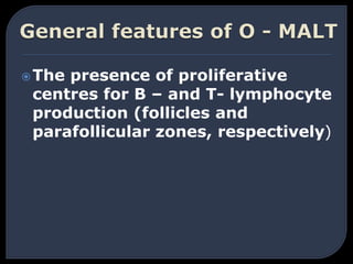
- 1. The presence of proliferative centres for B – and T- lymphocyte production (follicles and parafollicular zones, respectively)
- 2. Proximity to an epithelial surface, the lymphoid tissue being essentially situated within the mucosal lamina propria
- 3. The lack of a fibrous capsule
- 4. The provision of high – endothelium venules (HEVs) for immigration of lymphocytes.
- 5. The presence of efferent lymphatics but virtual absence of afferents.
- 7. Lies in the roof of the oral cavity Has two parts: • Hard (bony) palate anteriorly • Soft (muscular) palate posteriorly hard soft palate
- 8. Attached to the posterior border of the hard palate Covered on its upper and lower surfaces by mucous membrane Composed of: • Muscle fibers • An aponeurosis • Lymphoid tissue • Glands • Blood vessels • Nerves
- 9. Fibrous sheath Attached to posterior border of hard palate Is expanded tendon of tensor velli palatini Gives origin & insertion to palatine muscles
- 10. Tensor veli palatini • Origin: spine of sphenoid; auditory tube • Insertion: forms palatine aponeurosis • Action: Tenses soft palate
- 12. Levator veli palatini • Origin:petrous temporal bone, auditory tube, palatine aponeurosis • Insertion: palatine aponeurosis • Action: Raises soft palate
- 13. Musculus uvulae • Origin: posterior border of hard palate • Insertion: mucosa of uvula • Action: Elevates uvula
- 14. Palatoglossus • Origin: palatine aponeurosis • Insertion: side of tongue • Action: pulls root of tongue upward, narrowing oropharyngeal isthmus Palatopharyngeus • Origin: palatine aponeurosis • Insertion: posterior border of thyroid cartilage • Action: Elevates wall of the pharynx
- 15. Palatopharyngeus
- 16. Mostly by the maxillary nerve through its branches: • Greater palatine nerve • Lesser palatine nerve • Nasopalatine nerve Glossopharyngeal nerve supplies the region of the soft palate
- 17. All the muscles, except tensor veli palatini, are supplied by the: •Pharyngeal plexus Tensor veli palatini supplied by the: • Nerve to medial pterygoid, a branch of the mandibular division of the trigeminal nerve
- 18. Branches of the maxillary artery • Greater palatine • Lesser palatine • Sphenopalatine Ascending palatine, branch of the facial artery Ascending pharyngeal, branch of the external carotid artery
- 19. Cleft palate: •Unilateral •Bilateral •Median Paralysis of the soft palate •The pharyngeal isthmus can not be closed during swallowing and speech Pharyngeal isthmus
- 21. The lymphoid tissue in the pharyngeal aponeurosis aggregates in some areas forming tonsils: 1-one nasopharyngeal tonsil 2- two palatine tonsils 3- two lingual tonsils
- 23. Location The palatine tonsil is an ovoid mass of lymphoid tissue Tonsillar fossa in lateral wall of oropharynx
- 25. Boundaries of tonsillar fossa Anterior pillar- Palatoglossal arch Posterior pillar- Palatopharyngeal arch Apex- Soft palate where both arches meet Base – Dorsal surface of posterior one – third of tongue
- 26. External features 2 surfaces- medial, lateral(tonsillar bed) 2 Poles- upper, lower
- 27. Stratified squamous non keratinising epithelium Dips into the crypts The crypts are 12-15 in number crypta magna It represents the ventral part of second pharyngeal pouch External features-Medial surface
- 28. Fibrous capsule of the tonsil Loose areolar tissue The tonsillar bed External features-Lateral surface
- 29. Superior constrictor muscle Styloglossus muscle Glossopharyngeal nerve styloid process (if enlarged) Facial artery Medial pterygoid muscle Angle of mandible Submandibular salivary gland Tonsillar bed
- 31. External features-Poles Upper pole- extends into soft palate Lower pole- attached to tongue
- 33. The tonsil is supplied by 5 arteries: Tonsillar branch of facial artery (main supply) Ascending palatine branch of facial artery Ascending pharyngeal artery Dorsal linguae branch of lingual artery Descending palatine branch of maxillary artery Arterial supply
- 34. Blood supply from medial surface
- 35. The paratonsillar vein Pharyngeal venous plexus Venous drainage
- 36. Lymphatics from the tonsil pierce the superior constrictor and Drain into the upper cervical lymph nodes especially jugulodigastric (tonsillar) lymph node Lymphatic drainage
- 37. Lesser palatine branch of sphenopalatine ganglion Glossopharyngeal nerve Nerve supply
- 39. Acute tonsillitis Chronic tonsillitis Applied anatomy
- 42. Applied anatomy-Peritonsillar vein injury
- 43. Thank You