
Empfohlen
Empfohlen
Weitere ähnliche Inhalte
Was ist angesagt?
Was ist angesagt? (20)
Andere mochten auch
Ähnlich wie Wei wang
Ähnlich wie Wei wang (20)
Mehr von mganguly123
Mehr von mganguly123 (20)
Wei wang
- 1. International Journal of Pharmaceutics 203 (2000) 1 – 60 www.elsevier.com/locate/ijpharm Review Lyophilization and development of solid protein pharmaceuticals Wei Wang* Biotechnology, Bayer Corporation, 800 Dwight Way, Berkeley, CA 94701, USA Received 9 September 1999; received in revised form 21 December 1999; accepted 4 April 2000 Abstract Developing recombinant protein pharmaceuticals has proved to be very challenging because of both the complexity of protein production and purification, and the limited physical and chemical stability of proteins. To overcome the instability barrier, proteins often have to be made into solid forms to achieve an acceptable shelf life as pharmaceu- tical products. The most commonly used method for preparing solid protein pharmaceuticals is lyophilization (freeze-drying). Unfortunately, the lyophilization process generates both freezing and drying stresses, which can denature proteins to various degrees. Even after successful lyophilization with a protein stabilizer(s), proteins in solid state may still have limited long-term storage stability. In the past two decades, numerous studies have been conducted in the area of protein lyophilization technology, and instability/stabilization during lyophilization and long-term storage. Many critical issues have been identified. To have an up-to-date perspective of the lyophilization process and more importantly, its application in formulating solid protein pharmaceuticals, this article reviews the recent investigations and achievements in these exciting areas, especially in the past 10 years. Four interrelated topics are discussed: lyophilization and its denaturation stresses, cryo- and lyo-protection of proteins by excipients, design of a robust lyophilization cycle, and with emphasis, instability, stabilization, and formulation of solid protein pharmaceuticals. © 2000 Elsevier Science B.V. All rights reserved. Keywords: Aggregation; Cryoprotection; Denaturation; Excipient; Formulation; Freeze-drying; Glass transition; Stability; Lyopro- tection; Residual moisture 1. Introduction both the complexity of protein production and purification, and the limited physical and chemi- Developing recombinant protein pharmaceuti- cal stability of proteins. In fact, protein instability cals has proved to be very challenging because of is one of the two major reasons why protein pharmaceuticals are administered traditionally * Tel.: +1-510-7054755; fax: + 1-510-7055629. through injection rather than taken orally like E-mail address: wei.wang.b@bayer.com (W. Wang). most small chemical drugs (Wang, 1996). To over- 0378-5173/00/$ - see front matter © 2000 Elsevier Science B.V. All rights reserved. PII: S 0 3 7 8 - 5 1 7 3 ( 0 0 ) 0 0 4 2 3 - 3
- 2. 2 W. Wang / International Journal of Pharmaceutics 203 (2000) 1–60 come the instability barrier, proteins often have to cal and chemical instabilities and stabilization of be made into solid forms to achieve an acceptable proteins in aqueous and solid states (Manning et shelf life. al., 1989; Cleland et al., 1993); chemical instability The most commonly used method for preparing mechanisms of proteins in solid state (Lai and solid protein pharmaceuticals is lyophilization Topp, 1999); various factors affecting protein sta- (freeze-drying). However, this process generates a bility during freeze-thawing, freeze-drying, and variety of freezing and drying stresses, such as storage of solid protein pharmaceuticals solute concentration, formation of ice crystals, pH (Arakawa et al., 1993); and application of changes, etc. All of these stresses can denature lyophilization in protein drug development (Pikal, proteins to various degrees. Thus, stabilizers are 1990a,b; Skrabanja et al., 1994; Carpenter et al., often required in a protein formulation to protect 1997; Jennings, 1999). Nevertheless, it appears protein stability both during freezing and drying that several critical issues in the development of processes. solid protein pharmaceuticals have not been fully Even after successful lyophilization, the long- examined, including various instability factors, term storage stability of proteins may still be very stabilization, and formulation of solid protein limited, especially at high storage temperatures. In pharmaceuticals. several cases, protein stability in solid state has To have an up-to-date perspective of the been shown to be equal to, or even worse than, lyophilization process and more importantly, its that in liquid state, depending on the storage application in formulating solid protein pharma- temperature and formulation composition. For ceuticals, this article reviews the recent investiga- example, a major degradation pathway of human tions and achievements in these exciting areas, insulin-like growth factor I (hIGF-I) is oxidation especially in the past 10 years. Four interrelated of Met59 and the oxidation rate in a freeze-dried topics are discussed sequentially, lyophilization formulation in air-filled vials is roughly the same and its denaturation stresses; cryo- and lyo-pro- as that in a solution at either 25 or 30°C tection of proteins by excipients; design of a ro- (Fransson et al., 1996). Similarly, the oxidation bust lyophilization cycle; and with emphasis, rate of lyophilized interleukin 2 (IL-2) is the same instability, stabilization, and formulation of solid as that in a liquid formulation containing 1 mg protein pharmaceuticals. ml − 1 IL-2, 0.5% hydroxypropyl-b-cyclodextrin (HP-b-CD), and 2% sucrose during storage at 4°C (Hora et al., 1992b). At a high water content 2. Lyophilization and its denaturation stresses ( 50%), the degradation rate of insulin is higher in a lyophilized formulation than in a solution 2.1. Lyophilization process with similar pH-rate profiles in both states (Strickley and Anderson, 1996). The glucose-in- Lyophilization (freeze-drying) is the most com- duced formation of des-Ser relaxin in a mon process for making solid protein pharmaceu- lyophilized formulation is faster than in a solution ticals (Cleland et al., 1993; Fox, 1995). This during storage at 40°C (Li et al., 1996). These process consists of two major steps: freezing of a examples indicate that stabilizers are still required protein solution, and drying of the frozen solid in lyophilized formulations to increase long-term under vacuum. The drying step is further divided storage stability. into two phases: primary and secondary drying. In the past two decades, numerous studies have The primary drying removes the frozen water and been conducted in the areas of protein freezing the secondary drying removes the non-frozen and drying, and instability and stabilization of ‘bound’ water (Arakawa et al., 1993). The amount proteins during lyophilization and long-term stor- of non-frozen water for globular proteins is about age. Many critical issues have been identified in 0.3–0.35 g g − 1 protein, slightly less than the this period. These studies and achievements have proteins’ hydration shell (Rupley and Careri, been reviewed elsewhere with emphasis on physi- 1991; Kuhlman et al., 1997). More detailed analy-
- 3. W. Wang / International Journal of Pharmaceutics 203 (2000) 1–60 3 sis of each lyophilization step is provided in Sec- cluding formation of dendritic ice crystals, in- tion 4. creased ionic strength, changed pH, and phase Lyophilization generates a variety of stresses, separation; and (3) drying stress (removing of the which tend to destabilize or unfold/denature an protein hydration shell). unprotected protein. Different proteins tolerate freezing and/or drying stresses to various degrees. 2.2.1. Low temperature stress Freeze-thawing of ovalbumin at neutral pH did The first quantitative study on low-temperature not cause denaturation (Koseki et al., 1990). Re- denaturation of a model protein was conducted peated (three times) freeze-thawing of tissue-type presumably by Shikama and Yamazaki (1961). plasminogen activator (tPA) did not cause any They demonstrated a specific temperature range decrease in protein activity (Hsu et al., 1995). in which ox liver catalase was denatured during Some proteins can keep their activity both during freeze-thawing. Cold denaturation of catalase at freezing and drying processes, such as a1-an- 8.4 mg ml − 1 in 10 mM phosphate buffer (pH 7.0) titrypsin in phosphate – citrate buffer (Vemuri et started at − 6°C. Loss of catalase activity reached al., 1994), porcine pancreatic elastase without ex- 20% at − 12°C, remained at this level between cipients (Chang et al., 1993), and bovine pancre- − 12°C and near − 75°C, then decreased gradu- atic ribonuclease A (RNase A, 13.7 kD) in the ally from − 75 to − 120°C. There was almost no presence or absence of phosphate (Townsend and activity loss between − 129 and − 192°C. Similar DeLuca, 1990). results were also obtained for ovalbumin by However, many proteins cannot stand freezing Koseki et al. (1990). Incubation of frozen ovalbu- and/or drying stresses. Freeze-thawing caused loss min solution caused structural change of ovalbu- of activity of lactate dehydrogenase (LDH) min, as monitored by UV difference spectra, (Nema and Avis, 1992; Izutsu et al., 1994b; An- which increased with decreasing temperature be- dersson and Hatti-Kaul, 1999), 60% loss of L-as- tween −10 and − 40°C. Further decrease in paraginase (10 mg ml − 1) activity in 50 mM incubation temperature to − 80°C caused less sodium phosphate buffer (pH 7.4) (Izutsu et al., structural change, and no change at − 192°C. 1994a), and aggregation of recombinant Perlman and Nguyen (1992) reported that inter- hemoglobin (Kerwin et al., 1998). Freeze-drying feron-g(IFN-g) aggregation in a liquid mannitol caused 10% loss of the antigen-binding capacity formulation was more severe at −20°C than at of a mouse monoclonal antibody (MN12) (Ress- − 70, 5 and 15°C during storage. To prevent ing et al., 1992), more than 40% loss of bilirubin freezing-induced complication in studying cold oxidase (BO) activity in the presence of dextran or protein denaturation, cold and heat denaturation polyvinylalcohol (PVA) (Nakai et al., 1998), loss of RNase A has been conducted under high pres- of most b-galactosidase activity at 2 or 20 mg sure (3 kbar). Under this condition, RNase A ml − 1 (Izutsu et al., 1993, 1994a), complete loss of denatured below −22°C and above 40°C (Zhang phosphofructokinase (PFK) and LDH activity in et al., 1995). All these examples are clear indica- the absence of stabilizers (Carpenter et al., 1986, tion of low temperature denaturation rather than 1990; Prestrelski et al., 1993a; Anchordoquy and a freezing or thawing effect. Carpenter, 1996), and dissociation of Erwinia L- The nature of cold denaturation has not been asparaginase tetramer (135 kD) into four inactive satisfactorily delineated. Since solubility of non- subunits (34 kD each) in the absence of any polar groups in water increases with decreasing protectants (Adams and Ramsay, 1996). temperature due to increased hydration of the non-polar groups, solvophobic interaction in 2.2. Denaturation stresses during lyophilization proteins weakens with decreasing temperature (Dill et al., 1989; Graziano et al., 1997). The The lyophilization process generates a variety decreasing solvophobic interaction in proteins can of stresses to denature proteins. These include (1) reach a point where protein stability reaches zero, low temperature stress; (2) freezing stresses, in- causing cold denaturation (Jaenicke, 1990). While
- 4. 4 W. Wang / International Journal of Pharmaceutics 203 (2000) 1–60 normal or thermal denaturation is entropy-driven, centration upon freezing may drastically acceler- cold denaturation is enthalpy-driven (Dill et al., ate protease-catalyzed protein degradation. 1989; Shortle, 1996). Oligomeric proteins typically show cold denaturation, i.e. dissociation of sub- 2.2.3. Formation of ice-water interface unit oligomers, since association is considered to Freezing a protein solution generates an ice-wa- be a consequence of hydrophobic interaction ter interface. Proteins can be adsorbed to the (Jaenicke, 1990; Wisniewski, 1998). Theoretically, interface, loosening the native fold of proteins and the calculated free energy of unfolding (DGunf) for resulting in surface-induced denaturation proteins has a parabolic relationship with temper- (Strambini and Gabellieri, 1996). Rapid (quench) ature. This means that a temperature of maximum cooling generates a large ice-water interface while stability exists, and both high and low tempera- a smaller interface is induced by slow cooling ture can destabilize a protein (Jaenicke, 1990; (also see Section 4.2). Chang et al. (1996b) Kristjansson and Kinsella, 1991). ´ demonstrated that a single freeze–thaw cycle with quench cooling denatured six model proteins, in- 2.2.2. Concentration effect cluding ciliary neurotrophic factor (CNTF), gluta- Freezing a protein solution rapidly increases the mate dehydrogenase (GDH), interleukin-1 concentration of all solutes due to ice formation. receptor antagonist (IL-1ra), LDH, PFK, and For example, freezing a 0.9% NaCl solution to its tumor necrosis factor binding protein (TNFbp). eutectic temperature of −21°C can cause a 24- The denaturation effect of quench cooling was fold increase in its concentration (Franks, 1990). greater or equivalent to that after 11 cycles of The calculated concentration of small carbohy- slow cooling, suggesting surface-induced denatu- drates in the maximally freeze-concentrated ma- ration. This denaturation mechanism was sup- trices (MFCS) is as high as 80% (Roos, 1993). ported by a good correlation (r= 0.99) found Thus, all physical properties related to concentra- between the degree of freeze-induced denaturation tion may change, such as ionic strength and rela- and that of artificially surface-induced denatura- tive composition of solutes due to selective tion. The surface was introduced by shaking the crystallization. These changes may potentially protein solution containing hydrophobic Teflon destabilize a protein. beads. In a similar study, a correlation coefficient Generally, lowering the temperature reduces the of 0.93 was found between the tendency of freeze rate of chemical reactions. However, chemical denaturation and surface-induced denaturation reactions may actually accelerate in a partially for eight model proteins, including aldolase, basic frozen aqueous solution due to increased solute fibroblast growth factor (bFGF), GDH, IL-1ra, concentration (Pikal, 1999). Due to solute concen- LDH, maleate dehydrogenase (MDH), PFK, and tration, the rate of oligomerization of b-glutamic TNFbp (Kendrick et al., 1995b). However, there acid at −20°C was much faster than at 0 or 25°C was no significant correlation (r=0.78) between in the presence of a water-soluble carbodiimide, freeze denaturation and thermal denaturation 1-ethyl-3-(3-dimethylaminopropyl) carbodiimide temperature (Chang et al., 1996b). (EDAC) (Liu and Orgel, 1997). The increase in the rate of a chemical reaction 2.2.4. pH changes during freezing in a partially frozen state could reach several Many proteins are stable only in a narrow pH orders of magnitude relative to that in solution range, such as low molecular weight urokinase (Franks, 1990, 1994). (LMW-UK) at pH 6–7 (Vrkljan et al., 1994). At The reported oxygen concentration in a par- extreme pHs, increased electrostatic repulsion be- tially frozen solution at −3°C is as high as 1150 tween like charges in proteins tends to cause times that in solution at 0°C (Wisniewski, 1998). protein unfolding or denaturation (Goto and The increased oxygen concentration can readily Fink, 1989; Volkin and Klibanov, 1989; Dill, oxidize sulphydryl groups in proteins. If a protein 1990). Thus, the rate of protein aggregation is solution contains any contaminant proteases, con- strongly affected by pH, such as aggregation of
- 5. W. Wang / International Journal of Pharmaceutics 203 (2000) 1–60 5 interleukin 1b (IL-1b) (Gu et al., 1991), human separation due to polymers’ altered solubilities at relaxin (Li et al., 1995a), and bovine pancreatic low temperatures. Freezing-induced phase separa- RNase A (Townsend and DeLuca, 1990; Tsai et tion can easily occur in a solution containing two al., 1998). Moreover, the solution pH can signifi- incompatible polymers such as dextran and Ficoll cantly affect the rate of many chemical degrada- (Izutsu et al., 1996). During freezing of recombi- tions in proteins (Wang, 1999). nant hemoglobin in a phosphate buffer containing Freezing a buffered protein solution may selec- 4% (w/w) PEG 3350, 4% (w/w) dextran T500, and tively crystallize one buffering species, causing pH 150 mM NaCl, liquid–liquid phase separation changes. Na2HPO4 crystallizes more readily than occurred and created a large excess of interface, NaH2PO4 because the solubility of the disodium denaturing the protein (Heller et al., 1997). Addi- form is considerably lower than that of the tion of 5% sucrose or trehalose could not reverse monosodium form. Because of this, a sodium the denaturation effect in the system (Heller et al., phosphate buffer at pH 7 has a molar [NaH2PO4]/ 1999a). [Na2HPO4] ratio of 0.72, but this ratio increases Several strategies have been proposed to miti- to 57 at the ternary eutectic temperature during gate or prevent phase separation-induced protein freezing (Franks, 1990, 1993). This can lead to a denaturation during freezing. These include use of significant pH drop during freezing, which then alternative salts (Heller et al., 1999a), adjustment denatures pH-sensitive proteins. For example, of the relative composition of polymers to avoid freezing of a LDH solution caused protein denat- or to rapidly pass over a temperature region uration due to a pH drop from 7.5 to 4.5 upon where the system may result in liquid–liquid selective crystallization of Na2HPO4 (Anchordo- phase separation (Heller et al., 1999c), and chemi- quy and Carpenter, 1996). LDH is a pH-sensitive cal modification of the protein such as pegylation protein and a small drop in pH during freezing (Heller et al., 1999b). can partially denature the protein even in the presence of stabilizers such as sucrose and tre- 2.2.6. Dehydration stresses halose (Nema and Avis, 1992). The pH drop Proteins in an aqueous solution are fully hy- during freezing may also explain why freezing drated. A fully hydrated protein has a monolayer bovine and human IgG species in a sodium phos- of water covering the protein surface, which is phate buffer caused formation of more aggregates termed the hydration shell (Rupley and Careri, than in potassium phosphate buffer, because 1991). The amount of water in full hydration is potassium phosphate buffer does not show signifi- 0.3–0.35 g g − 1 protein (Rupley and Careri, 1991; cant pH changes during freezing (Sarciaux et al., Kuhlman et al., 1997). Generally, the water con- 1998). tent of a lyophilized protein product is less than The pH drop during freezing can potentially 10%. Therefore, lyophilization removes part of affect storage stability of lyophilized proteins. the hydration shell. Removal of the hydration Lyophilized IL-1ra in a formulation containing shell may disrupt the native state of a protein and phosphate buffer at pH 6.5 aggregated more cause denaturation. A hydrated protein, when rapidly than that containing citrate buffer at the exposed to a water-poor environment during de- same pH during storage at 8, 30 and 50°C (Chang hydration, tends to transfer protons to ionized et al., 1996c). Similarly, the pH drop of a succi- carboxyl groups and thus abolishes as many nate-containing formulation from 5 to 3 –4 during charges as possible in the protein (Rupley and freezing appeared to be the cause of less storage Careri, 1991). The decreased charge density may stability for lyophilized IFN-g than that contain- facilitate protein–protein hydrophobic interac- ing glycocholate buffer at the same pH (Lam et tion, causing protein aggregation. al., 1996). Water molecules can also be an integral part of an active site(s) in proteins. Removal of these 2.2.5. Phase separation during freezing functional water molecules during dehydration Freezing polymer solutions may cause phase easily inactivates proteins. For example, dehydra-
- 6. 6 W. Wang / International Journal of Pharmaceutics 203 (2000) 1–60 tion of lysozyme caused loss of activity apparently Lyophilization may induce several potential due to removal of those water molecules residing changes in the IR spectra of proteins. Disruption functionally in the active site (Nagendra et al., of hydrogen bonds in proteins during lyophiliza- 1998). tion generally leads to an increase in frequency Lastly, dehydration during lyophilization may and a decrease in intensity of hydroxyl stretching cause significant difference in moisture distribu- bands (Carpenter and Crowe, 1989). Unfolding of tion in different locations of a product cake. The proteins during lyophilization broadens and shifts uneven moisture distribution may lead to possible (to higher wave numbers) amide I component localized overdrying, which may exacerbate dehy- peaks (Prestrelski et al., 1993b; Allison et al., dration-induced protein denaturation (Pikal and 1996). Lyophilization often leads to an increase in Shah, 1997). b-sheet content with a concomitant decrease in a-helix content. Conversion of a-helix to b-sheet 2.3. Monitoring protein denaturation upon during lyophilization has been observed in many lyophilization proteins such as tetanus toxoid (TT) in 10 mM The most common method for monitoring sodium phosphate buffer (pH 7.3) (Costantino et protein denaturation upon lyophilization appears al., 1996), recombinant human albumin (rHA) in to be infrared (IR) spectroscopy, although other different buffer solutions at different pHs methods have been used such as mass spec- (Costantino et al., 1995a), hGH at pH 7.8, and troscopy (Bunk, 1997), and Raman spectroscopy seven model proteins in water, including bovine (Belton and Gil, 1994). In the following section, pancreatic trypsin inhibitor (BPTI), chymotrypsi- IR methodology is discussed in monitoring nogen, horse myoglobin (Mb), horse heart cy- protein denaturation upon lyophilization followed tochrome c (Cyt c), rHA, porcine insulin, and by a discussion on reversibility of protein RNase A (Griebenow and Klibanov, 1995). denaturation. An increase in b-sheet content during lyophilization is often an indication of protein 2.3.1. Infrared (IR) spectroscopy aggregation and/or increased intermolecular inter- IR (or FTIR) is probably the most extensively action (Yeo et al., 1994; Griebenow and used technique today for studying structural Klibanov, 1995; Overcashier et al., 1997). changes in proteins upon lyophilization (Susi and Lyophilization-induced increase in b-sheet content Byler, 1986; Dong et al., 1995; Carpenter et al., seems to be a rather general phenomenon as 1998, 1999). The lyophilization-induced structural lyophilization or air-drying of unordered poly-L- changes can be monitored conveniently in the lysine induced structural transition to a highly amide I, II, or III region. For lyophilized protein ordered b-sheet (Prestrelski et al., 1993b; Wolkers samples, residual water up to 10% (w/w) does not et al., 1998b). Such transition has also been ob- interfere significantly in the amide I region, a served in proteins during lyophilization such as frequently used sensitive region for determination human insulin in water (pH 7.1) (Pikal and Rigs- of secondary structures (Dong et al., 1995). How- bee, 1997). The b-sheet structure after lyophiliza- ever, IR studies on proteins in an aqueous solu- tion shows a higher degree of intermolecular tion need either subtraction of water absorption or solvent replacement with D2O (Goormaghtigh hydrogen bonding because polar groups must sat- et al., 1994). To make reliable subtraction, high isfy their H-bonding requirement by intra- or protein concentrations ( 10 mg ml − 1) are rec- intermolecular interaction upon removal of water. ommended to increase protein absorption signal, The intermolecular b-sheet is characterized by two and a CaF2 (or BaF2) cell with a path length of 10 major IR bands at about 1617 and 1697 cm − 1 in mm or less should be used to control the total solid state, which can be used to monitor protein sample absorbance within 1 (Cooper and Knut- denaturation (Allison et al., 1996). Similarly, the son, 1995). relative intensity of a-helix band also can be used
- 7. W. Wang / International Journal of Pharmaceutics 203 (2000) 1–60 7 in this regard (Yang et al., 1999; Heller et al., upon lyophilization (Carpenter et al., 1993), while 1999b). loss of BO activity in a PVA-containing formula- The extent of changes in overall IR spectrum of tion was at least partially reversible (Nakai et al., a protein upon lyophilization reflects the degree of 1998). Using IR spectroscopy, Prestrelski et al. protein denaturation. The changes relative to a (1993b) demonstrated that lyophilization-induced reference spectrum can be measured using a corre- structural changes were irreversible for bFGF, lation coefficient (r) as defined by Prestrelski et al. IFN-g, and bovine a-casein, but essentially re- (1993a), or the extent of spectral area overlap versible for G-CSF and bovine a-lactalbumin. (Heimburg and Marsh, 1993; Allison et al., 1996; The extensive aggregation and precipitation of Kendrick et al., 1996). Using the correlation co- IFN-g and casein upon rehydration confirmed the efficient, Prestrelski et al. (1993b) were able to irreversibility in structural changes. Therefore, measure the relative freeze-drying stability of sev- lyophilization of proteins may lead to three types eral model proteins, including bFGF, bovine a- of behavior, (1) no change in protein conforma- lactalbumin, bovine a-casein, IFN-g, and tion; (2) reversible denaturation; or (3) irreversible recombinant granulocyte colony-stimulating fac- denaturation. tor (rG-CSF) in the presence of different sugars. In many cases, IR-monitored structural changes Nevertheless, Griebenow and Klibanov (1995), during lyophilization seem to be reversible. after analyzing secondary structures of seven Griebenow and Klibanov (1995) demonstrated model proteins upon lyophilization, concluded that lyophilization (dehydration) caused signifi- that the correlation coefficient was not highly cant changes in the secondary structures of seven sensitive to structural alterations in proteins. In- model proteins in the amide III region (1220– stead, comparison of overlapping area-normalized 1330 cm − 1), including BPTI, chymotrypsinogen, second-derivative or deconvoluted spectra seemed Mb, Cyt c, rHA, insulin, and RNase A. The more reliable and objective. structure of almost all proteins became more or- Recently, IR has been used in real-time moni- dered upon lyophilization with a decrease in the toring of freezing and dehydration stresses on unordered structures. Nevertheless, all these struc- proteins during lyophilization. By this method, tural changes were reversible upon reconstitution. glucose at 10% was shown to protect lysozyme Other examples of reversible changes in the sec- both during the freezing and drying processes ondary structures of proteins upon lyophilization (Remmele et al., 1997). include rHA (Costantino et al., 1995a), Humicola lanuginosa lipase (Kreilgaard et al., 1999), IL-2 2.3.2. Re6ersibility of freezing- or (Prestrelski et al., 1995), and lysozyme (Allison et lyophilization-induced protein denaturation al., 1999). Many proteins denature to various extents upon freezing, especially at low concentrations ( B 0.1 mg ml − 1). Freezing-induced denaturation 3. Cryo- and lyo-protection of proteins by may or may not be reversible. Freezing lysozyme stabilizers or IL-1ra caused reversible denaturation (Kendrick et al., 1995a). In contrast, recombinant As discussed before, both freezing and dehydra- factor XIII (rFXIII, 166 kD) was irreversibly tion can induce protein denaturation. To protect a denatured upon freezing, and loss of native protein from freezing (cryoprotection) and/or de- rFXIII at 1 mg ml − 1 increased linearly with the hydration (lyoprotection) denaturation, a protein number of freeze –thaw cycles (Kreilgaard et al., stabilizer(s) may be used. These stabilizers are 1998b). also referenced as chemical additives (Li et al., Similarly, lyophilization-induced denaturation 1995b), co-solutes (Arakawa et al. 1993), co-sol- can be either reversible or irreversible. In the vents (Timasheff, 1993, 1998), or excipients absence of stabilizers, PFK at 25 mg ml − 1 at pH (Wong and Parascrampuria, 1997; Wang, 1999). 7.5 and 8.0 was fully and irreversibly inactivated In the following section, a variety of protein
- 8. 8 W. Wang / International Journal of Pharmaceutics 203 (2000) 1–60 stabilizers are presented for cryo- and lyo-protec- For example, freezing rabbit muscle LDH in wa- tion, followed by discussions of their possible ter caused 64% loss of protein activity, and in the stabilization mechanisms. presence of 5, 10 or 34.2% sucrose, the respective losses were 27, 12, and 0% (Nema and Avis, 3.1. Stabilizers for cryo- and lyo-protection 1992). Other sugars or polyols that can protect LDH during freeze-thawing to different degrees Nature protects life from freezing or osmotic include lactose, glycerol, xylitol, sorbitol, and shock by accumulating selected compounds to mannitol, at 0.5–1 M (Carpenter et al., 1990). high concentrations ( 1 M) within organisms. Increasing trehalose concentration gradually in- These accumulated compounds are known as cry- creased the recovery of PFK activity during oprotectants and osmolytes, which are preferen- freeze-thawing and the recovery reached a maxi- tially excluded from surfaces of proteins and act mum of 90% at about 300 mg ml − 1 (Carpenter et as structure stabilizers (Timasheff, 1993). How- al., 1990). A similar stabilizing trend was also ever, since the dehydration stress is different from observed for sucrose, maltose, glucose, or inositol those of freezing, many effective cryoprotectants (Carpenter et al., 1986). or protein stabilizers in solution do not stabilize Since freezing is part of the freeze-drying pro- proteins during dehydration (drying). Some even cess, high concentrations of sugars or polyols are destabilize proteins during lyophilization. For ex- often necessary for lyoprotection. These examples ample, CaCl2 stabilized elastase (20 mg ml − 1) in include the protection of chymotrypsinogen in the 10 mM sodium acetate (pH 5.0), but caused the presence of 300 mM sucrose (Allison et al., 1996), lyophilized protein cake to collapse and lose activ- complete inhibition of acidic fibroblast growth ity (Chang et al., 1993). factor (aFGF) aggregation by 2% sucrose (Volkin Similarly, effective lyophilization stabilizers (ly- and Middaugh, 1996), increase in glucose-6-phos- oprotectants) may or may not stabilize proteins phate dehydrogenase (G6PDH) activity from 40 effectively during freezing. Therefore, in cases to about 90% by 5.5% sugar mixture (glu- when a single stabilizer does not serve as both a cose:sucrose=1:10, w/w) (Sun et al., 1998), com- cryoprotectant and a lyoprotectant, two (or more) plete recovery of LDH by either 7% sucrose or 7% stabilizers may have to be used to protect proteins raffinose, a trisaccharide (Moreira et al., 1998), from denaturation during lyophilization. significant improvement of PFK recovery by 400 mM trehalose (Carpenter et al., 1993), and com- 3.1.1. Sugars/polyols plete protection of four restriction enzymes by Many sugars or polyols are frequently used 15% trehalose (Colaco et al., 1992). More exam- nonspecific protein stabilizers in solution and dur- ples can be found in Table 2. ing freeze-thawing and freeze-drying. They have Lower concentrations of sugars or polyols may been used both as effective cryoprotectants and or may not have any significant effect. At 5 to 100 remarkable lyoprotectants. In fact, their function mM, neither trehalose nor glucose could protect as lyoprotectants for proteins has long been LDH or PFK to a significant level during adopted by nature. Anhydrobiotic organisms (wa- lyophilization (Carpenter et al., 1993). To deter- ter content B1%) commonly contain high con- mine the minimum sugar concentration that offers centrations (up to 50%) of disaccharides, the maximum stabilization effect, Tanaka et al. particularly sucrose or trehalose, to protect them- (1991) studied the lyoprotective effect of saccha- selves (Crowe et al., 1992, 1998). rides on the denaturation of catalase during The level of stabilization afforded by sugars or lyophilization. They demonstrated that saccha- polyols generally depends on their concentrations. rides protected the protein by direct interaction A concentration of 0.3 M has been suggested to with the protein and a concentration of saccha- be the minimum to achieve significant stabiliza- rides sufficient to form a monomolecular layer on tion (Arakawa et al., 1993). This has been found the protein surface was the minimum to achieve to be true in many cases during freeze-thawing. the maximum stabilization. Therefore, the stabi-
- 9. W. Wang / International Journal of Pharmaceutics 203 (2000) 1–60 9 lization of catalase was found to depend not on buffer (pH 7.4) (Table 2), mannitol for LDH in 50 the bulk concentration of maltose but on the mM sodium phosphate buffer (pH 7.4) (Izutsu et weight ratio of maltose to catalase (Tanaka et al., al., 1994b), and myo-inositol for PFK during 1991). Maximum stabilization of catalase was at a freeze-thawing and freeze-drying (Table 2). Again, ratio of about 0.4. A recent study showed that the decreased protein recovery is probably due to maximum protection (about 75% recovery) of L- crystallization of these excipients at high asparaginase at 1.45 mg ml − 1 during lyophiliza- concentrations. tion was reached at a saccharide concentration of The level of protein protection afforded by about 0.5 mg ml − 1, which was about the calcu- different sugars or polyols can be either similar or lated monosaccharide concentration required to significantly different, depending on the formula- interact with all exposed highly polar residues of tion composition, concentration and physical the protein (Ward et al., 1999). At this concentra- properties of the stabilizer, and its compatibility tion, the weight ratio of saccharide to L-asparagi- with the protein. Ward et al. (1999) found that nase is 0.34, which is coincidentally very close to several saccharides, including trehalose, lactose, that of maltose to catalase. maltose, sucrose, glucose, and mannitol, displayed On the other hand, increasing sugar/polyol con- similar level of protection towards tetrameric L- centration to a certain level may eventually reach asparaginase (1.45 mg ml − 1) during lyophiliza- a limit of stabilization or even destabilize a tion at saccharide concentrations up to 0.1%. At protein during freeze-drying. For example, actin 2%, glucose or lactose protected L-asparaginase was maximally stabilized during lyophilization in from dissociation during freeze-drying, but man- the presence of 5% (w/v) sucrose and a further nitol did not, possibly due to its crystallization increase in sucrose concentration to 10% did not and loss of intimate interaction with the protein improve the protein stability significantly, which (Adams and Ramsay, 1996). Probably for the was apparently attributable to sticky, pliable, and same reason, mannitol at 88 mM inhibited the collapsed formulation structure (Allison et al., formation of insoluble hGH aggregates in phos- 1998). Increasing trehalose concentration to 150 phate buffer (pH 7.4) at a freezing rate of 50°C mg ml − 1 in a PFK formulation (at 50 mg ml − 1, min − 1, but accelerated hGH aggregation at lower pH 8.0) increased the freeze-drying recovery of freezing rates of 0.5 and 5°C min − 1 (Eckhardt et PFK activity to about 65%, but further increases al., 1991). In a different study, however, Tanaka in trehalose concentration caused a gradual de- et al. (1991) demonstrated that both mannitol and crease in recovery of the protein activity (Carpen- sorbitol could increase the recovery of catalase ter and Crowe, 1989). At a trehalose activity during lyophilization to a similar level as concentration of 400 mg ml − 1, basically no PFK that offorded by maltose. They also showed that activity was left after freeze-drying. Since tre- different sugars (maltose, glucose, and mal- halose at 400 mg ml − 1 protected about 90% of totriose) at 1 mg ml − 1 could increase the recovery the protein activity after freeze-thawing, the desta- of catalase activity to the same level (from 35 to bilization of PFK at high concentrations of tre- 90%), but maltopentaose, maltohexaose, and mal- halose occurred in the dehydration step, possibly toheptaose were not as effective (Tanaka et al., due to crystallization of trehalose, preventing req- 1991). The ineffectiveness of larger saccharides uisite hydrogen bonding to the dried protein (see suggests that protein stabilization by sugars may Section 3.2) (Carpenter and Crowe, 1989). A sim- depend on their glucoside chain lengths, and a ilar trend was observed in the stabilization of long chain length may interfere with intermolecu- several other proteins during lyophilization in the lar hydrogen-bonding between stabilizing sugars presence of increasing concentrations of excipi- and proteins. ents, including mannitol for L-asparaginase (10 mg In many cases, disaccharides appear to be the ml − 1) in 50 mM sodium phosphate buffer (pH most effective and universal stabilizers among 7.4) (Izutsu et al., 1994b), mannitol for b-galac- sugars and polyols (Arakawa et al., 1993; Carpen- tosidase (2 mg ml − 1) in 10 mM sodium phosphate ter et al., 1997). For example, the disaccharides
- 10. 10 W. Wang / International Journal of Pharmaceutics 203 (2000) 1–60 trehalose, sucrose, maltose, and lactose, were all 1998b), while sucrose was a better stabilizer than essentially equivalent to or more effective than trehalose during freeze-drying of Humicola lanugi- monosaccharides such as glucose in stabilizing nosa lipase, a hydrophobic protein (Kreilgaard et PFK during lyophilization (Crowe et al., 1993b). al., 1999). Trehalose at 20 mg mg − 1 protein was Trehalose at 400 mM increased the recovery of more effective than sucrose in stabilizing H+-AT- PFK activity to greater than 60% during Pase during lyophilization, but at 5 10 mg mg − 1 lyophilization whereas glucose at the same con- protein, sucrose was more effective (Sampedro et centration only recovered less than 5% of the al., 1998). In stabilizing G6PDH during protein activity (Carpenter et al., 1993). Similarly, lyophilization, both the glucose/trehalose (1:10, the activity of H+-ATPase upon lyophilization w/w) and glucose/sucrose (1:10, w/w) systems was increased from 4 to 100, 91, and 84% in the were shown to be equally effective (Sun and presence of disaccharides trehalose, maltose, and Davidson, 1998). sucrose, respectively, but only 72 and 37% in the Not all proteins can be stabilized by sugars/ presence of monosaccharides glucose and galac- polyols. This is still an unsolved puzzle (Carpenter tose at 20 mg sugar per mg protein (Sampedro et et al., 1999). For example, sucrose at concentra- al., 1998). tions from 0.1 to 0.5 M showed little effect on the Among disaccharides, sucrose and trehalose ap- aggregation of recombinant hemoglobin in PBS pear to be the most commonly used. In compari- during freeze–thaw cycles (Kerwin et al., 1998). son to sucrose, trehalose seems to be a preferable Addition of 5% sucrose in MN12 formulation did lyoprotectant for biomolecules, because it has a not show significant stabilizing effect during higher glass transition temperature (Crowe et al., lyophilization (Ressing et al., 1992). Trehalose at 1992, 1996). The higher glass transition tempera- 5% actually increased the loss of LDH activity in ture of trehalose arises at least partly from the water from 64 to 74% during freezing (Nema and formation of trehalose – protein – water microcrys- Avis, 1992). Although the pH of the trehalose tals, preventing water plasticizing the amorphous solution decreased during freezing, the pH change phase (Librizzi et al., 1999). Other properties of during freezing could not explain the destabiliza- trehalose are also considered to be advantageous, tion of LDH by trehalose because sucrose, which which include (1) less hygroscopicity, (2) an ab- stabilized LDH, also caused the same pH change. sence of internal hydrogen bonds, which allows Therefore, the type of sugar and its subunit orien- more flexible formation of hydrogen bonds with tation might have caused the difference in stabiliz- proteins, and (3) very low chemical reactivity ing LDH (Carpenter et al., 1986). (Roser, 1991). To support these arguments, Roser In a few cases, sugars have to be used with (1991) demonstrated that 35 air-dried restriction another excipient(s) to achieve satisfactory protein and DNA-modifying enzymes are maximally sta- stabilization. Carpenter et al. (1986) demonstrated bilized by 0.3 M trehalose in comparison to other that freezing rabbit skeletal muscle PFK in liquid non-reducing sugars, including sucrose, sorbitol, nitrogen for 30 s completely inactivated the mannitol, galactitol, etc. as well as reducing sug- protein. Inclusion of 1 mM ZnSO4 or 50 mM ars, including glucose, mannose, galactose, mal- sugars (trehalose, sucrose, or maltose) helped to tose, lactose, etc. These advantages of using retain less than 13 or 10% of the initial protein trehalose were later challenged by Levine and activity after freeze-thawing, while a combination Slade (1992), who contended that sucrose could of 1 mM ZnSO4 and 50 mM trehalose (sucrose or be equally effective in protecting biomolecules. In maltose) resulted in retention of more than 80% reality, the relative stabilization effect of these two protein activity. More than 85% of protein activ- sugars seems to be depend on both the protein ity was recovered when ZnSO4 was used with and sugar concentration. For example, trehalose glucose or inositol. Thus, sugars and metal ions at 30 mg ml − 1 was more effective in inhibiting had a synergistic effect in stabilizing PFK during IL-6 aggregation during lyophilization than su- freezing. Similarly, neither 10 mM sugar (tre- crose at the same concentration (Lueckel et al., halose, lactose or mannitol) nor 1% PEG could
- 11. W. Wang / International Journal of Pharmaceutics 203 (2000) 1–60 11 improve the lyophilization recovery (46%) of In addition to albumin, other polymers also LDH at 2 mg ml − 1 in phosphate buffer (pH 7.5). have been used. The level of protein stabilization However, combined use of 1% PEG and 10 mM afforded by these polymers depends on structure lactose completely protected the protein from in- and concentration of both the polymer and the activation (Prestrelski et al., 1993a). Also, com- protein. For example, dextran (5%), PVA (2.5%), bined use of 1% PEG and sugar ( 25 mM hydroxypropyl methylcellulose (HPMC) (1%), or trehalose or glucose) almost completely protected gelatin (0.5%) reduced the loss of rabbit muscle the activity of PFK during lyophilization (Car- LDH activity in water during freezing from 64 to penter et al., 1993). PEG in these formulations 24%, 24, 18, and 9%, respectively (Nema and served as a lyoprotectant, while the sugars were Avis, 1992). LDH activity was also protected used against dehydration denaturation. during lyophilization in the presence of different concentrations of polyethyleneimine (Andersson and Hatti-Kaul, 1999). While ovalbumin at 0.01% 3.1.2. Polymers had little effect on the stability of catalase (8.4 mg Polymers have been used to stabilize proteins in ml − 1 in 10 mM phosphate buffer, pH 7.0) during solution and during freeze-thawing and freeze- freezing, gelatin at the same concentration com- drying (Arakawa et al., 1993). One of the favor- pletely protected the protein activity (Shikama able polymers used in the history of protein drug and Yamazaki, 1961). Polyvinylpyrrolidone (PVP) development was serum albumin. It has been used (40 kD) increased both the freeze-thawing and both as a cryoprotectant and lyoprotectant. For freeze-drying recovery of LDH in a concentra- example, bovine serum albumin (BSA) at 1% tion-dependent manner (Anchordoquy and Car- completely protected the activity of rabbit muscle penter, 1996). Addition of 2% dextran (192 kD) LDH in water during freezing (Nema and Avis, into a sucrose-containing actin formulation sig- 1992). At much lower concentrations between nificantly increased the protein stability during 0.05 and 0.1% (w/v), human serum albumin lyophilization (Allison et al., 1998). Hydroxyethyl (HSA), due to its effective inhibition of protein cellulose (HEC) at 1% completely inhibited surface adsorption and general stabilization of lyophilization-induced aggregation of aFGF at proteins during lyophilization, was used in formu- 100 mg ml − 1 in PBS containing 33 mg ml − 1 lating freeze-dried hydrophobic cytokines, such as heparin, although reconstitution time was in- interleukin-1a (IL-1a), IL-1b, IL-3, and creased significantly (Volkin and Middaugh, macrophage colony stimulating factor (MCSF) 1996). (Dawson, 1992). Increasing BSA concentrations Stabilization of proteins by polymers can gener- to 0.05% gradually increased the activity recovery ally be attributed to one or more of these polymer of LDH at 25 mg ml − 1 from about 30 to 100% properties: preferential exclusion, surface activity, during freeze-thawing and to about 80% during steric hindrance of protein–protein interactions, freeze-drying (Anchordoquy and Carpenter, and/or increased solution viscosity limiting 1996). Many protein products on the market, protein structural movement. In recent years, ad- such as Betaseron®, Epogen®, Kogenate®, and ditional properties of polymers have been impli- Recombinate™ contain albumin (Physicians’ cated in stabilizing proteins during freeze-thawing Desk Reference, 1999). However, the ever-increas- and freeze-drying. Polymers such as dextran have ing concern about the potential contamination of been reported to stabilize proteins by raising the serum albumin with blood-borne pathogens limits glass transition temperature of a protein formula- its future application in protein products. There- tion significantly and by inhibiting crystallization fore, rHA has been recommended recently to of small stabilizing excipients such as sucrose replace serum albumin as a protein stabilizer (Skrabanja et al., 1994). PEG 3350 or dextran (Tarelli et al., 1998). Nevertheless, the ultimate T500 at 4% (w/w) has been found to inhibit a pH solution is to develop albumin-free formulations drop during freezing of a phosphate-buffered so- for protein pharmaceuticals. lution by inhibiting crystallization of disodium
- 12. 12 W. Wang / International Journal of Pharmaceutics 203 (2000) 1–60 phosphate (Heller et al., 1997). Probably by the 1996). For example, increasing initial concentra- same mechanism, BSA or PVP (40 kD) at 10% tion of rhFXIII from 1 to 10 mg ml − 1 increased dramatically inhibited the pH drop during freez- the recovery of native rhFXIII during repeated ing of a buffered LDH solution (Anchordoquy freeze-thawing (Kreilgaard et al., 1998b). The re- and Carpenter, 1996). At least partly due to this covery of LDH activity gradually increased from inhibition effect, both BSA and PVP increased the 6% at a protein concentration of 10 mg ml − 1 to freeze –thaw recovery of the protein in a concen- about 65% at concentrations above 175 mg ml − 1 tration-dependent manner. The inhibition of crys- after freeze-thawing (Carpenter et al., 1990). Up tallization of small molecules is apparently due to to about 90% LDH activity was recovered when polymer-induced viscosity increase (Slade et al., the concentration was increased to 500 mg ml − 1 1989). (Anchordoquy and Carpenter, 1996). Koseki et al. On the other hand, polymers may cause phase (1990) demonstrated that increasing the ovalbu- separation during freezing, adversely affecting min concentration in the range 0.5–2.5 mg ml − 1 protein stability (see Section 2.2). Certain poly- at pH 1.9 decreased the structural changes of the mers may destabilize proteins during lyophiliza- freeze-treated (−40°C) protein, as measured by tion due to steric hindrance, preventing efficient UV. hydrogen bonding with proteins. Dextran (40 kD) Similarly, the lyophilization recovery of PFK at concentrations of up to 100 mg ml − 1 failed to activity at 25 and 40 mg ml − 1 was 34 and 64%, inhibit dehydration-induced unfolding of respectively, in the presence of 200 mM trehalose lysozyme because of its inability to form adequate (Carpenter et al., 1987). Increasing the concentra- hydrogen bonding with the protein (Allison et al., tion of rabbit muscle LDH from 10 to 500 mg 1999). Similarly, this compound could hardly pre- ml − 1 gradually increased the activity recovery vent formation of b-sheets in poly-L-lysine during from less than 20% to about 60% during dehydration (Wolkers et al., 1998b). In fact, Dex- lyophilization (Anchordoquy and Carpenter, tran (162 kD) at 5% (w/v) was shown to destabi- 1996). Increasing the concentration of bovine and lize Humicola lanuginosa lipase during human IgG species markedly decreased lyophilization, as determined by IR (Kreilgaard et lyophilization-induced protein aggregation (Sar- al., 1999). ciaux et al., 1998). Certain proteins, however, do not show this concentration-dependent protec- 3.1.3. Protein itself tion. The percentage of lyophilization-induced de- Protein aggregation in solution is generally con- naturation of catalase in the absence of a centration-dependent. It has been suggested that stabilizer was determined to be about 65%, inde- increasing protein concentration to higher than pendent of the protein concentration in the range 0.02 mg ml − 1 may facilitate potential protein 1–5000 mg ml − 1 (Tanaka et al., 1991). aggregation (Ruddon and Bedows, 1997). Increas- The mechanisms of proteins’ self-stabilization ing protein concentration increases aggregation of during freezing and/or lyophilization have not many proteins in solution, such as LMW-UK in been clearly delineated. Proteins are polymers, the range 0.2–0.9 mg ml − 1 (Vrkljan et al., 1994), and therefore, at least some of the above-dis- IL-1b in the range 100 – 500 mg ml − 1 (Gu et al., cussed stabilization mechanisms for polymers may 1991), apomyoglobin in the range 4 – 12 mg ml − 1 be applicable to proteins’ self-stabilization. Re- in the presence of 2.4 M urea (De Young et al., cently, two hypotheses have been reiterated to 1993), and insulin (Brange et al., 1992a). explain the concentration-dependent protein sta- In contrast, proteins at higher concentrations bilization upon freezing (Allison et al., 1996). are often more resistant against both freezing- First, unfolding of proteins at high concentrations and lyophilization-induced protein denaturation/ during freezing may be temporarily inhibited by aggregation. The activity recovery of many labile steric repulsion of neighboring protein molecules. proteins after freeze-thawing correlates directly Second, the surface area of ice-water interface with initial protein concentration (Allison et al., formed upon freezing is finite, which limits the
- 13. W. Wang / International Journal of Pharmaceutics 203 (2000) 1–60 13 amount of protein to be accumulated and dena- the driving force of protein adsorption and/or tured at the interface. In addition, favorable aggregation at these interfaces. Low concentra- protein–protein interactions (possible formation tions of nonionic surfactants are often sufficient of dimers or multimers) may contribute to the to serve this purpose due to their relatively low increased protein stability at high concentrations, critical micelle concentrations (CMC) (Bam et al., as observed for thermophilic proteins (Mozhaev 1995). Other stabilization mechanisms were also and Martinek, 1984). proposed, such as assistance in protein refolding during thawing and protein binding, which may 3.1.4. Non-aqueous sol6ents inhibit protein–protein interactions (Carpenter et Non-aqueous solvents generally destabilize al., 1999). proteins in solution. At low concentrations certain Tween 80 is one of the commonly used surfac- non-aqueous solvents may have a stabilizing ef- tants for protein stabilization during freezing. fect. These stabilizing non-aqueous solvents in- Tween 80 at concentrations of ] 0.01% protected clude polyhydric alcohols such as PEGs, ethylene both LDH and GDH from denaturation during glycol, and glycerol and some polar and aprotic quench freezing and thawing (Chang et al., solvents such as dimethylsulphoxide (DMSO) and 1996b). The freeze–thaw recovery of LDH activ- dimethylformamide (DMF) (Volkin and ity was increased from 36 to 57 and 65% in the Klibanov, 1989; Carpenter et al., 1991). presence of 0.002 and 0.005% Tween 80, respec- In fact, polyhydric alcohols are among the com- tively (Nema and Avis, 1992). Maximum freeze– monly used and effective cryoprotectants. For thaw recovery (about 80%) of LDH activity was example, in the presence of 0.2 M PEG 400, the reached in the presence of 0.05% Tween 80. loss of rabbit muscle LDH activity upon freezing Tween 80 at concentrations from 0.005 to 0.01% was reduced from 64 to 15% (Nema and Avis, also protected several other proteins from freezing 1992). LDH can also be protected from freeze- denaturation, including TNFbp, IL-1ra, bFGF, thawing denaturation to different degrees by eth- MDH, aldolase, and PFK (Kendrick et al., ylene glycol or 2-methyl-2,4-pentanediol 1995b). (Carpenter et al., 1990). PEG at 1 – 10% (w/v) Other nonionic and ionic surfactants have also completely protected both LDH and PFK at 25 been reported in cryoprotection of proteins. The mg ml − 1 (at pH 7.5 and 8.0, respectively) during following surfactants protected LDH from freez- freeze-thawing, although they were not effective ing denaturation to various degrees, Brij 35; Brij stabilizers during freeze-drying (Carpenter et al., 30 (polyoxyethylene lauryl ether); Lubrol-px; Tri- 1993). Glycerol at 0.3% (v/v) prevented freezing ton X-10; Pluronic F127 (polyoxyethylene-poly- denaturation of ovalbumin (0.1%) (Koseki et al., oxypropylene copolymer); and SDS (Nema and 1990) and at 1 M, increased the recovery of Avis, 1992; Chang et al., 1996b). SDS at 0.5 mM catalase activity upon freezing from 80 to 95% increased the activity recovery of catalase (8.4 mg (Shikama and Yamazaki, 1961). ml − 1 in 10 mM phosphate buffer, pH 7.0) from Cryoprotection of proteins by these non- 80 to 90% upon freezing (Shikama and Yamazaki, aqueous solvents may be pH-dependent. Ethylene 1961). glycol stabilized RNase A at pH 2.3 but destabi- lized it at pH 5.5 (Arakawa et al., 1991). This is 3.1.6. Amino acids partly because proteins may tolerate freezing de- Certain amino acids can be used as cryoprotec- naturation to different degrees at different pHs. tants and/or lyoprotectants. For example, freezing rabbit skeletal muscle PFK in liquid nitrogen for 3.1.5. Surfactants 30 s inactivated the protein completely, and sev- The formation of ice-water interfaces during eral amino acids, including glycine, proline, or 4 freezing may cause surface denaturation of hydroxyproline, significantly increased the recov- proteins (see Section 2.2). Surfactants may drop ery of the protein activity (Carpenter et al., 1986). surface tension of protein solutions and reduce Glycine at low concentrations has been shown to
- 14. 14 W. Wang / International Journal of Pharmaceutics 203 (2000) 1–60 suppress the pH change in 10 or 100 mM sodium Recently, Ramos et al. (1997) demonstrated phosphate buffer during freezing (Pikal and Car- that 2-O-b-mannosylglycerate at 500 mM in- penter, 1998). Therefore, amino acids may protect creased the freeze-drying recovery of LDH activ- proteins from freezing denaturation at least partly ity (at 50 ug ml − 1) from 12 to 85% while by reducing the rate and extent of buffer salt trehalose only increased the recovery to 54%. crystallization. As lyoprotectants, several amino acids in- 3.2. Mechanisms of protein stabilization during creased the lyophilization recovery of LDH from lyophilization 22 to about 39–100%, including proline, L-serine, sodium glutamate, alanine, glycine, lysine hydro- Since freezing and drying stresses imposed on chloride, sarcosine, g-aminobutyric acid (Carpen- proteins during lyophilization are different, mech- ter et al., 1990). Glycine alone or in combination anisms of protein stabilization by excipients are with mannitol inhibited aggregation of an anti- not the same in the two stages of lyophilization. body-vinca conjugate during lyophilization (Roy et al., 1992). LDH activity was increased by 20% 3.2.1. Mechanisms of cryoprotection during vacuum-drying in the presence of pheny- One of the most widely accepted protein stabi- lalanine:arginine:H3PO4 (1:1:0.5 molar ratio) lization mechanisms in liquid state is preferential (Mattern et al., 1999). interaction. Preferential interaction means that a protein prefers to interact with either water or an 3.1.7. Miscellaneous excipients excipient(s) in an aqueous solution. In the pres- Salts and amines have been used as cryoprotec- ence of a stabilizing excipient, the protein prefers tants. LDH activity can be protected to various to interact with water (preferential hydration) and degrees upon freezing in the presence of potas- the excipient is preferentially excluded from the sium phosphate, sodium acetate, ammonium sul- domain of the protein (preferential exclusion). In fate, magnesium sulfate, sodium sulfate, this case, proportionally more water molecules trimethylamine N-oxide, or betaine (Carpenter et and fewer excipient molecules are found at the al., 1990). Increasing the potassium phosphate surface of the protein than in the bulk. Therefore, concentration from 10 mM to 1 M increased the preferential exclusion of an excipient is usually recovery of LDH upon freezing from less than associated with an increase in the surface tension 20% to more than 80% (Arakawa et al., 1993). of water. Detailed discussion of this stabilization Metal ions can protect certain proteins during mechanism can be found elsewhere (Arakawa et lyophilization. In the presence of 100 mM sugars al., 1991, 1993; Timasheff, 1993; Lin and such as trehalose, maltose, sucrose, glucose or Timasheff, 1996; Timasheff, 1998). galactose, some divalent metal ions improved the The preferential interaction mechanism applies recovery of PFK activity (at 40 mg ml − 1 in 1 mM equally well during freeze–thaw processes (Car- sodium borate, pH 7.8) during lyophilization in a penter et al., 1991; Arakawa et al., 1993; Crowe et concentration-dependent manner. The relative ef- al., 1993b). Protein stabilizers, which are excluded fectiveness of these metal ions was apparently in from protein surface in solution, can also stabilize the following order: Zn2 + Cu2 + Ca2 + , proteins during freezing. Nema and Avis (1992) Mn2 + Mg2 + (Carpenter et al., 1987). examined the stabilizing effect of 13 cryoprotec- The activity of LDH can be protected to differ- tants on the recovery of rabbit muscle LDH activ- ent degrees during lyophilization in the presence ity, including trehalose (5%), mannitol (5%), of some amphiphilic excipients, including HP-b- sucrose (5, 10, 34.2%), Brij 30 (polyoxyethylene CD, 3-[(3-cholamidepropyl)-dimethylammonio]-1- lauryl ether, 0.05%), Tween 80 (0.002– 1%), propanesulfate (CHAPS), sodium cholate, sucrose Pluronic F127 (1%), HPMC (1%), PVP (2.5%), monolaurate (Izutsu et al., 1995). Combinations PEG 400 (0.2 M), gelatin (0.5%), BSA (1%), of sucrose and these amphiphilic excipients in- b-cyclodextrin (0.9%) and dextran (5%). They creased the protein stability synergistically. found that the cryoptotectants that increased the
- 15. W. Wang / International Journal of Pharmaceutics 203 (2000) 1–60 15 stability of LDH in solution at room temperature activity at all during lyophilization (Ramos et al., also improved the recovery of protein activity 1997). after freeze– thaw. However, no apparent correla- One major mechanism of protein stabilization tion was found between the increase in surface by lyoprotectants is the formation of an amor- tension induced by the cryoprotectants and their phous glass during lyophilization (Roser, 1991; protective effect on protein recovery during Franks, 1994; Fox, 1995). Formation of a glass freeze –thaw cycles. Based on this study, several increases the viscosity to 1012 Pa s (1013 P) other stabilization mechanisms were postulated, (Angell, 1995). It is the extreme viscosity at the including modification of the size of ice crystals, glassy state, that increases protein stability by reduction (instead of elevation) of surface tension, slowing down interconversion of conformational and restriction of diffusion of reacting molecules. substates and conformational relaxation of a Supposedly, reduction of surface tension is how protein (Hagen et al., 1995, 1996). This stabiliza- most surfactants stabilize proteins during freezing. tion mechanism explains the retention of G6PDH Besides polymers, many cryoprotectants can in- activity during freeze-drying (Sun et al., 1998). crease the viscosity of a solution, restricting diffu- Amorphous materials are structurally more simi- sion of reacting molecules. In fact, the difference lar to a liquid than crystalline materials (Taylor in solution viscosity has explained why trehalose and Zografi, 1998b). Freeze-dried amorphous in- is apparently more effective than sucrose, maltose, sulin is far more stable than crystalline insulin glucose, or fructose in stabilizing liquid pyrophos- against deamidation and dimer formation at dif- phatase and G6DPH (Sola-Penna and Meyer-Fer- ferent water contents up to 15% (Pikal and Rigs- nandes, 1998). On top of this, concentration of all bee, 1997). Izutsu et al. (1995) studied the effect of solutes during freezing increases the solution vis- amphiphilic excipients on freeze-drying of LDH cosity rapidly. Therefore, the rate of a chemical and found that only those that remain amorphous reaction may increase initially due to concentra- in the solid state protected the enzyme during tion of all solutes but then drops gradually as the freeze-drying. These excipients, including HP-b- viscosity increases (Pikal, 1999). The rate of a CD, CHAPS, sodium cholate, sucrose monolau- chemical reaction is minimized at the glassy state rate, showed a concentration-dependent when the viscosity is increased to 1012 Pa·s (An- stabilization effect during freeze-drying. gell, 1995). In addition to viscosity increase, some A glass can be roughly divided into two types: of these cryoprotectants stabilize proteins by sup- fragile and strong. The viscosity of a fragile glass pressing pH changes during freezing (Anchordo- increases more deeply than a stronger glass for a quy and Carpenter, 1996). given temperature drop below the glass transition The preferential interaction mechanism does temperature (Angell, 1995). Therefore, excipients not fully explain protein cryoprotection by poly- forming fragile glasses are better stabilizing agents mers or by proteins themselves at high concentra- (Hatley, 1997). Both sucrose and trehalose can tions. These different mechanisms have been form a fragile glass (Hatley, 1997; Duddu et al., addressed in Section 3.1. 1997). Another interrelated stabilization mechanism is 3.2.2. Mechanisms of lyoprotection the water replacement hypothesis (Crowe et al., During lyophilization, the preferential interac- 1993a; Allison et al., 1996, 1998). This mechanism tion mechanism is no longer applicable because involves the formation of hydrogen bonds be- the hydration shell of proteins is removed (Car- tween a protein and an excipient(s) at the end of penter et al., 1993; Crowe et al., 1993b; Allison et the drying process to satisfy the hydrogen bond- al., 1996). Thus, many excipients that stabilize ing requirement of polar groups on the protein proteins in solution do not offer the same effect surface (Carpenter and Crowe, 1989; Carpenter et during lyophilization. For example, KCl at 500 al., 1990). These excipients preserve the native mM effectively protected LDH from thermal in- structures of proteins by serving as water substi- activation at 50°C, but did not protect the protein tutes (Carpenter et al., 1990; Arakawa et al., 1991;
- 16. 16 W. Wang / International Journal of Pharmaceutics 203 (2000) 1–60 Carpenter et al., 1993; Prestrelski et al., 1995). In because of their structural differences. Sucrose has this way, intra- or interprotein hydrogen bonding been found to form hydrogen bonds with lysozyme may be prevented during dehydration (Leslie et al., to a greater extent than trehalose (Allison et al., 1995; Cardona et al., 1997). Therefore, stabilization 1999) and with PVP than both trehalose and of proteins requires hydrogen bonding with an raffinose (Taylor and Zografi, 1998b). The differ- excipient(s) during freeze-drying or dehydration ence among sugars in stabilization of proteins may (Carpenter and Crowe, 1989; Arakawa et al., 1991; be partially due to the difference in the extent and Carpenter et al., 1991; Crowe et al., 1998). intimacy of hydrogen bond formation. Since an amorphous state of proteins and stabi- In addition to glass formation, many excipients, lizers allows maximal H-bonding between protein especially polymers, can stabilize proteins by in- and stabilizer molecules, crystallization of any creasing Tg of protein formulations, since higher amorphous protein stabilizers during lyophiliza- Tgs generally result in more stable protein formula- tion often causes protein destabilization due to tions during lyophilization. For example, inefficient hydrogen bonding. Mannitol can easily Costantino et al. (1998b) examined six stabilizers be crystallized and its crystallization is apparently (lactose, trehalose, cellobiose, mannitol, sorbitol, responsible for the destabilization of some proteins and methyl a-D-mannopyranoside) during during lyophilization. The aggregation of IL-6 lyophilization of rhGH and found that the higher during lyophilization could not be inhibited effec- the Tg of the stabilized formulation, the greater the tively in a formulation containing only mannitol degree of structural (such as a-helix) preservation (Lueckel et al., 1998b). In the presence of 1% PEG, in the co-lyophilizate with less protein aggregation. increasing the mannitol concentration above 10 In general, larger carbohydrates form a glass more mM reduced the activity of LDH and PFK, possi- readily with a higher Tg than smaller ones, but have bly due to crystallization of mannitol (Carpenter et more steric hindrance interfering with intimate al., 1993). Mannitol at 300 mM destabilized Humi- hydrogen bonding with a dried protein (Crowe et cola lanuginosa lipase during lyophilization and al., 1993b). Therefore, selection of such an excipient DSC analysis indicated that 85% mannitol was needs balancing both the formation of a glass with crystallized during lyophilization (Kreilgaard et al., 1999). a high Tg and intimacy of hydrogen bonding. Although it was debatable whether or not hydro- Other mechanisms of protein stabilization also gen bond was indeed formed between trehalose and seem operable. Sugars may stabilize proteins by lysozyme upon lyophilization (Belton and Gil, inhibiting crystallization of other excipients such as 1994), many studies have confirmed hydrogen PEGs during lyophilization (Izutsu et al., 1995), by bonding by IR spectroscopy between carbohy- inhibiting acute lyophilization-induced protein un- drates and freeze-dried proteins, such as lysozyme, folding such as rhIL-1ra (Chang et al., 1996a), or BSA, and PFK (Carpenter and Crowe, 1989; by preserving a protein’s internal mobility such as Crowe et al., 1993b; Remmele et al., 1997; Allison sperm whale Mb (Sastry and Agmon, 1997). et al. 1999) and bFGF, g-IFN, recombinant G- Polyelectrolytes can stabilize a protein during CSF, bovine a-lactalbumin, and bovine a-casein lyophilization by forming multiple electrostatic (Prestrelski et al., 1993b). The degree of structural interactions with the protein (Gibson, 1996). protection of lysozyme by sucrose and trehalose spectra was shown to correlate with the extent of hydrogen bonding between the sugars and the 4. Design of a robust lyophilization cycle — a protein (Allison et al., 1999). Hydrogen bonding step-by-step analysis has also been demonstrated between sucrose and other non-protein polymers, such as poly-L-lysine The purpose of designing a robust lyophiliza- (Wolkers et al., 1998b), and PVP (Taylor and tion cycle for protein pharmaceuticals is to obtain Zografi, 1998b). Different excipients may form a consistent, stable, and esthetically acceptable hydrogen bonds with proteins to different extents product. To achieve this goal, a number of
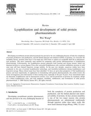
- 17. W. Wang / International Journal of Pharmaceutics 203 (2000) 1–60 17 parameters that directly determine or characterize eventually changes the solution from a viscous a lyophilization cycle need to be determined or liquid to a brittle glass, which contains about defined. These parameters should include glass 20–50% water (Pikal, 1990b; Hatley et al., 1996). transition temperature (T % )/collapse temperature g The temperature of this reversible transition for (Tcol), cooling rate, drying rate, and residual mois- the maximally freeze-concentrated solution is ture content. termed glass transition temperature, T % . This tem- g perature is also called the temperature of vitreous 4.1. Characterization of protein formulations prior transformation (Rey, 1999). T % is used to differen- g to lyophilization tiate this transition from the softening point of a true glass transition, Tg of a pure polymer. T % is g In addition to glass transition temperature (T % )/ g one of the most important parameters for opti- collapse temperature (Tcol), several other critical mization of a lyophilization process (Franks, temperatures, including crystallization tempera- 1990). ture (Tcry), eutectic temperature (Teut), and devit- The collapse temperature (Tcol) is the tempera- rification temperature (Tdev), should be ture at which the interstitial water in the frozen determined in order to design a robust lyophiliza- matrix becomes significantly mobile (Jennings, tion cycle. These temperatures are mostly deter- 1999). Tcol is closely related to T % . In fact, Tcol has g mined by thermal analysis such as DSC, electrical been considered to be equivalent to Tg of an resistance measurements, and direct microscopic amorphous system or to the eutectic melting tem- observation. perature of a crystalline system (Slade et al., 1989; Pikal, 1990a,b). Recent literature indicates that 4.1.1. Glass transition temperature (T % ) and g the Tcol of many small carbohydrates is consis- collapse temperature (Tcol) tently higher than their T % by about 12 K (Sun, g Ice formation during cooling of a protein solu- 1997). The discrepancy between T % and Tcol for g tion concentrates all solutes. Solute concentration polymers seems even larger (Roos and Karel, 1991). This is because the decrease in viscosity at T % may not be sufficient enough to cause struc- g tural collapse (Bindschaedler, 1999). For refer- ence, Table 1 lists T % s and Tcols of some g commonly used excipients and buffers. 4.1.2. Crystallization temperature (Tcry) When the temperature of an aqueous protein formulation drops below 0°C, water usually crys- tallizes out first. Then, the crystalline component, which usually has the least solubility in the formu- lation, may crystallize out. This temperature is termed crystallization temperature. 4.1.3. Eutectic crystallization/melting temperature (Teut) When the temperature of an aqueous protein formulation further decreases after crystallization of the least soluble component, this component and water crystallize out at the same time as a Fig. 1. A theoretical phase diagram showing ice formation, mixture. This temperature is termed eutectic crys- solute crystallization, eutectic point, and glass transition dur- tallization/melting temperature. The relationship ing freezing. between Teut and T % is shown in Fig. 1. Due to g
- 18. 18 Table 1 Glass transition and collapse temperatures (°C) of buffers, excipients and proteins Compounds T% g Tcol Tg References Buffering agents a Citric acid −54a, −53b 11b Chang and Randall, 1992; bLu and Zografi, 1997 Hepes −63 Chang and Randall, 1992 a Histidine −33a, −32b Chang and Randall, 1992; bOsterberg and Wadsten, 1999 8 Potassium −76 Franks, 1993 acetate Postassium −62 Franks, 1993 citrate Potassium −55 Chang and Randall, 1992 phosphate (KH2PO4) Sodium acetate −64 Chang and Randall, 1992 Sodium −52 Franks, 1993 bicarbonate Sodium citrate −41 Chang and Randall, 1992 Sodium −45 Chang and Randall, 1992 phosphate (NaH2PO4) Tris base −55 Franks, 1993 Tris–HCl −65 Chang and Randall, 1992 Excipients, low MW b-Alanine −65 Franks, 1993 Arabinose 3 Roos, 1993 Arginine 42 Mattern et al., 1999 Cellobiose 77 Franks, 1990; Costantino et al., 1998b a Fructose −42b 10a, 13b Roos, 1993; bWisniewski, 1998 Fucose 31 Roos, 1993 a Galactose −41a 31b, c38 Franks, 1990; bLevine and Slade, 1992; cRoos, 1993 a W. Wang / International Journal of Pharmaceutics 203 (2000) 1–60 Glucose −43a −41d 4c, 32b, 39a Franks, 1990; bLevine and Slade, 1992; cPrestrelski et al., 1995; dAdams and Ramsay, 1996 Glutamic acid −17 Chang and Randall, 1992 a Glycerol −65a −92b, −93a Franks, 1990; bBell et al., 1995 Glycine −37 Chang and Fischer, 1995 b a a Histidine −32 37 Mattern et al., 1999; bOsterberg and Wadsten, 1999 8 b a Lactose −30.5 101a, 114c Levine and Slade, 1992; bAdams and Ramsay, 1996; cTaylor and Zografi, 1998b Lysine 68 Mattern et al., 1999 a Maltose −30a 43a, 82b, Franks, 1990; bLevine and Slade, 1992; cTaylor and Zografi, 1998b 100c Maltotriose −23 Hatley and Franks, 1991 a Mannitol −33a,−27b 13c Meredith et al., 1996; bLueckel et al., 1998a; cKim et al., 1998
- 19. Table 1 (Continued) Compounds T% g Tcol Tg References a Mannose −41a 30a, 33b Franks, 1990; bLevine and Slade, 1992 Melibiose 91 Roos, 1993 Octulose 21 Wolkers et al., 1998a a Raffinose −26c 108b, 114a Taylor and Zografi, 1998b; bWolkers et al., 1998a; cWillemer, 1999 a Ribose −47b −10b, −13a Roos, 1993; bWisniewski, 1998 Sodium chloride B−60 Franks, 1993 a Sorbitol (glucitol) −44a,b −54c −3a Franks, 1990; bKerr et al., 1993; cAdams and Ramsay, 1996 Sucrose −32f −31d, −40h 31c, 61a, 72e, aLevine and Slade, 1992; bHancock et al., 1995; cPrestrelski et al., 1995; dAdams and 77b, 75g Ramsay, 1996; eCostantino et al., 1998c; fKasraian et al., 1998; fLueckel et al., 1998a; g Taylor and Zografi, 1998b; hOvercashier et al., 1999 Trehalose −29c, −34h 78a, 46b, 117d, aLevine and Slade, 1992; bPrestrelski et al., 1995; cAdams and Ramsay, 1996; dDuddu and 115c, 105f, 118g Dal Monte, 1997; eMiller et al., 1997; fLueckel et al., 1998a; gTaylor and Zografi, 1998b; h Overcashier et al., 1999 a Water −133a, −137b Bell et al., 1995; bMiller et al., 1997 b Xylitol −47b −23a, −39b Franks, 1990; aRoos, 1993 a Xylose −48a 9a, 14b Franks, 1990; bRoos, 1993 Excipients, high MW Cellulose 227 Hancock and Zografi, 1994 b-Cyclodextrin 108 Prestrelski et al., 1995 a Dextran (10 kD) −10b 91a Prestrelski et al., 1995; bWillemer, 1999 a Dextran (40 kD) 94a, 101b te Booy et al., 1992; bPrestrelski et al., 1995 Dextran (70 kD) −11 Adams and Ramsay, 1996 Ficoll −20 Pikal, 1990b; Izutsu et al., 1996 Gelatin −8 Pikal, 1990b Hydroxypropylmeth 155 Hancock and Zografi, 1994 yl-cellulose a Hydroxyethyl −12a −5b 110c Crowe et al., 1993b; bPikal, 1990b; cCrowe et al., 1998 starch Maltodextrin 860 169 Taylor and Zografi, 1998b Methocel −9 Willemer, 1999 W. Wang / International Journal of Pharmaceutics 203 (2000) 1–60 PEG (6 kD) −13 Willemer, 1999 Polydextrose −27 Kerr et al., 1993 a PVP K15 (10 kD) −27b 100a Bell et al., 1995; bIzutsu et al., 1996 PVP (40 kD) −24 Adams and Ramsay, 1996 PVP K30 (40 kD) 180 Bell et al., 1995 a PVP K90 (1000 185a, 176b Hancock et al., 1995; bTaylor and Zografi, 1998b kD) Sephadex G 200 −10 Willemer, 1999 Starch 227 Hancock and Zografi, 1994 Proteins a 19 BSA −13a, −11b Slade et al., 1989; bChang and Randall, 1992
- 20. 20 Table 1 (Continued) Compounds T% g Tcol Tg References a-Casein −12.5 Slade et al., 1989 Globulins (egg) −10.5 Slade et al., 1989 HSA −9.5 Pikal, 1990b a-Lactalbumin −10.5 Slade et al., 1989 (Sigma) LDH −9 Chang and Randall, 1992 a Lysozyme −16.5a, −13b Slade et al., 1989; bChang and Randall, 1992 rhuMAb HER2 −20 Overcashier et al., 1999 Myoglobin −15 Chang and Randall, 1992 a Ovalbumin −11a −10b Chang and Randall, 1992; bWillemer, 1999 RNase A −12 Chang and Randall, 1992 W. Wang / International Journal of Pharmaceutics 203 (2000) 1–60