Value Proposition canvas- Customer needs and pains
TEE Acquisition Guide
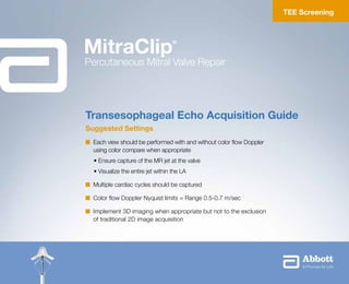
1. Suggested Settings
n Each view should be performed with and without color flow Doppler
using color compare when appropriate
• Ensure capture of the MR jet at the valve
• Visualize the entire jet within the LA
n Multiple cardiac cycles should be captured
n Color flow Doppler Nyquist limits = Range 0.5-0.7 m/sec
n Implement 3D imaging when appropriate but not to the exclusion
of traditional 2D image acquisition
Transesophageal Echo Acquisition Guide
TEE Screening
MitraClip®
Percutaneous Mitral Valve Repair
2. This material may be utilized for Clinical Site participants involved in Abbott Vascular sponsored trials or
disclosed to a user facility for educational purposes.
Indications for Use
MitraClip Clip Delivery System
The MitraClip Clip Delivery System is indicated for the percutaneous reduction of significant symptomatic mitral
regurgitation (MR ≥ 3+) due to primary abnormality of the mitral apparatus [degenerative MR] in patients who
have been determined to be at prohibitive risk for mitral valve surgery by a heart team, which includes a cardiac
surgeon experienced in mitral valve surgery and a cardiologist experienced in mitral valve disease, and in whom
existing comorbidities would not preclude the expected benefit from reduction of the mitral regurgitation.
Steerable Guide Catheter
The Steerable Guide Catheter is used for introducing various cardiovascular catheters into the left side of the
heart through the interatrial septum.
For Functional MR: Caution: Investigational device. Limited by Federal (US) law to investigational use only.
Indications
3. Central
4-chamber view with A2/P2 clearly visualized.
Advanced probe 1–3 cm. The LV cavity is
more completely visualized. For functional
MR, vertical coaptation length should be
measured. For degenerative MR, flail gap
should be measured, if present.
Inferior
4-chamber view with A3/P3 clearly visualized.
The probe is further advanced 1–3 cm.
The coronary sinus and tricuspid valve may
be seen.
Superior
5-chamber view with A1/P1 of the mitral valve
(MV) clearly visualized.
This view is obtained at the mid-esophageal
level. The aortic valve and left ventricular
outflow tract are clearly visualized. The LV
is foreshortened.
0º Views to Obtain
4. Midline
This view is obtained at the midline of the
valve to visualize P1, A2, and P3 scallops.
Posterior
This view is obtained at the posterior
side of the valve to visualize P1, P2,
and P3 scallops.
The posterior leaflet can be isolated by
torquing/rotating the probe counterclockwise
from midline.
Anterior
This view is obtained at the anterior side of
the valve to visualize A1, A2, and A3 scallops.
The anterior leaflet can be isolated by
torquing/rotating the probe clockwise from
the midline.
60–90º Views to Obtain
5. Medial
This view is obtained at the medial side of
the valve to visualize A3 and P3 scallops.
The medial aspect can be isolated by
torquing/rotating the probe clockwise
from central.
Lateral
This view is obtained at the lateral side of
the valve to visualize A1 and P1 scallops.
The lateral aspect can be isolated by
torquing/rotating the probe counterclockwise
from central.
Central
This view is of the central aspect of the
valve with A2 and P2 scallops clearly
visualized.
For degenerative MR, flail gap
should be measured, if present.
110–130º Views to Obtain
6. Right Upper Pulmonary Vein (90–120º)
Use color flow and PW Doppler. Place PW
Doppler sample volume 1–2 cm into PV.
Bicaval (80–110°)
SVC and IVC should be visible along
with atrial septum.
Left Upper Pulmonary Vein (0–30º)
Use color flow and PW Doppler. Place PW
Doppler sample volume 1–2 cm into PV.
Additional Views to Obtain
7. Transgastric Short Axis (0–20°)
Adjust angle to optimize SAX with both
anterior and posterior leaflets clearly visible.
Measure flail width if present. Use color flow
Doppler to demonstrate jet origin.
Short Axis at Base (15–45°)
This procedural view demonstrates a
cross-section of the aorta, atrial septum,
and right and left atria.
Additional Views to Obtain
3D En Face
3D images should be used to supplement
and confirm the initial diagnosis. 3D data
may be useful when available.
8. DMR Flail Gap
This should be taken in the view (LVOT or
4 chamber) where the flail gap is largest.
Flail Gap
Flail Width
DMR Flail Width
This measurement should be taken in the
transgastric short axis view where the flail
width is largest.
Anatomic Measurements I Degenerative Mitral Regurgitation (DMR)
9. Vertical
Coaptation
Length
FMR Vertical Coaptation Length
The measurement should be taken in
the 4-chamber view where the vertical
coaptation length is shortest.
Anatomic Measurements I Functional Mitral Regurgitation (FMR)
10. MitraClip Clip Delivery System
INDICATION FOR USE
The MitraClip Clip Delivery System is indicated for the
percutaneous reduction of significant symptomatic mitral
regurgitation (MR ≥ 3+) due to primary abnormality of the mitral
apparatus [degenerative MR] in patients who have been determined to be
at prohibitive risk for mitral valve surgery by a heart team, which includes
a cardiac surgeon experienced in mitral valve surgery and a cardiologist
experienced in mitral valve disease, and in whom existing comorbidities
would not preclude the expected benefit from reduction of the mitral
regurgitation.
CONTRAINDICATIONS
The MitraClip Clip Delivery System is contraindicated in DMR patients with
the following conditions:
• Patients who cannot tolerate procedural anticoagulation or post
procedural anti-platelet regimen
• Active endocarditis of the mitral valve
• Rheumatic mitral valve disease
• Evidence of intracardiac, inferior vena cava (IVC) or femoral venous
thrombus
WARNINGS
• DO NOT use MitraClip outside of the labeled indication. Treatment
of non-prohibitive risk DMR patients should be conducted in
accordance with standard hospital practices for surgical repair
and replacement.
• MitraClip is intended to reduce mitral regurgitation. The MitraClip
procedure is recommended to be performed when an experienced
heart team has determined that reduction of MR to ≤2+ is reasonably
expected following the MitraClip. If MR reduction to ≤2+ is not achieved,
the benefits of reduced symptoms and hospitalizations, improved quality
of life, and reverse LV remodeling expected from MitraClip may not occur.
• The MitraClip Device should be implanted with sterile techniques using
fluoroscopy and echocardiography (e.g., transesophageal [TEE] and
transthoracic [TTE]) in a facility with on-site cardiac surgery and
immediate access to a cardiac operating room.
• Read all instructions carefully. Failure to follow these
instructions, warnings and precautions may lead to device damage, user
injury or patient injury. Use universal
precautions for biohazards and sharps while handling the MitraClip
System to avoid user injury.
• Use of the MitraClip should be restricted to those physicians trained to
perform invasive endovascular and transseptal
procedures and those trained in the proper use of the system.
• The Clip Delivery System is provided sterile and designed for single use
only. Cleaning, re-sterilization and/or reuse may result in infections,
malfunction of the device or other serious injury or death.
• Inspect all product prior to use. DO NOT use if the package is opened
or damaged.
PRECAUTIONS
• Patient Selection:
n Prohibitive risk is determined by the clinical judgment of a heart team,
including a cardiac surgeon experienced in mitral valve surgery and a
cardiologist experienced in mitral valve disease, due to the presence of
one or more of the following documented surgical risk factors:
◆ 30-day STS predicted operative mortality risk score of
¤ ≥8% for patients deemed likely to undergo mitral valve replacement or
¤ ≥6% for patients deemed likely to undergo mitral valve repair
◆ Porcelain aorta or extensively calcified ascending aorta.
◆ Frailty (assessed by in-person cardiac surgeon consultation)
◆ Hostile chest
◆ Severe liver disease / cirrhosis (MELD Score 12)
◆ Severe pulmonary hypertension (systolic pulmonary artery pressure
2/3 systemic pressure)
◆ Unusual extenuating circumstance, such as right ventricular dysfunction
with severe tricuspid regurgitation, chemotherapy for malignancy,
major bleeding diathesis, immobility, AIDS, severe dementia, high risk
of aspiration, internal mammary artery (IMA) at high risk of injury, etc.
n Evaluable data regarding safety or effectiveness is not available for
prohibitive risk DMR patients with an LVEF 20% or an
LVESD 60mm. MitraClip should be used only when criteria for clip
suitability for DMR have been met.
• The major clinical benefits of MitraClip are reduction of MR to ≤2+
resulting in reduced hospitalizations, improved quality of life, reverse
LV remodeling and symptomatic relief in patients who have no other
therapeutic option. No mortality benefit following MitraClip therapy has
been demonstrated.
• The heart team should include a cardiac surgeon experienced in mitral
valve surgery and a cardiologist experienced in mitral valve disease and
may also include appropriate physicians to assess the adequacy of heart
failure treatment and valvular anatomy.
• The heart team may determine an in-person surgical consult is needed
to complete the assessment of prohibitive risk. The experienced mitral
valve surgeon and heart team should take into account the outcome
of this surgical consult when making the final determination of patient
risk status.
• For reasonable assurance of device effectiveness, pre-procedural
evaluation of the mitral valve and underlying pathologic anatomy and
procedural echocardiographic
assessment are essential.
• The inside of the outer pouch is not a sterile barrier. The inner pouch
within the outer pouch is the sterile barrier. Only the contents of the inner
pouch should be considered sterile. The outside surface of the inner
pouch is NOT sterile.
• Note the “Use by” date specified on the package.
POTENTIAL COMPLICATIONS AND
ADVERSE EVENTS
The following ANTICIPATED EVENTS have been identified as possible
complications of the MitraClip procedure.
Allergic reaction (anesthetic, contrast, Heparin, nickel alloy, latex);
Aneurysm or pseudo-aneurysm; Arrhythmias; Atrial fibrillation; Atrial septal
defect requiring intervention; Arterio-venous fistula; Bleeding; Cardiac
arrest; Cardiac perforation; Cardiac tamponade/Pericardial Effusion;
MitraClip erosion, migration or malposition; MitraClip Device thrombosis;
MitraClip System component(s) embolization; Coagulopathy; Conversion to
standard valve surgery; Death; Deep venous thrombus (DVT); Dislodgement
of previously implanted devices; Drug reaction to anti-platelet/anticoagulation
agents/contrast media; Dyspnea; Edema; Emboli (air, thrombus, MitraClip
Device); Emergency cardiac surgery; Endocarditis; Esophageal irritation;
Esophageal perforation or stricture; Failure to deliver MitraClip to the
intended site; Failure to retrieve MitraClip System components; Fever or
hyperthermia; Gastrointestinal bleeding or infarct; Hematoma; Hemolysis;
Hemorrhage requiring transfusion; Hypotension/hypertension; Infection and
pain at insertion site; Infection and pain at incision site; Injury to mitral
valve complicating or preventing later surgical repair; Lymphatic
complications; Mesenteric ischemia; Mitral stenosis; Mitral valve injury;
Multi-system organ failure; Myocardial infarction; Nausea/vomiting;
Peripheral ischemia; Prolonged angina; Prolonged ventilation; Pulmonary
congestion; Pulmonary thrombo-embolism; Renal insufficiency or failure;
Respiratory failure/atelectasis/pneumonia; Septicemia; Single leaflet device
attachment (SLDA); Skin injury or tissue changes due to exposure to ionizing
radiation; Stroke or transient ischemic attack (TIA); Urinary tract infection;
Vascular trauma, dissection or occlusion; Vessel spasm; Vessel perforation
or laceration; Worsening heart failure; Worsening mitral regurgitation;
Wound dehiscence
Steerable Guide Catheter
INDICATION FOR USE
The Steerable Guide Catheter is used for introducing various
cardiovascular catheters into the left side of the heart through
the interatrial septum.
CONTRAINDICATIONS
• Patients who cannot tolerate procedural anticoagulation or post procedural
anti-platelet regimen
• Evidence of intracardiac, inferior vena cava (IVC) or femoral venous
thrombus.
WARNINGS
• Read all instructions carefully. Failure to follow these instructions,
warning and precautions may lead to device damage, user injury or
patient injury. Use universal
precautions for biohazards and sharps to avoid user injury.
• Use the Steerable Guide Catheter with sterile techniques using
fluoroscopy and echocardiography (e.g., transesophageal [TEE] and
transthoracic [TTE]) in a facility with on-site cardiac surgery and
immediate access to a cardiac operating room.
• The Steerable Guide Catheter is designed for single use only. Cleaning,
re-sterilization and/or reuse may result in infections, malfunction of the
device or other serious injury or death.
• Patients with the following considerations in whom the Steerable Guide
Catheter is used may have an increased risk of having a serious adverse
event which may be avoided with preoperative evaluation and proper
device usage.
n Previous interatrial septal patch or prosthetic atrial septal defect (ASD)
closure device which could result in significant difficulty in visualization
or technical challenges during transseptal puncture and/or introducing
the SGC into the left atrium.
n Known or suspected unstable angina or myocardial
infarction within the last 12 weeks could increase the procedural
morbidity and mortality, due to increased hemodynamic stress
secondary to general anesthesia.
n Patients with active infection have an increased risk
of developing an intraoperative and/or postoperative
infection, such as sepsis or soft tissue abscess.
n Known or suspected left atrial myxoma could result in thromboembolism
and tissue injury due to difficulty with device positioning.
n Recent cerebrovascular event (CVA) may increase the procedural
morbidity associated with a transcatheter intervention, such as
recurrent stroke.
PRECAUTIONS
NOTE the “Use by” date specified on the package.
Inspect all product prior to use. Do not use if package is opened or
damaged.
The inside of the outer pouch is not a sterile barrier. The inner pouch within
the outer pouch is the sterile barrier. Only the contents of the inner pouch
should be considered sterile.
The outside surface of the inner pouch is NOT sterile.
Prior to use, please reference the Instructions for Use at
www.abbottvascular.com/ifu for more information on indications,
contraindications, warnings, precautions, and adverse events.
![This material may be utilized for Clinical Site participants involved in Abbott Vascular sponsored trials or
disclosed to a user facility for educational purposes.
Indications for Use
MitraClip Clip Delivery System
The MitraClip Clip Delivery System is indicated for the percutaneous reduction of significant symptomatic mitral
regurgitation (MR ≥ 3+) due to primary abnormality of the mitral apparatus [degenerative MR] in patients who
have been determined to be at prohibitive risk for mitral valve surgery by a heart team, which includes a cardiac
surgeon experienced in mitral valve surgery and a cardiologist experienced in mitral valve disease, and in whom
existing comorbidities would not preclude the expected benefit from reduction of the mitral regurgitation.
Steerable Guide Catheter
The Steerable Guide Catheter is used for introducing various cardiovascular catheters into the left side of the
heart through the interatrial septum.
For Functional MR: Caution: Investigational device. Limited by Federal (US) law to investigational use only.
Indications](data:image/gif;base64,R0lGODlhAQABAIAAAAAAAP///yH5BAEAAAAALAAAAAABAAEAAAIBRAA7)