SlideShare ist ein Scribd-Unternehmen logo
1 von 22
Downloaden Sie, um offline zu lesen
2012.10.30.


             Multimodal Imaging in Neurosciences Course
                                                            COURSE FAQ

                                                            Forthcoming lectures:
                                                            16. October – „IGT lecture”
                                                            23. October – NO LECTURE, holiday
                                                            30. October – MR Spectroscopy
                                                            6. November – PET + Final Test


                                                            Test:
                                                            -    Basic imaging techniques, what are they
                                                            -    5-10 easy, simple choice questions
                 Image guided therapies in                  -    If November 6. is not good for everyone, I will organize extra
                 neurological sciences and VR                    time for getting the short test done

                 Dr. András Jakab, MD, PhD
                                                            Study material:
                                                                 Lecture material will be distributed in PDF 2 wks before the
                                                                 test.


   Diagnostic neuroimaging modalities                                   What is multimodality?
CT – Computed Tomography       Structural MRI
                                                                Combining images and information from multiple
Brain anatomy                  Fine brain anatomy               imaging tools, devices
Stereotactic reference frame   Vascular structure
                                                                Anatomical alignment of images
                                                                Fusion display, co-analysis of multiple
Intra-operative imaging        Diffusion, perfusion MRI         information sources
modalities, open MRI, low-     Fine pathological
field                          information
                                                                 What is needed for multimodality?
Positron Emission multi-modal imaging for planning
             Using
                                                                CT, PET, MRI, SPECT, EEG, …
                                MR Spectroscopy
Tomographyimage-guided neurological interventions
             PET
                                Brain metabolism
Brain metabolism
                                Biochemical mapping
                                                                Hybrid devices – PET-CT, PET-MRI
Brain function
                                                                Image processing skills to create image fusions,
                                                                etc.
Electro encephalography,
                               Functional MR imaging fMRI
LORETTA,
                               Brain function
Magnetoencephalography


            Overview of the lecture
 1.   Image guided therapy – basics
      and the modalities used
 2.   Stereotactic functional
      neurosurgery
 3.   Radiosurgery and planning
      R di             d l    i                             1.
                                                            1 Image guided therapy -
 4.   Focused ultrasound                                    basics
      neurosurgery
 5.   VR applications in surgery


                                                                                                                                           1
2012.10.30.


                   Image guidance                                               AMIGO – Harvard, Boston
• Using preoperative images
  to define target volumes in
  a reference space
                                                                           Treatment planning w/   Intraoperative imaging with
• Using intraoperative                                                     preoperative imaging    MRI

  imaging to monitor
                                                                                                   Surgical robot for biopsies
  treatment
   – Intraoperative
     ultrasound
   – Intraoperative low-field
     MRI
   – Intraoperative high-field
                                 Intraoperative open MRI, Brigham
     open MRI                    and Women’s Hospital, Harvard,
                                 Boston (credits: Prof. Ferenc Jolesz)


       Intraoperative MR imaging
 •   Medtronic Polestar
 •   Low field (0.1-0.3T)
 •   Brain shift – a real problem
 •   Use of nonmagnetic tools
                                                                         2.
                                                                         2 Stereotaxy in functional
                                                                         neurosurgery


                                                                                      Why use stereotaxy in
            What is stereotactic?
                                                                                        neurosurgery?
 • Space, spatial
 • Coordinate frame                                                      • Very high (<1-2mm) precision needed
   of the brain:                                                         • Minimal invasive procedures
     – Intrinsic                                                            – Electrode implantation for ablation
     – extrinsic                                                            – Permanent electrode implantation (DBS)
                                                                            – Biopsy sampling from the brain
                                                                         • Procedures without opening the skull
                                                                            – Radiosurgery, LINAC, Gamma Knife,
                                                                              Cyberknife
                                                                            – Focused ultrasound surgery


                                                                                                                                          2
2012.10.30.


                Brain biopsy                           Imaging for brain biopsy
• Indications                                          • CT
   – Tumor suspect (enhancing CT, MRI, lesion)
                                                       • MRI (T1/T2+Contrast)
   – Inflammation, Neurodegeneration, unknown
                                                       • Pathology+Radiological
• Procedure (1-2 hours):                                 consultation – where to take
   – St
     Stereo f
            frame attachment
                   tt h    t                             the sample f
                                                          h       l from??
   – Computed tomography
   – Planning
   – General anaesthesia
   – Small craniotomy
   – Dexamethasone
   – Control scan(s)


      Functional neurosurgery                                Thalamotomy / pallidotomy
                                                       • Parkinson disease – associated tremor
• Alleviating symptoms by modifying                      alleviation
  functional areas of the brain, subcortical           • Essential tremor (unkown ethiology)
  areas
• Two main approaches:
   – Permanently cutting fiber pathways,
    destroying specific nuclei
                                Thermoablation
                                                                                                  Targets:
   – Retuning / stimulating fiber pathways, specific                                              VIM nucleus
     nuclei in the brain                                                                          Subthalamic
                         Deep brain stimulation                                                   nucleus, PTT
                                                                                                  Ventral Pallidum


                                                                         Thalamotomy
                                                        • Fantom pain (after amputation)
                                                        • Intractable tumor / stroke pain in limbs etc.
                                                        • CL / CM nucleus – central lateralis / centré median


                                                                                                                     3
2012.10.30.


                                                         Deep brain stimulation (DBS)
                                                     • FDA approval:
                                                       1997
                                                     • Implant in brain
                                                       + subcutaneous
                                                       stimulator
                                                       (~pacemaker)
                                                     • Retuning
                                                       functional
                                                       pathways
                                                       („arrhythmia”)


               Deep brain stimulation                                              Procedure
• Indications:                                      • Localization,
                                                      stereotaxy
  – Chronic pain, PD tremor,
    ET, dystonia                                    • Electrode trajectory:
  – Tourette, OCD, Major                            • Trials and
    depression
    d       i                                         monitoring
                                                    • Pre/postop imaging
  – Parkinsons
      •   Subthalamic nucleus
      •   Globus pallidus interna
      •   Zona incerta
      •   Pallidothalamic fibers


                                                                        Targeting scheme
                  Targeting scheme                   •   2. Defining targets on the patient’s coordinate frame (using CT+MRI)


  • 1. Target definition using stereotactic atlas   Manually finding AC,
    of the human thalamus and basal ganglia         PC points on scans

    (histology)                                     Finding landmarks of
                                                    the reference frame
                                                    (stereotactic frame
                                                    attached to the head)
  Getting x,y,z
  coordinates
  for your
  target

  Reference:
  AC-PC
  coordinate
  system


                                                                                                                                         4
2012.10.30.


                                                3. Radiosurgery


   Gamma ART 6000N Rotating Gamma System
   American Radiosurgery Inc., San Diego, CA,
                    USA                                      Challenges
                                                • Morphological imaging by MRI
                                                  – 3D isotrop voxel acquisitio
                                                     • Good tissue contrast
                                                     • Good signal-to-noise
•only 30 Co60 sources                                • Minimal image distortion
•Rotating sources
•Collimator size:
          4, 8, 14, 18 mm
                                                     • Optimal image resolution
•Stereotactic frame required
•0,3 mm accuracy
                                                  – MRI as reference system?
•Source half-life approx. 5 yrs
                                                • Robust CT method is necessary


       Challenges of multimodality
   – fMRI
   – DTI, fibertracking
   – MRS, MRSI
 • Postprocessing
   – Registration, image fusion (need fast)
 • PET, SPECT


                                                                                           5
2012.10.30.


       3D T1 weighted images                     3D T2 weighted images
•   1,2x1,2x1,2 mm
•   140 slices                   • 0,7x0,7x0,7 mm
•   6 minutes                    • 40-60 slices
•   Gd contrast agent            • 4 minutes


                                                  Our imaging protocolls
          3D TOF acquisition
•   1,2x1,2x1,2 mm
                                                      (+indications)
•   190 slices                   •            Metastasis, meningeoma
•   6 minutes                    •            AVM
•                g
    Gd contrast agent            •            Cavernoma
                                 •            Acustic neurinoma
                                 •            Hypophysis microadenoma


                AVM MRA                          Automatic radiologic image processing

                                                                                   Automated CT/MRI registration
                                                                     CT/T1/T2       (gammaknife, neurosurgery


                                                                                            automated
                                                                        T1         standardization, segmentation
                                                                                     (neurosurgery, neurology)
                                                      ImageSorter
                               DICOM server


                                                                                              automated
                                                                                    Tensor space calculations and
                                                                      DWI, T1               regularization
                                                                                      (neurosurgery, neurology)


                                                                                             automated
                                                                                             automated
                                                                    fMRI, T1, T2           SPM analysis
                                                                                            SPM analízis
                                                                                      (neurosurgery, neurology)


                                                                                            automated
                                                                     Pet & Mri          PET-MR registration
                                                                                         and roi analysis


                                                                                                                             6
2012.10.30.


  T1-CT registration for radiosurgery
           (gamma-knife)                                                                  Image registration, alignment
                                                                                                         gamma knife


                      - Automatic (maximalisation relative entropy)
                      - Manual correction (with internal „landmarks”)
                        To this time, manual correction was necessary in 60% of
                      the cases
                      - Optimalised automatic registration


                                                                                             T1-CT            Fiesta-CT        TOF-CT


Extracranial metastasis                                                                     Acoustic neurinoma treatment plan
     (epipharynx)
                                                                                  • 1 kezelési tervet betenni


                                                                                                     Acustic neurinoma


                                                                                    Pre-treatment        6 months post    12 months post


                                                                                                                                           7
2012.10.30.


                                                                                                                       Trigeminal neuralgia
       Treating trigeminal neuralgia                                                                                  CT and FIESTA imaging


                                                                                                                     Validation of the T1-CT optimized
 Dataflow                     Service available via a DICOM-server
                                                                                                                            registration method
                                                                                                       REFERENCE IMAGES                                                   AUTOMATICALLY
                                                                                                                                                                        REGISTERED IMAGES


     MNI and M3I tools:                                                                                 S01
                                                       BrainCAD:
 •Dicom-minc conversion
                                                • 2D fusion
 •Segmentation                                                                                                                            Comparison using normalized
                                                                                                        S02
 •Automatic registration      SPM-analysis • 3D fusion                                                                                          relative entropy
                                                • Fiber visualization
 •Spatial standardization
 •Automatic region analysis
                                                                                                        S03


   AUTOMATIC                          INTERACTIVE                                                        Sn
                                                                                PRESENTATION           MRI data revision by experts
IMAGE PROCESSING                   IMAGE PROCESSING                             (neuronavigation)   (occassionaly manual registrations)


                         Further fancy options                                                                                   CT-DTI registration
   PET/SPECT images
                                      T2, PD etc.


 SPM images


                                                    Precise T1-CT registration


                                     Structures defined in the space of a brain atlas
 DTI parametric images


                                                                                                                                                                                            8
2012.10.30.


CT-DTI registration


                               9
2012.10.30.


                                                               Proton therapy


    DTI + MRI data in the CT frame. Metastatic
                 disease, 55/M
    ( Gamma Radiosurgery Centre, Debrecen )


                                                       The proton advantage:
              Medulloblastoma
PHOTONS       “dose bath”                                         Nasopharynx
                                                       Photons (IMRT)       Protons


                                                   Dose bath
                            100%

                            60%
PROTONS

                            10%


     The proton advantage:
          Paraspinal
    Photons                            Protons


     Dose bath


                                                 4.
                                                 4 Focused ultrasound
                                                 neurosurgery


                                                                                              10
2012.10.30.


Energy Conversion and Transport in Biosystems (09.12.10)
                                                                     Welcome
Non-invasive                                                            Beat Werner
                                                                        MR-Center
Interventions                                                           University Children’s Hospital Zurich

with                                                                    www.kispi.uzh.ch/mr
                                                                        beat.werner@kispi.uzh.ch
                                                                        beat werner@kispi uzh ch
Focused                                                                 dipl. phys. ETH

Ultrasound                                                              MR-Physics
Beat Werner                                                             HIFU surgery since 2005,
MR-Center                                                               research project as part of NCCR Co-Me
University Children‘s Hospital
Zurich                                                                                         Slide credits: Beat Werner, Ernst Martin


 Intro: Image guided interventions                                   Intro: Image guided interventions
                                                                        Image Guided Interventions
                                                                           Improve accuracy
                                                                           Decrease intervention risks
                                                                           Minimally invasive / Non-invasive Interventions


                        Slide credits: Beat Werner, Ernst Martin   Planning and navigation Complex interventions in cranio-maxillo-facial surgery.
                                                                                              Slide credits: Beat Werner, Ernst Martin


 Intro: Image guided interventions                                   Intro: Image guided interventions
   "Multimodal" Imaging                                                 Enabling technologies used here:
      Multimodal: US, CT, MR, ...                                          MR
      Multidomain: Anatomical, physiological, statistical                       Closed-loop intervention control
      information                                                               US field calibration
      Multi-Timescales: Static (Pre-/intra-intervention),                  Focused Ultrasound
      Realtime                                                                  Mechanical / Thermal therapy
      Augmented reality: condition, fuse, model                                 (Targeted) drug delivery
      Enhanced visualization: 2D, 3D, ..., force feedback                       (Targeted) drug activation
                                                                           Nano-Particles
                                                                                Molecular markers
                                                                                Contrast agents
                                                                                Drug carriers
  Computer-assisted support in ORL surgery Werner, Ernst Martin
                         Slide credits: Beat                                    Therapeutic devices Beat Werner, Ernst Martin
                                                                                          Slide credits:


                                                                                                                                                     11
2012.10.30.


Motivation: Non-invasive HIFU Surgery
            Non-                                             Motivation: Clinical value of
                                                             MR guided Focused US Surgery
                                                              Excellent soft tissue contrast for
                                                              target localization
                                                              Local therapy
                                                              Non invasive
                                                              Non-invasive
                                                              No dose accumulation effects
                                                              No long term toxicity
                                                              Immediate result –
                                                              no radiation necrosis
                                                              Treatments can be repeated
                                                              Closed-loop image guidance
                                                              Real time monitoring
                  Slide credits: Beat Werner, Ernst Martin


Imaging <-> Focused Ultrasound
        <-                                                   Phased-
                                                             Phased-Array Transducers
 Imaging (Diagnostics):
 want wide field of view with                                   Phased-Array Transducer = Array of Transducers
 uniform low acoustic intensity                                 Each transducer element controlled individually
                                                                Transducer phases define superposition pattern of
                                                                waves -> Electronic steering


                    HIFU (Intervention / Therapy):           Constructive           Destructive                    Shift
                    want small focal spot with
                  Slide credits: Beat intensity
                    high acoustic Werner, Ernst Martin                            Slide credits: Beat Werner, Ernst Martin


Beam Forming / Focusing                                      Phased-
                                                             Phased-Array Transducers
                                                                Linear Array      ->               Plane Wave
   Geometric focusing         Lens focusing


 Electronic beam forming Electronic steering                    Spherical Array   ->               Focus


                  Slide credits: Beat Werner, Ernst Martin                        Slide credits: Beat Werner, Ernst Martin


                                                                                                                             12
2012.10.30.


Phased-
Phased-Array Transducers                                                                       Phased-
                                                                                               Phased-Array Transducers

                                                                                               Spherical Array
   Spherical Array


    Pressure Distribution                       Temperature Distribution
                                                                                               Electronic Steering


                               Slide credits: Beat Werner, Ernst Martin                                              Slide credits: Beat Werner, Ernst Martin


  Biological Effects                                                                           Medical applications
      The effects of Ultrasound on biological tissue                                             Lithotrypsy
      are a field of active research                                                             Physiotherapy
      Many effects are not well understood                                                       HIFU Surgery (Tissue ablation)
      Effects include:                                                                           Blood-Brain-Barrier-Opening
               thermal
               th     l                                             non-thermal
                                                                    non thermal                  Cell sonoporation                           Targeted
                                                                                                                                                g
              mechanisms               ultrasound                   mechanisms                                                               drug delivery
                                                                                                 Local drug activation
  energy        non-cavitation mechanisms                              cavitation mechanisms     Vessel occlusion
absorption
                                                                                                 Thrombolysis
temperature
  increase           radiation force      non-inertial cavitation        inertial cavitation     Nerve activation / blockage


 ablation                        enhanced Delivery                           lithothripsy

                               Slide credits: Beat Werner, Ernst Martin                                              Slide credits: Beat Werner, Ernst Martin


Development of HIFU Technology                                                                 Development of HIFU Technology

  1880             Piezoelectric effect (P. & J. Curie)
  1918             Sonar (Langevin)                                                            W.Fry, circa 1960, with 4-beam HIFU system for neuro-
                                                                                                                       4-                     neuro-
  1927             Effects on biological tissues (Looms, Wood)                                                                        surgery in his
  1942             First HIFU lesions in animal brains (J.Lynn, T.Putnam)                                                             bioacoustics
  1950–
  1950–2000        Pioneer work on (a) HIFU effects on tissue (brain                                                                  laboratory.
                   tumors) and (b) LIU for soft tissue visualization (W Fry
                                                                     (W.Fry
                   & F.Fry).
  1951–
  1951–1960        Technical development at MGH (Cosman)
  1951–
  1951–1967        HIFU stereotactic neurosurgery against pain, psycho-
                                                                psycho-
                   neuroses, anxiety, depression and epilepsy (Lindstrom)
                   and Radiosurgery (Leksell) gamma knife
  Mid 1970s
      1970s        First stereotactic HIFU brain surgery with open cranium
                   (F.Fry & R.Heimburger).


                               Slide credits: Beat Werner, Ernst Martin                                              Slide credits: Beat Werner, Ernst Martin


                                                                                                                                                                13
2012.10.30.


Development of HIFU Technology                                    Image guidance
                                    First US-image guided
                                    HIFU system to treat           US-Imaging
                                    brain cancer patients.            Cheap
                                    W.Fry in the 1970s
                                                                      Limited feedback
                                    F.Fry and R.Heimburger 1974
                                                                   MR
                                                                        Expensive
                                                                        High-resolution imaging
                                                                        Closed loop process


                     Slide credits: Beat Werner, Ernst Martin                        Slide credits: Beat Werner, Ernst Martin


Development of HIFU Technology                                    HIFU Surgery for Uterine Fibroids
Key technologies:
1. Phased array transducers
2. Acoustic field modelling
3. MR imaging for accurate real time monitoring


Early 1990s
   l 1990s
       990    Ultrasound phased arrays (Hynynen)
                l        d h d          (        )
Mid–1990s
Mid–1990s     MRI thermometry (Jolesz)
2001          First integrated MRgHIFU system (InSightec)
2001          Fibroadenoma of Breast and Uterine Fibroids
2004          FDA approval for Uterine Fibroid Application
…
2008          Functional Neurosurgery

                     Slide credits: Beat Werner, Ernst Martin                        Slide credits: Beat Werner, Ernst Martin


InSightec Exablate 2000                                           Treatment process for
                                                                  Uterine Fibroids
 InSightec Haifa, Israel                                          benign (non-cancerous) tumors
     Technology originally GE                                     most common pelvic tumors in women
 FDA approved                                                     20-40% in women of reproductive age
     First & only worldwide                                       Classical procedure: Hysterectomy
 GE MR-Scanners
 Ca. 200 systems installed


                     Slide credits: Beat Werner, Ernst Martin                        Slide credits: Beat Werner, Ernst Martin


                                                                                                                                14
2012.10.30.


Patient interface                                                   Transducer
                                                                   16 element array                   Mechanical positioning
                                                   MR-coil
    Patient                                                          vertical electronic steering        4 axes
                                                                     focal depth 12cm – 19cm             Computer controlled


Coupling                                       Target


                                               Ultrasound

                                                                                                        MR-compatible
                                     Transducer                                                            Ceramic step motors
                       Slide credits: Beat Werner, Ernst Martin                          Slide credits: Beat Werner, Ernst Martin


Safety checks                                                       Treatment planning

   Air bubbles?                                                        Delineate
   Metallic clips?                                                     region of
   Scars?                                                              treatment
   Bowels?                                                             System creates
                                                                       treatment plan
                                                                                 p
   Bladder?
   Bl dd ?                                                             (array of
   Nerves?                                                             sonications
                                                                       to be done)
                                                                       Plan can be
                                                                       changed
                                                                       manually
                                                                       by operator


                       Slide credits: Beat Werner, Ernst Martin                          Slide credits: Beat Werner, Ernst Martin


Real-
Real-time temperature monitoring                                    Treatment assessment
   MR-Thermometry
   Proton resonance frequency shift ~ Temperature
   Phase difference ~ Temperature difference
1.1 seconds    4.5 seconds       7.9 seconds        11.3 seconds


                                                                       Blue dots = Cell                   Treatment control by
                                                                       ablation as calculated             contrast enhanced MR
                                                                       by thermal dose

                                                                       Very good agreement
                       Slide credits: Beat Werner, Ernst Martin                          Slide credits: Beat Werner, Ernst Martin


                                                                                                                                    15
2012.10.30.


     Closed loop control
      MRgFUS =             Magnetic Resonance guided
                           Focused UltraSound


Planing                    Safety           Real-Time              Assement
                           checks           Monitoring


                               Closed Loop
                                Slide credits: Beat Werner, Ernst Martin                                                           Slide credits: Beat Werner, Ernst Martin


   Clinical applications and research                                           Uterine fibroids
                                                                                                                                       T1w contrast
                                                                                                                                       enhanced image
                                                                                                                                       before treatment
                         Brain Tumors      Brain Functional


                      Breast Cancer                                           T2w planning
                       Liver Tumors                                           image with
                                            Uterine Fibroids                  dose overlay
                             Prostate


                      Bone Tumors

                                                                                                                                            T1w contrast enhanced image
                                Slide credits: Beat Werner, Ernst Martin      Courtesy of Sheba Medical Center, Tel Aviv, Israel                immediately post-treatment
                                                                                                                                   Slide credits: Beat Werner, Ernst Martin


   Prostate                                                                     Liver tumors

  Treatment effects in canine prostate


    Thermal dose               T1w contrast              TTC stained
    estimate                   enhanced image            tissue                             Sagittal, Axial and coronal post treatment dose
                               imaging
                                                                                            maps (in blue) and post treatment T1W+C
 Courtesy InSightec                                                           Courtesy of St. Mary’s Hospital, London, UK
                                Slide credits: Beat Werner, Ernst Martin                                                           Slide credits: Beat Werner, Ernst Martin


                                                                                                                                                                              16
2012.10.30.


                                                                             Caution-Investigational Device
                                                                              Limited by United States Law     IDE


  Breast Cancer                                                                                                        Bone tumors (Pain palliation)
                                                                                     to Investigational Use.


                                                                                                                                      10.0
                                                                                                                                                           Screening
                                                                                                                                       9.0
                                                                                                                                                            Average
                                                                                                                                       8.0
                                                                                                                                                 7
                                                                                                                                       7.0


                                                                                                                          VAS score
                                                                                                                                       6.0

                                                                                                                                       5.0
                                                                                                                                       4.0                                         1 Month
                                                                                                                                                                                  Follow Up
                                                                                                                                       3.0
                                                                                                                                                                         1.5                              3 Months
                                                                                                                                       2.0
    Pre treatment T1w+C                                    2 weeks post treatment                                                                    1.5                                                  Follow Up
                                                                                                                                       1.0
                                                                                                                                                                0                                                     0
    MIP image                                              T1w+C MIP image                                                             0.0
                                                                                                                                             0             10       20   30      40     50      60   70       80      90

                                                                                                                                                                          Days post treatment
Courtesy of Breastopia Namba Hospital, Japan                                                                         Courtesy InSightec
                                               Slide credits: Beat Werner, Ernst Martin                                                                                        Slide credits: Beat Werner, Ernst Martin


   Non-
   Non-invasive neuro-surgery
                neuro-                                                                                                   Skull: Anisotropic Aberration
   in the MR-suite
          MR-


                                                                                                                        Focused waves …                                                      … are distrorted after skull


                                                                                                                        Phase corrected waves …                                               … are refocused after skull
                                                                                                                     Courtesy of ESPCI, Paris, F


  InSightec ExAblate 4000                                                                                              Patient Interface
   3D-Positioner                                                  Frontend                                                             Degassed water used for acoustic coupling &
                                                                  Amplifier                                                            cooling
                                                              InSightec ExAblate 4000 /
                                                              3.0T GE Signa HDx
                                                                                                                                       Stereotactic frame for patient immobillization
                                                              Hemispheric 1024-element
                                                              phased-array transducer                                                                                    Transducer                  Sealing membrane
                                                              (650kHz)
                                                              Stereotactic frame
                                                              Water cooling of skull
                                                              surface
                                                              CT-based acoustic modeling
                                                               PRS-Thermometry
       Transducer
                                                              Installation June 2006

                                                                                                                                                                               Water                        Stereotactic frame


                                                                                                                                                                                                                                   17
2012.10.30.


MRI-
MRI-HIFU Console                                    Clinical phase I study
                                                     Centro-Lateral Thalamotomy against
                                                     chronic, therapy resistant, neuropathic
                                                     pain
                                                     Established procedure: RF-ablation
                                                      o minimally invasive
                                                      o n (pain) > 100 interventions


                                                                              New procedure: Non-invasive TcMRgHIFU
                                                                               o Minimize intervention risks (Collateral
                                                                                 damage, infections, bleeding, tissue
                                                                                 shift)
                                                                               o Enhanced efficacy (no trajectory
                                                                                 restrictions, anatomically adapted
                                                                                 volume ablation, image guidance)
                                                                               o Outpatient process


  Bildfolge der Behandlung (sonication procedure)     Bildfolge der Behandlung (sonication procedure)


       X+20’: Stereotactic frame                             X+50: Positioning


       X+1h20: Imaging                                       X+3h: Verification sonications


                                                                                                                           18
2012.10.30.


         X+3h: Verification sonications        X+4h: Ablation


Dosemap 17 CEM 43°C
               43°


Dosemap 240 CEM 43°C
                43°


                                               X+6h: End of treatment


                                          Patient #4
                                                  #4                          Patient #2
                                                                                      #2
                                          immediately after                   48 h after
                                          the intervention                    the intervention


                                           Trigeminal neuralgia            Chronic lumbar pain syndrome
                                                                           following disc hernia op L4/L5
                                                                                                    L4/L5
         X+7h: Happy End                                      Slide credits: Beat Werner, Ernst Martin


                                                                                                            19
2012.10.30.


    Patient #4
            #4                           Patient #2
                                                 #2                  Patient #4
                                                                             #4                               Patient #2
                                                                                                                      #2
    immediately after                    48 h after                  immediately after                        48 h after
    the intervention                     the intervention            the intervention                         the intervention


             T1WI + Gd                       T1WI + Gd                        DTI                                    DTI


Combined Tumor therapy:                                             Blood Brain Barrier
HIFU-
HIFU-Ablation & LIFU BBBD                                            Brain protected by BBB:
                    Tumorvolume                                          Structural and functional barrier in the vessel walls
                                                                         Controls transport and diffusion from the
                                                 HIFU-
                                                 HIFU-Ablation           vasculature to the central nervous system
                                                                         Severely limits ability to deliver drugs to the brain


McDannold et al.
Neurosurgery 2010


  BBB-
  BBB-Opening with Low Intensity
  Focused Ultrasound (LIFU)
  and Adjuvant Chemotherapy
                         Slide credits: Beat Werner, Ernst Martin                            Slide credits: Beat Werner, Ernst Martin


Reversible BBB-Disruption using
           BBB-                                                     TcMRgFUS BBB-Disruption
                                                                             BBB-
FUS and Microbubble UCA                                                                                      US freq. 220kHz – 1MHz
    Microbubbles                         Lipid
                                         monolayer                                                           Acc.Power < 1W
       Phospholipid / Hexafluorid (2-4um)                                                                    Sonication 10 – 200s
       Commercial CE / FDA                                                                                   Duty cycle 1%
                                                                                                             Tail vein injection
                                                                                                                  MR-contrast agent /
                                                                                                                  drug
                                                                                                                  Micro bubbles
                                                                              Inj. Drug


                                                                                          Inj. Micro bubbles
                                                                                                                                        Histo
                                                                      MRI


                                                                                                                           MRI


    Reversible opening of BBB by                                                              Low Intensity US
       Cavitation                                                                          (Bursts of 10ms/1000ms)

       Shear stress (Acoustic streaming,
       radiation force, ...)
                     Slide credits: Beat Werner, Ernst Martin


                                                                                                                                                20
2012.10.30.


  TcMRgFUS BBB-Disruption
           BBB-                                       TcMRgFUS BBB-Disruption
                                                               BBB-
                                                       Duration
   MR-Scanner:      GE 3.0T                               several hours
   Transducer:      Imasonic (Aperture 8cm, f# 0.8)
                                                       Application
   Microbubbles:    Bracco
                                                          Neuro-
Positioner
P iti                           MR-Coil
                                MR C il                   Phamacology
                                                          Tumors
                                                          Alzheimer
                                                          Neuron
                                                          regeneration


Transducer          Watertank   Mouse


  Targeted Drug Delivery
   Targeted Drug Delivery                             Targeted Drug Delivery
                                                        Target specific
                                                        Carries contrast agents (Dye, fluorescent, magnetic, ...)
                                                        Carries drugs
                                                        Remote activation


    Bubble constructs
       Add specificity
       Add payload
       Remote activation


  Targeted Drug Delivery                              Gene Delivery
    Releasing drug by Ultrasound
        Heat
        Cavitation


                                                                                                                    21
2012.10.30.


       Gene Activation: hsp-80
                        hsp-                                             Summary
                                                                          Image guided FUS is a new modality for non-invasive
                                                                          interventions deep in soft tissue
                                                                          Thermal ablation clinically established
                                                                          Intense research on treatment strategies based on
                                                                          mechanical effects
                                                                          Very promising results in animal models for targeted
                                                                          drug delivery based on transient BBBD / Sonoporation
                                                                          Intense research on development of nano-constructs
                                                                          for imaging and therapy
                                                                          Clinical studies to come soon


                              Slide credits: Beat Werner, Ernst Martin                    Slide credits: Beat Werner, Ernst Martin


                           Clot Lyses for Stroke
      Outlook
                                                                              Thank you !
                                                        Neurological
                                                        Disorders
Targeted Drug Delivery


                          In-vitro T2W MRI before
                          [l] and after[r] sonication


                              Slide credits: Beat Werner, Ernst Martin                    Slide credits: Beat Werner, Ernst Martin


  Thank you for your attention!
  Presentation credits:

  Dr. András Jakab, M.D. Ph.D.
  Dr. Ervin Berényi, M.D. Ph.D.
  Dr. Miklós Emri (Nuclear Medicine Institute, UD)
  Prof. Ernst Martin – Uni. Zürich (Focused Ultrasound)
  Beat Werner – Uni. Zürich (Focused Ultrasound)


                                                                                                                                     22

Weitere ähnliche Inhalte

Was ist angesagt?

Reliability and validity of fMRI
Reliability and validity of fMRIReliability and validity of fMRI
Reliability and validity of fMRIElham Faghihzadeh
 
Let’s master the digital toolkit to harness lifelong neuroplasticity
Let’s master the digital toolkit to harness lifelong neuroplasticityLet’s master the digital toolkit to harness lifelong neuroplasticity
Let’s master the digital toolkit to harness lifelong neuroplasticitySharpBrains
 
Translational Research in Functional Optical Brain Imaging
Translational Research in Functional Optical  Brain ImagingTranslational Research in Functional Optical  Brain Imaging
Translational Research in Functional Optical Brain ImagingConquer Collaborative
 
MICCAI - Workshop on High Performance and Distributed Computing for Medical I...
MICCAI - Workshop on High Performance and Distributed Computing for Medical I...MICCAI - Workshop on High Performance and Distributed Computing for Medical I...
MICCAI - Workshop on High Performance and Distributed Computing for Medical I...Joel Saltz
 
Teaching Techniques: Neurotechnologies the way of the future (Stotler, 2019)
Teaching Techniques: Neurotechnologies the way of the future (Stotler, 2019)Teaching Techniques: Neurotechnologies the way of the future (Stotler, 2019)
Teaching Techniques: Neurotechnologies the way of the future (Stotler, 2019)Jacob Stotler
 
Alotaiby2014 article eeg_seizure_detectionandpredicti
Alotaiby2014 article eeg_seizure_detectionandpredictiAlotaiby2014 article eeg_seizure_detectionandpredicti
Alotaiby2014 article eeg_seizure_detectionandpredictiMuhammad Rizwan
 
Issues in brainmapping...Textbook of brainmapping
Issues in brainmapping...Textbook of brainmappingIssues in brainmapping...Textbook of brainmapping
Issues in brainmapping...Textbook of brainmappingProfessor Yasser Metwally
 
Smart Brain Wave Sensor for Paralyzed- A Real Time Implementation
Smart Brain Wave Sensor for Paralyzed- A Real Time ImplementationSmart Brain Wave Sensor for Paralyzed- A Real Time Implementation
Smart Brain Wave Sensor for Paralyzed- A Real Time ImplementationSiraj Ahmed
 
Thesis section: Current status of quantitative EGG...Professor Yasser Metwally
Thesis section: Current status of quantitative EGG...Professor Yasser MetwallyThesis section: Current status of quantitative EGG...Professor Yasser Metwally
Thesis section: Current status of quantitative EGG...Professor Yasser MetwallyProfessor Yasser Metwally
 
Study on Different Human Emotions Using Back Propagation Method
Study on Different Human Emotions Using Back Propagation MethodStudy on Different Human Emotions Using Back Propagation Method
Study on Different Human Emotions Using Back Propagation Methodijiert bestjournal
 
15 Trends In Neurotechnologies That Will Change The World
15 Trends In Neurotechnologies That Will Change The World15 Trends In Neurotechnologies That Will Change The World
15 Trends In Neurotechnologies That Will Change The WorldNikita Lukianets
 
Осадчий А.Е. Анализ многомерных магнито- и электроэнцефалографических данных ...
Осадчий А.Е. Анализ многомерных магнито- и электроэнцефалографических данных ...Осадчий А.Е. Анализ многомерных магнито- и электроэнцефалографических данных ...
Осадчий А.Е. Анализ многомерных магнито- и электроэнцефалографических данных ...bigdatabm
 
Charting the human thalamus - basic contepts and recent developments
Charting the human thalamus - basic contepts and recent developmentsCharting the human thalamus - basic contepts and recent developments
Charting the human thalamus - basic contepts and recent developmentsDr. Jakab András
 
Parkinsons disease diagnosis using
Parkinsons disease diagnosis usingParkinsons disease diagnosis using
Parkinsons disease diagnosis usingijcsa
 
Toward Tractable AGI: Challenges for System Identification in Neural Circuitry
Toward Tractable AGI: Challenges for System Identification in Neural CircuitryToward Tractable AGI: Challenges for System Identification in Neural Circuitry
Toward Tractable AGI: Challenges for System Identification in Neural CircuitryRandal Koene
 
Fmri and neural imaging technology has advanced our understanding of how the ...
Fmri and neural imaging technology has advanced our understanding of how the ...Fmri and neural imaging technology has advanced our understanding of how the ...
Fmri and neural imaging technology has advanced our understanding of how the ...Ozella Brundidge
 
Neurobionics and robotic neurorehabilitations
Neurobionics and robotic neurorehabilitationsNeurobionics and robotic neurorehabilitations
Neurobionics and robotic neurorehabilitationsNeurologyKota
 

Was ist angesagt? (20)

Reliability and validity of fMRI
Reliability and validity of fMRIReliability and validity of fMRI
Reliability and validity of fMRI
 
Let’s master the digital toolkit to harness lifelong neuroplasticity
Let’s master the digital toolkit to harness lifelong neuroplasticityLet’s master the digital toolkit to harness lifelong neuroplasticity
Let’s master the digital toolkit to harness lifelong neuroplasticity
 
Onaral IITL_Brainstorm25feb10v4
Onaral IITL_Brainstorm25feb10v4Onaral IITL_Brainstorm25feb10v4
Onaral IITL_Brainstorm25feb10v4
 
Translational Research in Functional Optical Brain Imaging
Translational Research in Functional Optical  Brain ImagingTranslational Research in Functional Optical  Brain Imaging
Translational Research in Functional Optical Brain Imaging
 
MICCAI - Workshop on High Performance and Distributed Computing for Medical I...
MICCAI - Workshop on High Performance and Distributed Computing for Medical I...MICCAI - Workshop on High Performance and Distributed Computing for Medical I...
MICCAI - Workshop on High Performance and Distributed Computing for Medical I...
 
Teaching Techniques: Neurotechnologies the way of the future (Stotler, 2019)
Teaching Techniques: Neurotechnologies the way of the future (Stotler, 2019)Teaching Techniques: Neurotechnologies the way of the future (Stotler, 2019)
Teaching Techniques: Neurotechnologies the way of the future (Stotler, 2019)
 
Alotaiby2014 article eeg_seizure_detectionandpredicti
Alotaiby2014 article eeg_seizure_detectionandpredictiAlotaiby2014 article eeg_seizure_detectionandpredicti
Alotaiby2014 article eeg_seizure_detectionandpredicti
 
Issues in brainmapping...Textbook of brainmapping
Issues in brainmapping...Textbook of brainmappingIssues in brainmapping...Textbook of brainmapping
Issues in brainmapping...Textbook of brainmapping
 
Smart Brain Wave Sensor for Paralyzed- A Real Time Implementation
Smart Brain Wave Sensor for Paralyzed- A Real Time ImplementationSmart Brain Wave Sensor for Paralyzed- A Real Time Implementation
Smart Brain Wave Sensor for Paralyzed- A Real Time Implementation
 
Thesis section: Current status of quantitative EGG...Professor Yasser Metwally
Thesis section: Current status of quantitative EGG...Professor Yasser MetwallyThesis section: Current status of quantitative EGG...Professor Yasser Metwally
Thesis section: Current status of quantitative EGG...Professor Yasser Metwally
 
Study on Different Human Emotions Using Back Propagation Method
Study on Different Human Emotions Using Back Propagation MethodStudy on Different Human Emotions Using Back Propagation Method
Study on Different Human Emotions Using Back Propagation Method
 
15 Trends In Neurotechnologies That Will Change The World
15 Trends In Neurotechnologies That Will Change The World15 Trends In Neurotechnologies That Will Change The World
15 Trends In Neurotechnologies That Will Change The World
 
Осадчий А.Е. Анализ многомерных магнито- и электроэнцефалографических данных ...
Осадчий А.Е. Анализ многомерных магнито- и электроэнцефалографических данных ...Осадчий А.Е. Анализ многомерных магнито- и электроэнцефалографических данных ...
Осадчий А.Е. Анализ многомерных магнито- и электроэнцефалографических данных ...
 
Charting the human thalamus - basic contepts and recent developments
Charting the human thalamus - basic contepts and recent developmentsCharting the human thalamus - basic contepts and recent developments
Charting the human thalamus - basic contepts and recent developments
 
Rpaper
RpaperRpaper
Rpaper
 
104 108
104 108104 108
104 108
 
Parkinsons disease diagnosis using
Parkinsons disease diagnosis usingParkinsons disease diagnosis using
Parkinsons disease diagnosis using
 
Toward Tractable AGI: Challenges for System Identification in Neural Circuitry
Toward Tractable AGI: Challenges for System Identification in Neural CircuitryToward Tractable AGI: Challenges for System Identification in Neural Circuitry
Toward Tractable AGI: Challenges for System Identification in Neural Circuitry
 
Fmri and neural imaging technology has advanced our understanding of how the ...
Fmri and neural imaging technology has advanced our understanding of how the ...Fmri and neural imaging technology has advanced our understanding of how the ...
Fmri and neural imaging technology has advanced our understanding of how the ...
 
Neurobionics and robotic neurorehabilitations
Neurobionics and robotic neurorehabilitationsNeurobionics and robotic neurorehabilitations
Neurobionics and robotic neurorehabilitations
 

Andere mochten auch

Durham ir approaches
Durham ir approachesDurham ir approaches
Durham ir approachesIsha Rabani
 
Transarterial chemoembolization in patients with hepatocellular carcinoma
Transarterial chemoembolization in patients with hepatocellular carcinomaTransarterial chemoembolization in patients with hepatocellular carcinoma
Transarterial chemoembolization in patients with hepatocellular carcinomambouattour
 
Tanzania Agriculture Commodity Exchange
Tanzania Agriculture Commodity ExchangeTanzania Agriculture Commodity Exchange
Tanzania Agriculture Commodity Exchangejumanne rajabu
 
Imaging modalities
Imaging modalitiesImaging modalities
Imaging modalitiesRad Tech
 
Therapeutic Procedures
Therapeutic ProceduresTherapeutic Procedures
Therapeutic Proceduresjben501
 
Image guided radiation therapy (2011)
Image guided radiation therapy (2011)Image guided radiation therapy (2011)
Image guided radiation therapy (2011)Parminder S. Basran
 
Image guided radiation therapy (IGRT)
Image guided radiation therapy (IGRT)Image guided radiation therapy (IGRT)
Image guided radiation therapy (IGRT)Anees Muhammed
 

Andere mochten auch (8)

Durham ir approaches
Durham ir approachesDurham ir approaches
Durham ir approaches
 
Transarterial chemoembolization in patients with hepatocellular carcinoma
Transarterial chemoembolization in patients with hepatocellular carcinomaTransarterial chemoembolization in patients with hepatocellular carcinoma
Transarterial chemoembolization in patients with hepatocellular carcinoma
 
Tanzania Agriculture Commodity Exchange
Tanzania Agriculture Commodity ExchangeTanzania Agriculture Commodity Exchange
Tanzania Agriculture Commodity Exchange
 
PTBD
PTBDPTBD
PTBD
 
Imaging modalities
Imaging modalitiesImaging modalities
Imaging modalities
 
Therapeutic Procedures
Therapeutic ProceduresTherapeutic Procedures
Therapeutic Procedures
 
Image guided radiation therapy (2011)
Image guided radiation therapy (2011)Image guided radiation therapy (2011)
Image guided radiation therapy (2011)
 
Image guided radiation therapy (IGRT)
Image guided radiation therapy (IGRT)Image guided radiation therapy (IGRT)
Image guided radiation therapy (IGRT)
 

Ähnlich wie Multimodal Neuroimaging Course Overview

Techniques in measuring Brain Function
Techniques in measuring Brain FunctionTechniques in measuring Brain Function
Techniques in measuring Brain FunctionOrlando Pistan, MAEd
 
Week 3. Neurosurgical planning with multimodal imaging
Week 3. Neurosurgical planning with multimodal imagingWeek 3. Neurosurgical planning with multimodal imaging
Week 3. Neurosurgical planning with multimodal imagingDr. Jakab András
 
President gogue no movie 2011 10 28
President gogue no movie 2011 10 28President gogue no movie 2011 10 28
President gogue no movie 2011 10 28aucompbiker
 
MEDICAL IMAGE PROCESSING
MEDICAL IMAGE PROCESSING MEDICAL IMAGE PROCESSING
MEDICAL IMAGE PROCESSING MOUMITA GHOSH
 
Application of Neuronavigation in Brain Surgery
Application of Neuronavigation in Brain SurgeryApplication of Neuronavigation in Brain Surgery
Application of Neuronavigation in Brain SurgeryDr Fakir Mohan Sahu
 
DUSHYANT VERMA - BRAIN TECHNIQUES FOR MIND MAPPING.pptx
DUSHYANT VERMA - BRAIN TECHNIQUES FOR MIND MAPPING.pptxDUSHYANT VERMA - BRAIN TECHNIQUES FOR MIND MAPPING.pptx
DUSHYANT VERMA - BRAIN TECHNIQUES FOR MIND MAPPING.pptxdushyantverma25
 
MRI Image Segmentation Using Gradient Based Watershed Transform In Level Set ...
MRI Image Segmentation Using Gradient Based Watershed Transform In Level Set ...MRI Image Segmentation Using Gradient Based Watershed Transform In Level Set ...
MRI Image Segmentation Using Gradient Based Watershed Transform In Level Set ...IJERA Editor
 
An Ameliorate Technique for Brain Lumps Detection Using Fuzzy C-Means Clustering
An Ameliorate Technique for Brain Lumps Detection Using Fuzzy C-Means ClusteringAn Ameliorate Technique for Brain Lumps Detection Using Fuzzy C-Means Clustering
An Ameliorate Technique for Brain Lumps Detection Using Fuzzy C-Means ClusteringIRJET Journal
 
Introduction to Magnetic resonance imaging (mri)
Introduction to Magnetic resonance imaging (mri)Introduction to Magnetic resonance imaging (mri)
Introduction to Magnetic resonance imaging (mri)CHINENYE OZOTA
 
MRI Atlas ISBN 978-3-525-40424-9
MRI Atlas ISBN 978-3-525-40424-9 MRI Atlas ISBN 978-3-525-40424-9
MRI Atlas ISBN 978-3-525-40424-9 Ich
 
研究方法&工具
研究方法&工具研究方法&工具
研究方法&工具景淳 許
 
Brain Abnormality Categorization
Brain Abnormality CategorizationBrain Abnormality Categorization
Brain Abnormality CategorizationIRJET Journal
 
biopsyc methods.ppt
biopsyc  methods.pptbiopsyc  methods.ppt
biopsyc methods.pptPavithra L N
 
Artificial vision using embedded system
Artificial vision using embedded systemArtificial vision using embedded system
Artificial vision using embedded systemJegannath Alagendran
 
Brain Tumor Diagnosis using Image De Noising with Scale Invariant Feature Tra...
Brain Tumor Diagnosis using Image De Noising with Scale Invariant Feature Tra...Brain Tumor Diagnosis using Image De Noising with Scale Invariant Feature Tra...
Brain Tumor Diagnosis using Image De Noising with Scale Invariant Feature Tra...ijtsrd
 

Ähnlich wie Multimodal Neuroimaging Course Overview (20)

call for papers, research paper publishing, where to publish research paper, ...
call for papers, research paper publishing, where to publish research paper, ...call for papers, research paper publishing, where to publish research paper, ...
call for papers, research paper publishing, where to publish research paper, ...
 
Techniques in measuring Brain Function
Techniques in measuring Brain FunctionTechniques in measuring Brain Function
Techniques in measuring Brain Function
 
Week 3. Neurosurgical planning with multimodal imaging
Week 3. Neurosurgical planning with multimodal imagingWeek 3. Neurosurgical planning with multimodal imaging
Week 3. Neurosurgical planning with multimodal imaging
 
Whole brain optical imaging
Whole brain optical imagingWhole brain optical imaging
Whole brain optical imaging
 
President gogue no movie 2011 10 28
President gogue no movie 2011 10 28President gogue no movie 2011 10 28
President gogue no movie 2011 10 28
 
MEDICAL IMAGE PROCESSING
MEDICAL IMAGE PROCESSING MEDICAL IMAGE PROCESSING
MEDICAL IMAGE PROCESSING
 
Application of Neuronavigation in Brain Surgery
Application of Neuronavigation in Brain SurgeryApplication of Neuronavigation in Brain Surgery
Application of Neuronavigation in Brain Surgery
 
DUSHYANT VERMA - BRAIN TECHNIQUES FOR MIND MAPPING.pptx
DUSHYANT VERMA - BRAIN TECHNIQUES FOR MIND MAPPING.pptxDUSHYANT VERMA - BRAIN TECHNIQUES FOR MIND MAPPING.pptx
DUSHYANT VERMA - BRAIN TECHNIQUES FOR MIND MAPPING.pptx
 
MRI Image Segmentation Using Gradient Based Watershed Transform In Level Set ...
MRI Image Segmentation Using Gradient Based Watershed Transform In Level Set ...MRI Image Segmentation Using Gradient Based Watershed Transform In Level Set ...
MRI Image Segmentation Using Gradient Based Watershed Transform In Level Set ...
 
B04530612
B04530612B04530612
B04530612
 
An Ameliorate Technique for Brain Lumps Detection Using Fuzzy C-Means Clustering
An Ameliorate Technique for Brain Lumps Detection Using Fuzzy C-Means ClusteringAn Ameliorate Technique for Brain Lumps Detection Using Fuzzy C-Means Clustering
An Ameliorate Technique for Brain Lumps Detection Using Fuzzy C-Means Clustering
 
MRI
MRIMRI
MRI
 
Introduction to Magnetic resonance imaging (mri)
Introduction to Magnetic resonance imaging (mri)Introduction to Magnetic resonance imaging (mri)
Introduction to Magnetic resonance imaging (mri)
 
MRI Atlas ISBN 978-3-525-40424-9
MRI Atlas ISBN 978-3-525-40424-9 MRI Atlas ISBN 978-3-525-40424-9
MRI Atlas ISBN 978-3-525-40424-9
 
Paper presentation report
Paper presentation reportPaper presentation report
Paper presentation report
 
研究方法&工具
研究方法&工具研究方法&工具
研究方法&工具
 
Brain Abnormality Categorization
Brain Abnormality CategorizationBrain Abnormality Categorization
Brain Abnormality Categorization
 
biopsyc methods.ppt
biopsyc  methods.pptbiopsyc  methods.ppt
biopsyc methods.ppt
 
Artificial vision using embedded system
Artificial vision using embedded systemArtificial vision using embedded system
Artificial vision using embedded system
 
Brain Tumor Diagnosis using Image De Noising with Scale Invariant Feature Tra...
Brain Tumor Diagnosis using Image De Noising with Scale Invariant Feature Tra...Brain Tumor Diagnosis using Image De Noising with Scale Invariant Feature Tra...
Brain Tumor Diagnosis using Image De Noising with Scale Invariant Feature Tra...
 

Mehr von Dr. Jakab András

Nemiszervek anatómiája - OLKD hallgatók részére
Nemiszervek anatómiája - OLKD hallgatók részéreNemiszervek anatómiája - OLKD hallgatók részére
Nemiszervek anatómiája - OLKD hallgatók részéreDr. Jakab András
 
Központi idegrendszer anatómiája OLKD-soknak, 2012
Központi idegrendszer anatómiája OLKD-soknak, 2012Központi idegrendszer anatómiája OLKD-soknak, 2012
Központi idegrendszer anatómiája OLKD-soknak, 2012Dr. Jakab András
 
Hasi zsigerek OLKDA anatómia konzultáció
Hasi zsigerek OLKDA anatómia konzultációHasi zsigerek OLKDA anatómia konzultáció
Hasi zsigerek OLKDA anatómia konzultációDr. Jakab András
 
Week 5. Basics and clinical uses of MR spectroscopy.
Week 5. Basics and clinical uses of MR spectroscopy.Week 5. Basics and clinical uses of MR spectroscopy.
Week 5. Basics and clinical uses of MR spectroscopy.Dr. Jakab András
 
05 - Légzőszervek anatómiája OLKDAsoknak
05 - Légzőszervek anatómiája OLKDAsoknak05 - Légzőszervek anatómiája OLKDAsoknak
05 - Légzőszervek anatómiája OLKDAsoknakDr. Jakab András
 
02 - Funkcionális Anatómia (OLKD, I. évf.): Izomtan, ízületek
02 - Funkcionális Anatómia (OLKD, I. évf.): Izomtan, ízületek02 - Funkcionális Anatómia (OLKD, I. évf.): Izomtan, ízületek
02 - Funkcionális Anatómia (OLKD, I. évf.): Izomtan, ízületekDr. Jakab András
 
01 - Funkcionális Anatómia (OLKD, I. évf.): Bevezetés
01 - Funkcionális Anatómia (OLKD, I. évf.): Bevezetés01 - Funkcionális Anatómia (OLKD, I. évf.): Bevezetés
01 - Funkcionális Anatómia (OLKD, I. évf.): BevezetésDr. Jakab András
 
Agykutatás hete 2012 - Dr. Jakab András - "Digitális agykutatás a III. évezre...
Agykutatás hete 2012 - Dr. Jakab András - "Digitális agykutatás a III. évezre...Agykutatás hete 2012 - Dr. Jakab András - "Digitális agykutatás a III. évezre...
Agykutatás hete 2012 - Dr. Jakab András - "Digitális agykutatás a III. évezre...Dr. Jakab András
 
Connectivity-augmented Surgical Targeting: Individualization of a 3D Atlas of...
Connectivity-augmented Surgical Targeting: Individualization of a 3D Atlas of...Connectivity-augmented Surgical Targeting: Individualization of a 3D Atlas of...
Connectivity-augmented Surgical Targeting: Individualization of a 3D Atlas of...Dr. Jakab András
 
A gerincvelő funkcionális anatómiája - OLKDA
A gerincvelő funkcionális anatómiája - OLKDAA gerincvelő funkcionális anatómiája - OLKDA
A gerincvelő funkcionális anatómiája - OLKDADr. Jakab András
 
A központi idegrendszer funkcionális anatómiája I
A központi idegrendszer funkcionális anatómiája IA központi idegrendszer funkcionális anatómiája I
A központi idegrendszer funkcionális anatómiája IDr. Jakab András
 
1 Diffúziós jelenségeken alapuló képalkotás
1 Diffúziós jelenségeken alapuló képalkotás1 Diffúziós jelenségeken alapuló képalkotás
1 Diffúziós jelenségeken alapuló képalkotásDr. Jakab András
 

Mehr von Dr. Jakab András (20)

Nemiszervek anatómiája - OLKD hallgatók részére
Nemiszervek anatómiája - OLKD hallgatók részéreNemiszervek anatómiája - OLKD hallgatók részére
Nemiszervek anatómiája - OLKD hallgatók részére
 
Központi idegrendszer anatómiája OLKD-soknak, 2012
Központi idegrendszer anatómiája OLKD-soknak, 2012Központi idegrendszer anatómiája OLKD-soknak, 2012
Központi idegrendszer anatómiája OLKD-soknak, 2012
 
Hasi zsigerek OLKDA anatómia konzultáció
Hasi zsigerek OLKDA anatómia konzultációHasi zsigerek OLKDA anatómia konzultáció
Hasi zsigerek OLKDA anatómia konzultáció
 
Week 5. Basics and clinical uses of MR spectroscopy.
Week 5. Basics and clinical uses of MR spectroscopy.Week 5. Basics and clinical uses of MR spectroscopy.
Week 5. Basics and clinical uses of MR spectroscopy.
 
05 - Légzőszervek anatómiája OLKDAsoknak
05 - Légzőszervek anatómiája OLKDAsoknak05 - Légzőszervek anatómiája OLKDAsoknak
05 - Légzőszervek anatómiája OLKDAsoknak
 
02 - Funkcionális Anatómia (OLKD, I. évf.): Izomtan, ízületek
02 - Funkcionális Anatómia (OLKD, I. évf.): Izomtan, ízületek02 - Funkcionális Anatómia (OLKD, I. évf.): Izomtan, ízületek
02 - Funkcionális Anatómia (OLKD, I. évf.): Izomtan, ízületek
 
01 - Funkcionális Anatómia (OLKD, I. évf.): Bevezetés
01 - Funkcionális Anatómia (OLKD, I. évf.): Bevezetés01 - Funkcionális Anatómia (OLKD, I. évf.): Bevezetés
01 - Funkcionális Anatómia (OLKD, I. évf.): Bevezetés
 
Agykutatás hete 2012 - Dr. Jakab András - "Digitális agykutatás a III. évezre...
Agykutatás hete 2012 - Dr. Jakab András - "Digitális agykutatás a III. évezre...Agykutatás hete 2012 - Dr. Jakab András - "Digitális agykutatás a III. évezre...
Agykutatás hete 2012 - Dr. Jakab András - "Digitális agykutatás a III. évezre...
 
Neuroplasticity
NeuroplasticityNeuroplasticity
Neuroplasticity
 
Connectivity-augmented Surgical Targeting: Individualization of a 3D Atlas of...
Connectivity-augmented Surgical Targeting: Individualization of a 3D Atlas of...Connectivity-augmented Surgical Targeting: Individualization of a 3D Atlas of...
Connectivity-augmented Surgical Targeting: Individualization of a 3D Atlas of...
 
Flirt
FlirtFlirt
Flirt
 
Dtifit
DtifitDtifit
Dtifit
 
Fdt3
Fdt3Fdt3
Fdt3
 
Fdt2
Fdt2Fdt2
Fdt2
 
Fdt1
Fdt1Fdt1
Fdt1
 
Bet2
Bet2Bet2
Bet2
 
A gerincvelő funkcionális anatómiája - OLKDA
A gerincvelő funkcionális anatómiája - OLKDAA gerincvelő funkcionális anatómiája - OLKDA
A gerincvelő funkcionális anatómiája - OLKDA
 
A központi idegrendszer funkcionális anatómiája I
A központi idegrendszer funkcionális anatómiája IA központi idegrendszer funkcionális anatómiája I
A központi idegrendszer funkcionális anatómiája I
 
1 Diffúziós jelenségeken alapuló képalkotás
1 Diffúziós jelenségeken alapuló képalkotás1 Diffúziós jelenségeken alapuló képalkotás
1 Diffúziós jelenségeken alapuló képalkotás
 
Telemedicina
TelemedicinaTelemedicina
Telemedicina
 

Multimodal Neuroimaging Course Overview

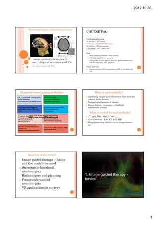
  • 1. 2012.10.30. Multimodal Imaging in Neurosciences Course COURSE FAQ Forthcoming lectures: 16. October – „IGT lecture” 23. October – NO LECTURE, holiday 30. October – MR Spectroscopy 6. November – PET + Final Test Test: - Basic imaging techniques, what are they - 5-10 easy, simple choice questions Image guided therapies in - If November 6. is not good for everyone, I will organize extra neurological sciences and VR time for getting the short test done Dr. András Jakab, MD, PhD Study material: Lecture material will be distributed in PDF 2 wks before the test. Diagnostic neuroimaging modalities What is multimodality? CT – Computed Tomography Structural MRI Combining images and information from multiple Brain anatomy Fine brain anatomy imaging tools, devices Stereotactic reference frame Vascular structure Anatomical alignment of images Fusion display, co-analysis of multiple Intra-operative imaging Diffusion, perfusion MRI information sources modalities, open MRI, low- Fine pathological field information What is needed for multimodality? Positron Emission multi-modal imaging for planning Using CT, PET, MRI, SPECT, EEG, … MR Spectroscopy Tomographyimage-guided neurological interventions PET Brain metabolism Brain metabolism Biochemical mapping Hybrid devices – PET-CT, PET-MRI Brain function Image processing skills to create image fusions, etc. Electro encephalography, Functional MR imaging fMRI LORETTA, Brain function Magnetoencephalography Overview of the lecture 1. Image guided therapy – basics and the modalities used 2. Stereotactic functional neurosurgery 3. Radiosurgery and planning R di d l i 1. 1 Image guided therapy - 4. Focused ultrasound basics neurosurgery 5. VR applications in surgery 1
  • 2. 2012.10.30. Image guidance AMIGO – Harvard, Boston • Using preoperative images to define target volumes in a reference space Treatment planning w/ Intraoperative imaging with • Using intraoperative preoperative imaging MRI imaging to monitor Surgical robot for biopsies treatment – Intraoperative ultrasound – Intraoperative low-field MRI – Intraoperative high-field Intraoperative open MRI, Brigham open MRI and Women’s Hospital, Harvard, Boston (credits: Prof. Ferenc Jolesz) Intraoperative MR imaging • Medtronic Polestar • Low field (0.1-0.3T) • Brain shift – a real problem • Use of nonmagnetic tools 2. 2 Stereotaxy in functional neurosurgery Why use stereotaxy in What is stereotactic? neurosurgery? • Space, spatial • Coordinate frame • Very high (<1-2mm) precision needed of the brain: • Minimal invasive procedures – Intrinsic – Electrode implantation for ablation – extrinsic – Permanent electrode implantation (DBS) – Biopsy sampling from the brain • Procedures without opening the skull – Radiosurgery, LINAC, Gamma Knife, Cyberknife – Focused ultrasound surgery 2
  • 3. 2012.10.30. Brain biopsy Imaging for brain biopsy • Indications • CT – Tumor suspect (enhancing CT, MRI, lesion) • MRI (T1/T2+Contrast) – Inflammation, Neurodegeneration, unknown • Pathology+Radiological • Procedure (1-2 hours): consultation – where to take – St Stereo f frame attachment tt h t the sample f h l from?? – Computed tomography – Planning – General anaesthesia – Small craniotomy – Dexamethasone – Control scan(s) Functional neurosurgery Thalamotomy / pallidotomy • Parkinson disease – associated tremor • Alleviating symptoms by modifying alleviation functional areas of the brain, subcortical • Essential tremor (unkown ethiology) areas • Two main approaches: – Permanently cutting fiber pathways, destroying specific nuclei Thermoablation Targets: – Retuning / stimulating fiber pathways, specific VIM nucleus nuclei in the brain Subthalamic Deep brain stimulation nucleus, PTT Ventral Pallidum Thalamotomy • Fantom pain (after amputation) • Intractable tumor / stroke pain in limbs etc. • CL / CM nucleus – central lateralis / centré median 3
  • 4. 2012.10.30. Deep brain stimulation (DBS) • FDA approval: 1997 • Implant in brain + subcutaneous stimulator (~pacemaker) • Retuning functional pathways („arrhythmia”) Deep brain stimulation Procedure • Indications: • Localization, stereotaxy – Chronic pain, PD tremor, ET, dystonia • Electrode trajectory: – Tourette, OCD, Major • Trials and depression d i monitoring • Pre/postop imaging – Parkinsons • Subthalamic nucleus • Globus pallidus interna • Zona incerta • Pallidothalamic fibers Targeting scheme Targeting scheme • 2. Defining targets on the patient’s coordinate frame (using CT+MRI) • 1. Target definition using stereotactic atlas Manually finding AC, of the human thalamus and basal ganglia PC points on scans (histology) Finding landmarks of the reference frame (stereotactic frame attached to the head) Getting x,y,z coordinates for your target Reference: AC-PC coordinate system 4
  • 5. 2012.10.30. 3. Radiosurgery Gamma ART 6000N Rotating Gamma System American Radiosurgery Inc., San Diego, CA, USA Challenges • Morphological imaging by MRI – 3D isotrop voxel acquisitio • Good tissue contrast • Good signal-to-noise •only 30 Co60 sources • Minimal image distortion •Rotating sources •Collimator size: 4, 8, 14, 18 mm • Optimal image resolution •Stereotactic frame required •0,3 mm accuracy – MRI as reference system? •Source half-life approx. 5 yrs • Robust CT method is necessary Challenges of multimodality – fMRI – DTI, fibertracking – MRS, MRSI • Postprocessing – Registration, image fusion (need fast) • PET, SPECT 5
  • 6. 2012.10.30. 3D T1 weighted images 3D T2 weighted images • 1,2x1,2x1,2 mm • 140 slices • 0,7x0,7x0,7 mm • 6 minutes • 40-60 slices • Gd contrast agent • 4 minutes Our imaging protocolls 3D TOF acquisition • 1,2x1,2x1,2 mm (+indications) • 190 slices • Metastasis, meningeoma • 6 minutes • AVM • g Gd contrast agent • Cavernoma • Acustic neurinoma • Hypophysis microadenoma AVM MRA Automatic radiologic image processing Automated CT/MRI registration CT/T1/T2 (gammaknife, neurosurgery automated T1 standardization, segmentation (neurosurgery, neurology) ImageSorter DICOM server automated Tensor space calculations and DWI, T1 regularization (neurosurgery, neurology) automated automated fMRI, T1, T2 SPM analysis SPM analízis (neurosurgery, neurology) automated Pet & Mri PET-MR registration and roi analysis 6
  • 7. 2012.10.30. T1-CT registration for radiosurgery (gamma-knife) Image registration, alignment gamma knife - Automatic (maximalisation relative entropy) - Manual correction (with internal „landmarks”) To this time, manual correction was necessary in 60% of the cases - Optimalised automatic registration T1-CT Fiesta-CT TOF-CT Extracranial metastasis Acoustic neurinoma treatment plan (epipharynx) • 1 kezelési tervet betenni Acustic neurinoma Pre-treatment 6 months post 12 months post 7
  • 8. 2012.10.30. Trigeminal neuralgia Treating trigeminal neuralgia CT and FIESTA imaging Validation of the T1-CT optimized Dataflow Service available via a DICOM-server registration method REFERENCE IMAGES AUTOMATICALLY REGISTERED IMAGES MNI and M3I tools: S01 BrainCAD: •Dicom-minc conversion • 2D fusion •Segmentation Comparison using normalized S02 •Automatic registration SPM-analysis • 3D fusion relative entropy • Fiber visualization •Spatial standardization •Automatic region analysis S03 AUTOMATIC INTERACTIVE Sn PRESENTATION MRI data revision by experts IMAGE PROCESSING IMAGE PROCESSING (neuronavigation) (occassionaly manual registrations) Further fancy options CT-DTI registration PET/SPECT images T2, PD etc. SPM images Precise T1-CT registration Structures defined in the space of a brain atlas DTI parametric images 8
  • 10. 2012.10.30. Proton therapy DTI + MRI data in the CT frame. Metastatic disease, 55/M ( Gamma Radiosurgery Centre, Debrecen ) The proton advantage: Medulloblastoma PHOTONS “dose bath” Nasopharynx Photons (IMRT) Protons Dose bath 100% 60% PROTONS 10% The proton advantage: Paraspinal Photons Protons Dose bath 4. 4 Focused ultrasound neurosurgery 10
  • 11. 2012.10.30. Energy Conversion and Transport in Biosystems (09.12.10) Welcome Non-invasive Beat Werner MR-Center Interventions University Children’s Hospital Zurich with www.kispi.uzh.ch/mr beat.werner@kispi.uzh.ch beat werner@kispi uzh ch Focused dipl. phys. ETH Ultrasound MR-Physics Beat Werner HIFU surgery since 2005, MR-Center research project as part of NCCR Co-Me University Children‘s Hospital Zurich Slide credits: Beat Werner, Ernst Martin Intro: Image guided interventions Intro: Image guided interventions Image Guided Interventions Improve accuracy Decrease intervention risks Minimally invasive / Non-invasive Interventions Slide credits: Beat Werner, Ernst Martin Planning and navigation Complex interventions in cranio-maxillo-facial surgery. Slide credits: Beat Werner, Ernst Martin Intro: Image guided interventions Intro: Image guided interventions "Multimodal" Imaging Enabling technologies used here: Multimodal: US, CT, MR, ... MR Multidomain: Anatomical, physiological, statistical Closed-loop intervention control information US field calibration Multi-Timescales: Static (Pre-/intra-intervention), Focused Ultrasound Realtime Mechanical / Thermal therapy Augmented reality: condition, fuse, model (Targeted) drug delivery Enhanced visualization: 2D, 3D, ..., force feedback (Targeted) drug activation Nano-Particles Molecular markers Contrast agents Drug carriers Computer-assisted support in ORL surgery Werner, Ernst Martin Slide credits: Beat Therapeutic devices Beat Werner, Ernst Martin Slide credits: 11
  • 12. 2012.10.30. Motivation: Non-invasive HIFU Surgery Non- Motivation: Clinical value of MR guided Focused US Surgery Excellent soft tissue contrast for target localization Local therapy Non invasive Non-invasive No dose accumulation effects No long term toxicity Immediate result – no radiation necrosis Treatments can be repeated Closed-loop image guidance Real time monitoring Slide credits: Beat Werner, Ernst Martin Imaging <-> Focused Ultrasound <- Phased- Phased-Array Transducers Imaging (Diagnostics): want wide field of view with Phased-Array Transducer = Array of Transducers uniform low acoustic intensity Each transducer element controlled individually Transducer phases define superposition pattern of waves -> Electronic steering HIFU (Intervention / Therapy): Constructive Destructive Shift want small focal spot with Slide credits: Beat intensity high acoustic Werner, Ernst Martin Slide credits: Beat Werner, Ernst Martin Beam Forming / Focusing Phased- Phased-Array Transducers Linear Array -> Plane Wave Geometric focusing Lens focusing Electronic beam forming Electronic steering Spherical Array -> Focus Slide credits: Beat Werner, Ernst Martin Slide credits: Beat Werner, Ernst Martin 12
  • 13. 2012.10.30. Phased- Phased-Array Transducers Phased- Phased-Array Transducers Spherical Array Spherical Array Pressure Distribution Temperature Distribution Electronic Steering Slide credits: Beat Werner, Ernst Martin Slide credits: Beat Werner, Ernst Martin Biological Effects Medical applications The effects of Ultrasound on biological tissue Lithotrypsy are a field of active research Physiotherapy Many effects are not well understood HIFU Surgery (Tissue ablation) Effects include: Blood-Brain-Barrier-Opening thermal th l non-thermal non thermal Cell sonoporation Targeted g mechanisms ultrasound mechanisms drug delivery Local drug activation energy non-cavitation mechanisms cavitation mechanisms Vessel occlusion absorption Thrombolysis temperature increase radiation force non-inertial cavitation inertial cavitation Nerve activation / blockage ablation enhanced Delivery lithothripsy Slide credits: Beat Werner, Ernst Martin Slide credits: Beat Werner, Ernst Martin Development of HIFU Technology Development of HIFU Technology 1880 Piezoelectric effect (P. & J. Curie) 1918 Sonar (Langevin) W.Fry, circa 1960, with 4-beam HIFU system for neuro- 4- neuro- 1927 Effects on biological tissues (Looms, Wood) surgery in his 1942 First HIFU lesions in animal brains (J.Lynn, T.Putnam) bioacoustics 1950– 1950–2000 Pioneer work on (a) HIFU effects on tissue (brain laboratory. tumors) and (b) LIU for soft tissue visualization (W Fry (W.Fry & F.Fry). 1951– 1951–1960 Technical development at MGH (Cosman) 1951– 1951–1967 HIFU stereotactic neurosurgery against pain, psycho- psycho- neuroses, anxiety, depression and epilepsy (Lindstrom) and Radiosurgery (Leksell) gamma knife Mid 1970s 1970s First stereotactic HIFU brain surgery with open cranium (F.Fry & R.Heimburger). Slide credits: Beat Werner, Ernst Martin Slide credits: Beat Werner, Ernst Martin 13
  • 14. 2012.10.30. Development of HIFU Technology Image guidance First US-image guided HIFU system to treat US-Imaging brain cancer patients. Cheap W.Fry in the 1970s Limited feedback F.Fry and R.Heimburger 1974 MR Expensive High-resolution imaging Closed loop process Slide credits: Beat Werner, Ernst Martin Slide credits: Beat Werner, Ernst Martin Development of HIFU Technology HIFU Surgery for Uterine Fibroids Key technologies: 1. Phased array transducers 2. Acoustic field modelling 3. MR imaging for accurate real time monitoring Early 1990s l 1990s 990 Ultrasound phased arrays (Hynynen) l d h d ( ) Mid–1990s Mid–1990s MRI thermometry (Jolesz) 2001 First integrated MRgHIFU system (InSightec) 2001 Fibroadenoma of Breast and Uterine Fibroids 2004 FDA approval for Uterine Fibroid Application … 2008 Functional Neurosurgery Slide credits: Beat Werner, Ernst Martin Slide credits: Beat Werner, Ernst Martin InSightec Exablate 2000 Treatment process for Uterine Fibroids InSightec Haifa, Israel benign (non-cancerous) tumors Technology originally GE most common pelvic tumors in women FDA approved 20-40% in women of reproductive age First & only worldwide Classical procedure: Hysterectomy GE MR-Scanners Ca. 200 systems installed Slide credits: Beat Werner, Ernst Martin Slide credits: Beat Werner, Ernst Martin 14
  • 15. 2012.10.30. Patient interface Transducer 16 element array Mechanical positioning MR-coil Patient vertical electronic steering 4 axes focal depth 12cm – 19cm Computer controlled Coupling Target Ultrasound MR-compatible Transducer Ceramic step motors Slide credits: Beat Werner, Ernst Martin Slide credits: Beat Werner, Ernst Martin Safety checks Treatment planning Air bubbles? Delineate Metallic clips? region of Scars? treatment Bowels? System creates treatment plan p Bladder? Bl dd ? (array of Nerves? sonications to be done) Plan can be changed manually by operator Slide credits: Beat Werner, Ernst Martin Slide credits: Beat Werner, Ernst Martin Real- Real-time temperature monitoring Treatment assessment MR-Thermometry Proton resonance frequency shift ~ Temperature Phase difference ~ Temperature difference 1.1 seconds 4.5 seconds 7.9 seconds 11.3 seconds Blue dots = Cell Treatment control by ablation as calculated contrast enhanced MR by thermal dose Very good agreement Slide credits: Beat Werner, Ernst Martin Slide credits: Beat Werner, Ernst Martin 15
  • 16. 2012.10.30. Closed loop control MRgFUS = Magnetic Resonance guided Focused UltraSound Planing Safety Real-Time Assement checks Monitoring Closed Loop Slide credits: Beat Werner, Ernst Martin Slide credits: Beat Werner, Ernst Martin Clinical applications and research Uterine fibroids T1w contrast enhanced image before treatment Brain Tumors Brain Functional Breast Cancer T2w planning Liver Tumors image with Uterine Fibroids dose overlay Prostate Bone Tumors T1w contrast enhanced image Slide credits: Beat Werner, Ernst Martin Courtesy of Sheba Medical Center, Tel Aviv, Israel immediately post-treatment Slide credits: Beat Werner, Ernst Martin Prostate Liver tumors Treatment effects in canine prostate Thermal dose T1w contrast TTC stained estimate enhanced image tissue Sagittal, Axial and coronal post treatment dose imaging maps (in blue) and post treatment T1W+C Courtesy InSightec Courtesy of St. Mary’s Hospital, London, UK Slide credits: Beat Werner, Ernst Martin Slide credits: Beat Werner, Ernst Martin 16
  • 17. 2012.10.30. Caution-Investigational Device Limited by United States Law IDE Breast Cancer Bone tumors (Pain palliation) to Investigational Use. 10.0 Screening 9.0 Average 8.0 7 7.0 VAS score 6.0 5.0 4.0 1 Month Follow Up 3.0 1.5 3 Months 2.0 Pre treatment T1w+C 2 weeks post treatment 1.5 Follow Up 1.0 0 0 MIP image T1w+C MIP image 0.0 0 10 20 30 40 50 60 70 80 90 Days post treatment Courtesy of Breastopia Namba Hospital, Japan Courtesy InSightec Slide credits: Beat Werner, Ernst Martin Slide credits: Beat Werner, Ernst Martin Non- Non-invasive neuro-surgery neuro- Skull: Anisotropic Aberration in the MR-suite MR- Focused waves … … are distrorted after skull Phase corrected waves … … are refocused after skull Courtesy of ESPCI, Paris, F InSightec ExAblate 4000 Patient Interface 3D-Positioner Frontend Degassed water used for acoustic coupling & Amplifier cooling InSightec ExAblate 4000 / 3.0T GE Signa HDx Stereotactic frame for patient immobillization Hemispheric 1024-element phased-array transducer Transducer Sealing membrane (650kHz) Stereotactic frame Water cooling of skull surface CT-based acoustic modeling PRS-Thermometry Transducer Installation June 2006 Water Stereotactic frame 17
  • 18. 2012.10.30. MRI- MRI-HIFU Console Clinical phase I study Centro-Lateral Thalamotomy against chronic, therapy resistant, neuropathic pain Established procedure: RF-ablation o minimally invasive o n (pain) > 100 interventions New procedure: Non-invasive TcMRgHIFU o Minimize intervention risks (Collateral damage, infections, bleeding, tissue shift) o Enhanced efficacy (no trajectory restrictions, anatomically adapted volume ablation, image guidance) o Outpatient process Bildfolge der Behandlung (sonication procedure) Bildfolge der Behandlung (sonication procedure) X+20’: Stereotactic frame X+50: Positioning X+1h20: Imaging X+3h: Verification sonications 18
  • 19. 2012.10.30. X+3h: Verification sonications X+4h: Ablation Dosemap 17 CEM 43°C 43° Dosemap 240 CEM 43°C 43° X+6h: End of treatment Patient #4 #4 Patient #2 #2 immediately after 48 h after the intervention the intervention Trigeminal neuralgia Chronic lumbar pain syndrome following disc hernia op L4/L5 L4/L5 X+7h: Happy End Slide credits: Beat Werner, Ernst Martin 19
  • 20. 2012.10.30. Patient #4 #4 Patient #2 #2 Patient #4 #4 Patient #2 #2 immediately after 48 h after immediately after 48 h after the intervention the intervention the intervention the intervention T1WI + Gd T1WI + Gd DTI DTI Combined Tumor therapy: Blood Brain Barrier HIFU- HIFU-Ablation & LIFU BBBD Brain protected by BBB: Tumorvolume Structural and functional barrier in the vessel walls Controls transport and diffusion from the HIFU- HIFU-Ablation vasculature to the central nervous system Severely limits ability to deliver drugs to the brain McDannold et al. Neurosurgery 2010 BBB- BBB-Opening with Low Intensity Focused Ultrasound (LIFU) and Adjuvant Chemotherapy Slide credits: Beat Werner, Ernst Martin Slide credits: Beat Werner, Ernst Martin Reversible BBB-Disruption using BBB- TcMRgFUS BBB-Disruption BBB- FUS and Microbubble UCA US freq. 220kHz – 1MHz Microbubbles Lipid monolayer Acc.Power < 1W Phospholipid / Hexafluorid (2-4um) Sonication 10 – 200s Commercial CE / FDA Duty cycle 1% Tail vein injection MR-contrast agent / drug Micro bubbles Inj. Drug Inj. Micro bubbles Histo MRI MRI Reversible opening of BBB by Low Intensity US Cavitation (Bursts of 10ms/1000ms) Shear stress (Acoustic streaming, radiation force, ...) Slide credits: Beat Werner, Ernst Martin 20
  • 21. 2012.10.30. TcMRgFUS BBB-Disruption BBB- TcMRgFUS BBB-Disruption BBB- Duration MR-Scanner: GE 3.0T several hours Transducer: Imasonic (Aperture 8cm, f# 0.8) Application Microbubbles: Bracco Neuro- Positioner P iti MR-Coil MR C il Phamacology Tumors Alzheimer Neuron regeneration Transducer Watertank Mouse Targeted Drug Delivery Targeted Drug Delivery Targeted Drug Delivery Target specific Carries contrast agents (Dye, fluorescent, magnetic, ...) Carries drugs Remote activation Bubble constructs Add specificity Add payload Remote activation Targeted Drug Delivery Gene Delivery Releasing drug by Ultrasound Heat Cavitation 21
  • 22. 2012.10.30. Gene Activation: hsp-80 hsp- Summary Image guided FUS is a new modality for non-invasive interventions deep in soft tissue Thermal ablation clinically established Intense research on treatment strategies based on mechanical effects Very promising results in animal models for targeted drug delivery based on transient BBBD / Sonoporation Intense research on development of nano-constructs for imaging and therapy Clinical studies to come soon Slide credits: Beat Werner, Ernst Martin Slide credits: Beat Werner, Ernst Martin Clot Lyses for Stroke Outlook Thank you ! Neurological Disorders Targeted Drug Delivery In-vitro T2W MRI before [l] and after[r] sonication Slide credits: Beat Werner, Ernst Martin Slide credits: Beat Werner, Ernst Martin Thank you for your attention! Presentation credits: Dr. András Jakab, M.D. Ph.D. Dr. Ervin Berényi, M.D. Ph.D. Dr. Miklós Emri (Nuclear Medicine Institute, UD) Prof. Ernst Martin – Uni. Zürich (Focused Ultrasound) Beat Werner – Uni. Zürich (Focused Ultrasound) 22