SlideShare ist ein Scribd-Unternehmen logo
1 von 4
Downloaden Sie, um offline zu lesen
International Journal of Pharmaceutical Science Invention
ISSN (Online): 2319 – 6718, ISSN (Print): 2319 – 670X
www.ijpsi.org Volume 2 Issue 7‖ July2013 ‖ PP.23-26
www.ijpsi.org 23 | P a g e
Brain Tumor Segmentation from MRI images and volume
calculation of Tumor
1
Aman Chandra Kaushik, 2
Vandana Sharma
1
Department of Bioinformatics, University Institute of Engineering & Technology
Chhatrapati Shahu Ji Maharaj University, Kanpur-208024, Uttar Pradesh, India
ABSTRACT: Oncogene causing Cancer is mainly a tumor oriented disorder which has been found in various
parts of the body, specialized into its particular types. The study of uncontrolled histological growth due to
apoptosis has become a very important field of Cancer biology. Extracting quantitative data from MRI imaging
source helps to understand the functions of different outgrowth clefts. However, such approaches are applicable
in the case of Benign Cancer, where the type of Cancer has been initialized to approach inside the body. Insilico
image processing of the MRI can be considered as a high throughput for the convenience to analyze the
outgrowth region. Differentiation in between the higher proximity and lower proximity has been analyzed by the
MRIIS (Magnetic Resonance Image InSilico) which is built up on MATLAB. Extensive experiments are done to
analyze the performance of intensity-based techniques for homogeneous tumors on brain magnetic resonance
(MR) images. The analysis shows that the state-of-art methods fail to segment homogeneous tumors against
similar background or when these tumors show partial diversity toward the background. Therefore, the
proposed method content-based active contour (CBAC) uses both intensity and texture information present
within the active contour to overcome above-stated problems capturing large range in an image. It also
proposes a novel use of Gray-Level Co-occurrence Matrix to define texture space for tumor segmentation in
MATLAB.In proposed work Region Growing method is used for segmenting ROI, and then by using Edge
detection for boundary segmentation volume of tumor will be calculated.First task will be Region growing
segmentation used for segmenting ROI (region of interest).
KEY WORDS: Region Growing Algorithm, Thresholding, Level Set Method
I. INTRODUCTION
In proposed work Region Growing method is used for segmenting ROI, and then by using Edge
detection for boundary segmentation volume of tumor will be calculated. First task will be Region growing
segmentation used for segmenting ROI (region of interest). In the region growing segmentation, the first aim is
to determine the initial seed points. A seed point is the starting point for region growing and its selection is vital
for the segmentation result. Seed selection could be done manually or automatically. In this work seed selection
will done automatically. After getting ROI next step will be edge detection operation for segmenting boundary.
The level set approach used to achieve an exact edges of tumor boundary which is useful in accurate estimation
of its volume. In the region growing segmentation, the first aim is to determine the initial seed points. A seed
point is the starting point for region growing and its selection is vital for the segmentation result. Seed selection
could be done manually or automatically. In this work seed selection will done semi automatically.
A semi-interactive seed point selection which is neither fully automatic nor fully manual was
introduced. The principle is a user views the image and based on the personal judgment, choose the seed point
by a mouse-based point and click mechanism. Some matlab functions are employed in order to obtain an initial
seed point .
After getting ROI next step will be edge detection operation for segmenting boundary.
The level set approach used to achieve an exact edges of tumor boundary which is useful in accurate
estimation of its volume.
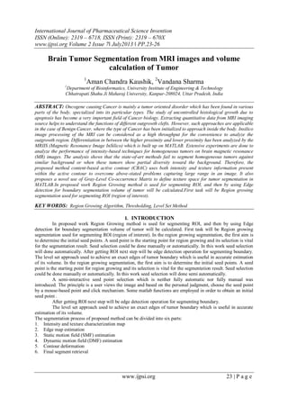
The segmentation process of proposed method can be divided into six parts:
1. Intensity and texture characterization map
2. Edge map estimation
3. Static motion field (SMF) estimation
4. Dynamic motion field (DMF) estimation
5. Contour deformation
6. Final segment retrieval
Brain Tumor Segmentation from MRI

www.ijpsi.org 24 | P a g e
II. PROBLEMS
‱ The problem addressed here is the semiautomatic brain tumor extraction from MRI using segmentation.
‱ The input for the proposed system will be the series of slices taken from different MR modalities of the
same individual in the same session.
‱ The output will be a binary segmentation of brain tumours, where each pixel in the input images is labeled
as either normal or abnormal.
‱ Finally the area and volume of the tumour will be measured which can be used for the treatment trials.
Main three Algorithm
‱ Region Growing Algorithm
‱ Thresholding
‱ Level Set Method
1. Region Growing Algorithm
The first region-growing method was the seeded region growing method. This method takes a set of
seeds as input along with the image. The seeds mark each of the objects to be segmented. The regions are
iteratively grown by comparing all unallocated neighbouring pixels to the regions. The difference between a
pixel's intensity value and the region's mean, , is used as a measure of similarity. The pixel with the smallest
difference measured this way is allocated to the respective region.
Partition image directly into regions, without first looking for boundaries or thresholds
Two main approaches:
1. “Bottom-up”: Region Growing
2. “Top-down”: Region Splitting and Merging
 Start from a seed point(a point that belongs to the region you want to segment)
Algorithm:
We have a predicate
a) Start from seed point
b) Look at its neighborhood
c) For each point p in nhood:
d) If P(p) == TRUE then include pin region L
e) Else p is a border
f) In each iteration, look at neighbors of each point in L, to save computation, “order” the points in L and
don‟t check the points that have high score (e.g. the ones that are in the middle of the region).
2. Thresholding
The simplest method of image segmentation is called the thresholding method. This method is based on
a clip-level (or a threshold value) to turn a gray-scale image into a binary image. The key of this method is to
select the threshold value (or values when multiple-levels are selected).
Play with seed points & thresholds
‱ Criteria forthreshold selection:
‱ Noise level
‱ Borders (how welldefinedare they? )
‱ Topology (e.g. holes)
‱ In short, it‟s often trial and error

3. Level Set Method
It can be used to efficiently address the problem of curve/surface/etc. propagation in an implicit
manner. The central idea is to represent the evolving contour using a signed function, where its zero level
corresponds to the actual contour. Then, according to the motion equation of the contour, one can easily derive a
similar flow for the implicit surface that when applied to the zero-level will reflect the propagation of the
contour.
The level set approach can be used as a powerful tool for 3D segmentation of a tumor to achieve an accurate
estimation of its volume. A major challenge of such algorithms is to set the equation parameters, especially the
speed function.
Brain Tumor Segmentation from MRI

www.ijpsi.org 25 | P a g e
III. MATERIAL AND METHOD
1- Software Requirements –
Matlab: Matrix Laboratory is a special –purpose computer program optimized to perform engineering and
scientific calculations. The MATLAB program implements the matlab language and provides a very extensive
library of predefined functions to make technical programming task easier and more efficient. It provides
accuracy in results.
IV. METHODOLOGY
For automatic seed selection (Seed based region growing)
The concepts of regional maximum and local maximum are used to obtain an initial seed point
automatically. The algorithm can be outlined in 6 steps;
Step 1: Find the regional maxima of the image (f) within eight connected neighborhoods. This will return the
binary image that identifies the location of the regional maxima in f. The Regional maxima identified are set to
„1‟ and all others are set to „0‟.
Step 2: Get the pixel intensity value of the regional maxima. Set the matrix as nx1 size, where n is the number
of regional maxima.
Step 3: Eliminate the plateau pixels by the following process.
a. Let X be a nx1 matrix which contains n regional maxima.
b. Arrange the pixel values of X in ascending order.
c. Determine the minimum successive adjacent differences in X and denote then as
b) ∆X ,i= 1,2,3,....n -1
a. Apply the formula= X + random (n) x min { ∆ Xi}
Step 4: Use the dilation operator with a suitable structuring element and appropriate radius. Based on the radius,
the maximum of the consecutive pixels are determined. This is repeated until all pixels are considered as pivots.
This will return a new matrix Y where each point is replaced by the maximum values obtained.
Step 5: The location of local maxima in the original set X are determined by comparing the points where’=Y.
Step 6: Evaluate the average of local maxima and set the average as the initial seed point.
Brain Tumor Segmentation from MRI

www.ijpsi.org 26 | P a g e
V. 5. RESULTS & DISCUSSION
MATLAB OUTPUT
VI. REFERENCES
[1]. K.Jumaat, R. Mahmud, S. S. Yasiran, proposed Region and boundary segmentation of micro calcifications using seed-based
region growing and mathematical morphology.Procedia Social and Behavioral Sciences 8 (2010) 634–639. International
Conference on Mathematics Education Research 2010 (ICMER 2010) 2010 Published by Elsevier.
[2]. N. Saha, N. Ray, R. Greiner, A. Murtha, H. Zhang proposed Quick detection of brain tumors and edemas: A bounding box
method using Symmetry. Computerized Medical Imaging and Graphics 36 (2012) 95– 107.
[3]. J. Sachdeva, V. Kumar, I. Gupta, N. Khandelwal, C. K.Ahuja , proposed A novel content-based active contour model for brain
tumor segmentation. Magnetic Resonance Imaging xx (2012) Elsevier.
[4]. Q-H. Huang, Su-Y. Lee, L-Z. Liu, M-H. Lu, L-W. Jin, A-H. Li proposed A robust graph-based segmentation method for breast
tumors in ultrasound images.Ultrasonics 52 (2011) 266–275.
[5]. S. Taheri , S.H. Ong , V.F.H. Chong proposed Level-set segmentation of brain tumors using a threshold-based speed function.
Image and Vision Computing 28 (2009) 26–37.

Weitere Àhnliche Inhalte

Was ist angesagt?

SEGMENTATION OF MAGNETIC RESONANCE BRAIN TUMOR USING INTEGRATED FUZZY K-MEANS...
SEGMENTATION OF MAGNETIC RESONANCE BRAIN TUMOR USING INTEGRATED FUZZY K-MEANS...SEGMENTATION OF MAGNETIC RESONANCE BRAIN TUMOR USING INTEGRATED FUZZY K-MEANS...
SEGMENTATION OF MAGNETIC RESONANCE BRAIN TUMOR USING INTEGRATED FUZZY K-MEANS...ijcsit
 
Segmentation techniques for extraction and description of tumour region from ...
Segmentation techniques for extraction and description of tumour region from ...Segmentation techniques for extraction and description of tumour region from ...
Segmentation techniques for extraction and description of tumour region from ...Swarada Kanap
 
A Survey on: Hyper Spectral Image Segmentation and Classification Using FODPSO
A Survey on: Hyper Spectral Image Segmentation and Classification Using FODPSOA Survey on: Hyper Spectral Image Segmentation and Classification Using FODPSO
A Survey on: Hyper Spectral Image Segmentation and Classification Using FODPSOrahulmonikasharma
 
International Journal of Engineering Research and Development (IJERD)
International Journal of Engineering Research and Development (IJERD)International Journal of Engineering Research and Development (IJERD)
International Journal of Engineering Research and Development (IJERD)IJERD Editor
 
LEARNING FINGERPRINT RECONSTRUCTION: FROM MINUTIAE TO IMAGE
 LEARNING FINGERPRINT RECONSTRUCTION: FROM MINUTIAE TO IMAGE LEARNING FINGERPRINT RECONSTRUCTION: FROM MINUTIAE TO IMAGE
LEARNING FINGERPRINT RECONSTRUCTION: FROM MINUTIAE TO IMAGENexgen Technology
 
MRI Tissue Segmentation using MICO
MRI Tissue Segmentation using MICOMRI Tissue Segmentation using MICO
MRI Tissue Segmentation using MICOPooja G N
 
icrtitcs1014
icrtitcs1014icrtitcs1014
icrtitcs1014Priya Shetty
 
Brain tumor detection by scanning MRI images (using filtering techniques)
Brain tumor detection by scanning MRI images (using filtering techniques)Brain tumor detection by scanning MRI images (using filtering techniques)
Brain tumor detection by scanning MRI images (using filtering techniques)Vivek reddy
 
Fuzzy based hyperspectral image
Fuzzy based hyperspectral imageFuzzy based hyperspectral image
Fuzzy based hyperspectral imageijistjournal
 
Iaetsd early detection of breast cancer
Iaetsd early detection of breast cancerIaetsd early detection of breast cancer
Iaetsd early detection of breast cancerIaetsd Iaetsd
 
Image Registration
Image RegistrationImage Registration
Image RegistrationAngu Ramesh
 
A Dualistic Sub-Image Histogram Equalization Based Enhancement and Segmentati...
A Dualistic Sub-Image Histogram Equalization Based Enhancement and Segmentati...A Dualistic Sub-Image Histogram Equalization Based Enhancement and Segmentati...
A Dualistic Sub-Image Histogram Equalization Based Enhancement and Segmentati...inventy
 
Medical Image segmentation using Image Mining concepts
Medical Image segmentation using Image Mining conceptsMedical Image segmentation using Image Mining concepts
Medical Image segmentation using Image Mining conceptsEditor IJMTER
 
Literature Survey on Detection of Brain Tumor from MRI Images
Literature Survey on Detection of Brain Tumor from MRI Images Literature Survey on Detection of Brain Tumor from MRI Images
Literature Survey on Detection of Brain Tumor from MRI Images IOSR Journals
 
Image Segmentation using Otsu's Method - Computer Graphics (UCS505) Project R...
Image Segmentation using Otsu's Method - Computer Graphics (UCS505) Project R...Image Segmentation using Otsu's Method - Computer Graphics (UCS505) Project R...
Image Segmentation using Otsu's Method - Computer Graphics (UCS505) Project R...Akshit Arora
 
An overview of automatic brain tumor detection frommagnetic resonance images
An overview of automatic brain tumor detection frommagnetic resonance imagesAn overview of automatic brain tumor detection frommagnetic resonance images
An overview of automatic brain tumor detection frommagnetic resonance imagesMangesh Lingampalle
 
Brain Tumor Detection using CNN
Brain Tumor Detection using CNNBrain Tumor Detection using CNN
Brain Tumor Detection using CNNMohammadRakib8
 

Was ist angesagt? (19)

SEGMENTATION OF MAGNETIC RESONANCE BRAIN TUMOR USING INTEGRATED FUZZY K-MEANS...
SEGMENTATION OF MAGNETIC RESONANCE BRAIN TUMOR USING INTEGRATED FUZZY K-MEANS...SEGMENTATION OF MAGNETIC RESONANCE BRAIN TUMOR USING INTEGRATED FUZZY K-MEANS...
SEGMENTATION OF MAGNETIC RESONANCE BRAIN TUMOR USING INTEGRATED FUZZY K-MEANS...
 
Segmentation techniques for extraction and description of tumour region from ...
Segmentation techniques for extraction and description of tumour region from ...Segmentation techniques for extraction and description of tumour region from ...
Segmentation techniques for extraction and description of tumour region from ...
 
A Survey on: Hyper Spectral Image Segmentation and Classification Using FODPSO
A Survey on: Hyper Spectral Image Segmentation and Classification Using FODPSOA Survey on: Hyper Spectral Image Segmentation and Classification Using FODPSO
A Survey on: Hyper Spectral Image Segmentation and Classification Using FODPSO
 
International Journal of Engineering Research and Development (IJERD)
International Journal of Engineering Research and Development (IJERD)International Journal of Engineering Research and Development (IJERD)
International Journal of Engineering Research and Development (IJERD)
 
LEARNING FINGERPRINT RECONSTRUCTION: FROM MINUTIAE TO IMAGE
 LEARNING FINGERPRINT RECONSTRUCTION: FROM MINUTIAE TO IMAGE LEARNING FINGERPRINT RECONSTRUCTION: FROM MINUTIAE TO IMAGE
LEARNING FINGERPRINT RECONSTRUCTION: FROM MINUTIAE TO IMAGE
 
MRI Tissue Segmentation using MICO
MRI Tissue Segmentation using MICOMRI Tissue Segmentation using MICO
MRI Tissue Segmentation using MICO
 
JBSC_online
JBSC_onlineJBSC_online
JBSC_online
 
icrtitcs1014
icrtitcs1014icrtitcs1014
icrtitcs1014
 
Brain tumor detection by scanning MRI images (using filtering techniques)
Brain tumor detection by scanning MRI images (using filtering techniques)Brain tumor detection by scanning MRI images (using filtering techniques)
Brain tumor detection by scanning MRI images (using filtering techniques)
 
Fuzzy based hyperspectral image
Fuzzy based hyperspectral imageFuzzy based hyperspectral image
Fuzzy based hyperspectral image
 
An Image Segmentation and Classification for Brain Tumor Detection using Pill...
An Image Segmentation and Classification for Brain Tumor Detection using Pill...An Image Segmentation and Classification for Brain Tumor Detection using Pill...
An Image Segmentation and Classification for Brain Tumor Detection using Pill...
 
Iaetsd early detection of breast cancer
Iaetsd early detection of breast cancerIaetsd early detection of breast cancer
Iaetsd early detection of breast cancer
 
Image Registration
Image RegistrationImage Registration
Image Registration
 
A Dualistic Sub-Image Histogram Equalization Based Enhancement and Segmentati...
A Dualistic Sub-Image Histogram Equalization Based Enhancement and Segmentati...A Dualistic Sub-Image Histogram Equalization Based Enhancement and Segmentati...
A Dualistic Sub-Image Histogram Equalization Based Enhancement and Segmentati...
 
Medical Image segmentation using Image Mining concepts
Medical Image segmentation using Image Mining conceptsMedical Image segmentation using Image Mining concepts
Medical Image segmentation using Image Mining concepts
 
Literature Survey on Detection of Brain Tumor from MRI Images
Literature Survey on Detection of Brain Tumor from MRI Images Literature Survey on Detection of Brain Tumor from MRI Images
Literature Survey on Detection of Brain Tumor from MRI Images
 
Image Segmentation using Otsu's Method - Computer Graphics (UCS505) Project R...
Image Segmentation using Otsu's Method - Computer Graphics (UCS505) Project R...Image Segmentation using Otsu's Method - Computer Graphics (UCS505) Project R...
Image Segmentation using Otsu's Method - Computer Graphics (UCS505) Project R...
 
An overview of automatic brain tumor detection frommagnetic resonance images
An overview of automatic brain tumor detection frommagnetic resonance imagesAn overview of automatic brain tumor detection frommagnetic resonance images
An overview of automatic brain tumor detection frommagnetic resonance images
 
Brain Tumor Detection using CNN
Brain Tumor Detection using CNNBrain Tumor Detection using CNN
Brain Tumor Detection using CNN
 

Andere mochten auch

TU Patiala Aman Mehta -What marketing strategies are appropriate at each stag...
TU Patiala Aman Mehta -What marketing strategies are appropriate at each stag...TU Patiala Aman Mehta -What marketing strategies are appropriate at each stag...
TU Patiala Aman Mehta -What marketing strategies are appropriate at each stag...Bhaskar Jyoti Bora
 
marketing mix and stp of glucon-d
marketing mix and stp of glucon-dmarketing mix and stp of glucon-d
marketing mix and stp of glucon-d9959995802
 
Pharmaceutical Portfolio & Product Life Cycle Management
Pharmaceutical Portfolio & Product Life Cycle ManagementPharmaceutical Portfolio & Product Life Cycle Management
Pharmaceutical Portfolio & Product Life Cycle Managementsbryant89
 
Heinz company overview mba
Heinz company overview mbaHeinz company overview mba
Heinz company overview mbaAkash Halankar
 
PRODUCT LIFE CYCLE....A STUDY ON PHARMA & NON-PHARMA EXAMPLE
PRODUCT LIFE CYCLE....A STUDY ON PHARMA & NON-PHARMA EXAMPLEPRODUCT LIFE CYCLE....A STUDY ON PHARMA & NON-PHARMA EXAMPLE
PRODUCT LIFE CYCLE....A STUDY ON PHARMA & NON-PHARMA EXAMPLEPooja Mangal
 
Pharmaceutical Lifecycle Management Challenges - Slides from Pharmaceutical P...
Pharmaceutical Lifecycle Management Challenges - Slides from Pharmaceutical P...Pharmaceutical Lifecycle Management Challenges - Slides from Pharmaceutical P...
Pharmaceutical Lifecycle Management Challenges - Slides from Pharmaceutical P...IHS
 

Andere mochten auch (7)

TU Patiala Aman Mehta -What marketing strategies are appropriate at each stag...
TU Patiala Aman Mehta -What marketing strategies are appropriate at each stag...TU Patiala Aman Mehta -What marketing strategies are appropriate at each stag...
TU Patiala Aman Mehta -What marketing strategies are appropriate at each stag...
 
marketing mix and stp of glucon-d
marketing mix and stp of glucon-dmarketing mix and stp of glucon-d
marketing mix and stp of glucon-d
 
Pharmaceutical Portfolio & Product Life Cycle Management
Pharmaceutical Portfolio & Product Life Cycle ManagementPharmaceutical Portfolio & Product Life Cycle Management
Pharmaceutical Portfolio & Product Life Cycle Management
 
Heinz company overview mba
Heinz company overview mbaHeinz company overview mba
Heinz company overview mba
 
PRODUCT LIFE CYCLE....A STUDY ON PHARMA & NON-PHARMA EXAMPLE
PRODUCT LIFE CYCLE....A STUDY ON PHARMA & NON-PHARMA EXAMPLEPRODUCT LIFE CYCLE....A STUDY ON PHARMA & NON-PHARMA EXAMPLE
PRODUCT LIFE CYCLE....A STUDY ON PHARMA & NON-PHARMA EXAMPLE
 
Pharmaceutical Lifecycle Management Challenges - Slides from Pharmaceutical P...
Pharmaceutical Lifecycle Management Challenges - Slides from Pharmaceutical P...Pharmaceutical Lifecycle Management Challenges - Slides from Pharmaceutical P...
Pharmaceutical Lifecycle Management Challenges - Slides from Pharmaceutical P...
 
Buying Decision Process
Buying Decision ProcessBuying Decision Process
Buying Decision Process
 

Ähnlich wie International Journal of Pharmaceutical Science Invention (IJPSI)

Comparison of Image Segmentation Algorithms for Brain Tumor Detection
Comparison of Image Segmentation Algorithms for Brain Tumor DetectionComparison of Image Segmentation Algorithms for Brain Tumor Detection
Comparison of Image Segmentation Algorithms for Brain Tumor DetectionIJMTST Journal
 
Segmentation and Classification of MRI Brain Tumor
Segmentation and Classification of MRI Brain TumorSegmentation and Classification of MRI Brain Tumor
Segmentation and Classification of MRI Brain TumorIRJET Journal
 
D05222528
D05222528D05222528
D05222528IOSR-JEN
 
Brain Tumor Classification using Support Vector Machine
Brain Tumor Classification using Support Vector MachineBrain Tumor Classification using Support Vector Machine
Brain Tumor Classification using Support Vector MachineIRJET Journal
 
Multiple Analysis of Brain Tumor Detection based on FCM
Multiple Analysis of Brain Tumor Detection based on FCMMultiple Analysis of Brain Tumor Detection based on FCM
Multiple Analysis of Brain Tumor Detection based on FCMIRJET Journal
 
A HYBRID APPROACH BASED SEGMENTATION TECHNIQUE FOR BRAIN TUMOR IN MRI IMAGES
A HYBRID APPROACH BASED SEGMENTATION TECHNIQUE FOR BRAIN TUMOR IN MRI IMAGESA HYBRID APPROACH BASED SEGMENTATION TECHNIQUE FOR BRAIN TUMOR IN MRI IMAGES
A HYBRID APPROACH BASED SEGMENTATION TECHNIQUE FOR BRAIN TUMOR IN MRI IMAGESsipij
 
Detection of Skin Cancer using SVM
Detection of Skin Cancer using SVMDetection of Skin Cancer using SVM
Detection of Skin Cancer using SVMIRJET Journal
 
Detection of leaf diseases and classification using digital image processing
Detection of leaf diseases and classification using digital image processingDetection of leaf diseases and classification using digital image processing
Detection of leaf diseases and classification using digital image processingNaeem Shehzad
 
MALIGNANT AND BENIGN BRAIN TUMOR SEGMENTATION AND CLASSIFICATION USING SVM WI...
MALIGNANT AND BENIGN BRAIN TUMOR SEGMENTATION AND CLASSIFICATION USING SVM WI...MALIGNANT AND BENIGN BRAIN TUMOR SEGMENTATION AND CLASSIFICATION USING SVM WI...
MALIGNANT AND BENIGN BRAIN TUMOR SEGMENTATION AND CLASSIFICATION USING SVM WI...sipij
 
Multiple Analysis of Brain Tumor Detection Based on FCM
Multiple Analysis of Brain Tumor Detection Based on FCMMultiple Analysis of Brain Tumor Detection Based on FCM
Multiple Analysis of Brain Tumor Detection Based on FCMIRJET Journal
 
Face Recognition System using Self Organizing Feature Map and Appearance Base...
Face Recognition System using Self Organizing Feature Map and Appearance Base...Face Recognition System using Self Organizing Feature Map and Appearance Base...
Face Recognition System using Self Organizing Feature Map and Appearance Base...ijtsrd
 
IRJET- Brain Tumor Detection using Digital Image Processing
IRJET- Brain Tumor Detection using Digital Image ProcessingIRJET- Brain Tumor Detection using Digital Image Processing
IRJET- Brain Tumor Detection using Digital Image ProcessingIRJET Journal
 
Comparitive study of brain tumor detection using morphological operators
Comparitive study of brain tumor detection using morphological operatorsComparitive study of brain tumor detection using morphological operators
Comparitive study of brain tumor detection using morphological operatorseSAT Journals
 
Q UANTUM C LUSTERING -B ASED F EATURE SUBSET S ELECTION FOR MAMMOGRAPHIC I...
Q UANTUM  C LUSTERING -B ASED  F EATURE SUBSET  S ELECTION FOR MAMMOGRAPHIC I...Q UANTUM  C LUSTERING -B ASED  F EATURE SUBSET  S ELECTION FOR MAMMOGRAPHIC I...
Q UANTUM C LUSTERING -B ASED F EATURE SUBSET S ELECTION FOR MAMMOGRAPHIC I...ijcsit
 
MRI_processing
MRI_processingMRI_processing
MRI_processingLitu Rout
 
Spot Edge Detection in cDNA Microarray Images using Window based Bi-Dimension...
Spot Edge Detection in cDNA Microarray Images using Window based Bi-Dimension...Spot Edge Detection in cDNA Microarray Images using Window based Bi-Dimension...
Spot Edge Detection in cDNA Microarray Images using Window based Bi-Dimension...idescitation
 
SEGMENTATION AND CLASSIFICATION OF BRAIN TUMOR CT IMAGES USING SVM WITH WEIGH...
SEGMENTATION AND CLASSIFICATION OF BRAIN TUMOR CT IMAGES USING SVM WITH WEIGH...SEGMENTATION AND CLASSIFICATION OF BRAIN TUMOR CT IMAGES USING SVM WITH WEIGH...
SEGMENTATION AND CLASSIFICATION OF BRAIN TUMOR CT IMAGES USING SVM WITH WEIGH...csandit
 
SLIC Superpixel Based Self Organizing Maps Algorithm for Segmentation of Micr...
SLIC Superpixel Based Self Organizing Maps Algorithm for Segmentation of Micr...SLIC Superpixel Based Self Organizing Maps Algorithm for Segmentation of Micr...
SLIC Superpixel Based Self Organizing Maps Algorithm for Segmentation of Micr...IJAAS Team
 

Ähnlich wie International Journal of Pharmaceutical Science Invention (IJPSI) (20)

Comparison of Image Segmentation Algorithms for Brain Tumor Detection
Comparison of Image Segmentation Algorithms for Brain Tumor DetectionComparison of Image Segmentation Algorithms for Brain Tumor Detection
Comparison of Image Segmentation Algorithms for Brain Tumor Detection
 
Segmentation and Classification of MRI Brain Tumor
Segmentation and Classification of MRI Brain TumorSegmentation and Classification of MRI Brain Tumor
Segmentation and Classification of MRI Brain Tumor
 
D05222528
D05222528D05222528
D05222528
 
Brain Tumor Classification using Support Vector Machine
Brain Tumor Classification using Support Vector MachineBrain Tumor Classification using Support Vector Machine
Brain Tumor Classification using Support Vector Machine
 
Multiple Analysis of Brain Tumor Detection based on FCM
Multiple Analysis of Brain Tumor Detection based on FCMMultiple Analysis of Brain Tumor Detection based on FCM
Multiple Analysis of Brain Tumor Detection based on FCM
 
A HYBRID APPROACH BASED SEGMENTATION TECHNIQUE FOR BRAIN TUMOR IN MRI IMAGES
A HYBRID APPROACH BASED SEGMENTATION TECHNIQUE FOR BRAIN TUMOR IN MRI IMAGESA HYBRID APPROACH BASED SEGMENTATION TECHNIQUE FOR BRAIN TUMOR IN MRI IMAGES
A HYBRID APPROACH BASED SEGMENTATION TECHNIQUE FOR BRAIN TUMOR IN MRI IMAGES
 
A HYBRID APPROACH BASED SEGMENTATION TECHNIQUE FOR BRAIN TUMOR IN MRI IMAGES
A HYBRID APPROACH BASED SEGMENTATION TECHNIQUE FOR BRAIN TUMOR IN MRI IMAGESA HYBRID APPROACH BASED SEGMENTATION TECHNIQUE FOR BRAIN TUMOR IN MRI IMAGES
A HYBRID APPROACH BASED SEGMENTATION TECHNIQUE FOR BRAIN TUMOR IN MRI IMAGES
 
Detection of Skin Cancer using SVM
Detection of Skin Cancer using SVMDetection of Skin Cancer using SVM
Detection of Skin Cancer using SVM
 
Detection of leaf diseases and classification using digital image processing
Detection of leaf diseases and classification using digital image processingDetection of leaf diseases and classification using digital image processing
Detection of leaf diseases and classification using digital image processing
 
MALIGNANT AND BENIGN BRAIN TUMOR SEGMENTATION AND CLASSIFICATION USING SVM WI...
MALIGNANT AND BENIGN BRAIN TUMOR SEGMENTATION AND CLASSIFICATION USING SVM WI...MALIGNANT AND BENIGN BRAIN TUMOR SEGMENTATION AND CLASSIFICATION USING SVM WI...
MALIGNANT AND BENIGN BRAIN TUMOR SEGMENTATION AND CLASSIFICATION USING SVM WI...
 
Multiple Analysis of Brain Tumor Detection Based on FCM
Multiple Analysis of Brain Tumor Detection Based on FCMMultiple Analysis of Brain Tumor Detection Based on FCM
Multiple Analysis of Brain Tumor Detection Based on FCM
 
Face Recognition System using Self Organizing Feature Map and Appearance Base...
Face Recognition System using Self Organizing Feature Map and Appearance Base...Face Recognition System using Self Organizing Feature Map and Appearance Base...
Face Recognition System using Self Organizing Feature Map and Appearance Base...
 
IRJET- Brain Tumor Detection using Digital Image Processing
IRJET- Brain Tumor Detection using Digital Image ProcessingIRJET- Brain Tumor Detection using Digital Image Processing
IRJET- Brain Tumor Detection using Digital Image Processing
 
Comparitive study of brain tumor detection using morphological operators
Comparitive study of brain tumor detection using morphological operatorsComparitive study of brain tumor detection using morphological operators
Comparitive study of brain tumor detection using morphological operators
 
F0522833
F0522833F0522833
F0522833
 
Q UANTUM C LUSTERING -B ASED F EATURE SUBSET S ELECTION FOR MAMMOGRAPHIC I...
Q UANTUM  C LUSTERING -B ASED  F EATURE SUBSET  S ELECTION FOR MAMMOGRAPHIC I...Q UANTUM  C LUSTERING -B ASED  F EATURE SUBSET  S ELECTION FOR MAMMOGRAPHIC I...
Q UANTUM C LUSTERING -B ASED F EATURE SUBSET S ELECTION FOR MAMMOGRAPHIC I...
 
MRI_processing
MRI_processingMRI_processing
MRI_processing
 
Spot Edge Detection in cDNA Microarray Images using Window based Bi-Dimension...
Spot Edge Detection in cDNA Microarray Images using Window based Bi-Dimension...Spot Edge Detection in cDNA Microarray Images using Window based Bi-Dimension...
Spot Edge Detection in cDNA Microarray Images using Window based Bi-Dimension...
 
SEGMENTATION AND CLASSIFICATION OF BRAIN TUMOR CT IMAGES USING SVM WITH WEIGH...
SEGMENTATION AND CLASSIFICATION OF BRAIN TUMOR CT IMAGES USING SVM WITH WEIGH...SEGMENTATION AND CLASSIFICATION OF BRAIN TUMOR CT IMAGES USING SVM WITH WEIGH...
SEGMENTATION AND CLASSIFICATION OF BRAIN TUMOR CT IMAGES USING SVM WITH WEIGH...
 
SLIC Superpixel Based Self Organizing Maps Algorithm for Segmentation of Micr...
SLIC Superpixel Based Self Organizing Maps Algorithm for Segmentation of Micr...SLIC Superpixel Based Self Organizing Maps Algorithm for Segmentation of Micr...
SLIC Superpixel Based Self Organizing Maps Algorithm for Segmentation of Micr...
 

KĂŒrzlich hochgeladen

Factors to Consider When Choosing Accounts Payable Services Providers.pptx
Factors to Consider When Choosing Accounts Payable Services Providers.pptxFactors to Consider When Choosing Accounts Payable Services Providers.pptx
Factors to Consider When Choosing Accounts Payable Services Providers.pptxKatpro Technologies
 
FULL ENJOY 🔝 8264348440 🔝 Call Girls in Diplomatic Enclave | Delhi
FULL ENJOY 🔝 8264348440 🔝 Call Girls in Diplomatic Enclave | DelhiFULL ENJOY 🔝 8264348440 🔝 Call Girls in Diplomatic Enclave | Delhi
FULL ENJOY 🔝 8264348440 🔝 Call Girls in Diplomatic Enclave | Delhisoniya singh
 
08448380779 Call Girls In Civil Lines Women Seeking Men
08448380779 Call Girls In Civil Lines Women Seeking Men08448380779 Call Girls In Civil Lines Women Seeking Men
08448380779 Call Girls In Civil Lines Women Seeking MenDelhi Call girls
 
How to Troubleshoot Apps for the Modern Connected Worker
How to Troubleshoot Apps for the Modern Connected WorkerHow to Troubleshoot Apps for the Modern Connected Worker
How to Troubleshoot Apps for the Modern Connected WorkerThousandEyes
 
A Domino Admins Adventures (Engage 2024)
A Domino Admins Adventures (Engage 2024)A Domino Admins Adventures (Engage 2024)
A Domino Admins Adventures (Engage 2024)Gabriella Davis
 
WhatsApp 9892124323 ✓Call Girls In Kalyan ( Mumbai ) secure service
WhatsApp 9892124323 ✓Call Girls In Kalyan ( Mumbai ) secure serviceWhatsApp 9892124323 ✓Call Girls In Kalyan ( Mumbai ) secure service
WhatsApp 9892124323 ✓Call Girls In Kalyan ( Mumbai ) secure servicePooja Nehwal
 
Enhancing Worker Digital Experience: A Hands-on Workshop for Partners
Enhancing Worker Digital Experience: A Hands-on Workshop for PartnersEnhancing Worker Digital Experience: A Hands-on Workshop for Partners
Enhancing Worker Digital Experience: A Hands-on Workshop for PartnersThousandEyes
 
How to Remove Document Management Hurdles with X-Docs?
How to Remove Document Management Hurdles with X-Docs?How to Remove Document Management Hurdles with X-Docs?
How to Remove Document Management Hurdles with X-Docs?XfilesPro
 
My Hashitalk Indonesia April 2024 Presentation
My Hashitalk Indonesia April 2024 PresentationMy Hashitalk Indonesia April 2024 Presentation
My Hashitalk Indonesia April 2024 PresentationRidwan Fadjar
 
The 7 Things I Know About Cyber Security After 25 Years | April 2024
The 7 Things I Know About Cyber Security After 25 Years | April 2024The 7 Things I Know About Cyber Security After 25 Years | April 2024
The 7 Things I Know About Cyber Security After 25 Years | April 2024Rafal Los
 
Unblocking The Main Thread Solving ANRs and Frozen Frames
Unblocking The Main Thread Solving ANRs and Frozen FramesUnblocking The Main Thread Solving ANRs and Frozen Frames
Unblocking The Main Thread Solving ANRs and Frozen FramesSinan KOZAK
 
Human Factors of XR: Using Human Factors to Design XR Systems
Human Factors of XR: Using Human Factors to Design XR SystemsHuman Factors of XR: Using Human Factors to Design XR Systems
Human Factors of XR: Using Human Factors to Design XR SystemsMark Billinghurst
 
Pigging Solutions in Pet Food Manufacturing
Pigging Solutions in Pet Food ManufacturingPigging Solutions in Pet Food Manufacturing
Pigging Solutions in Pet Food ManufacturingPigging Solutions
 
Automating Business Process via MuleSoft Composer | Bangalore MuleSoft Meetup...
Automating Business Process via MuleSoft Composer | Bangalore MuleSoft Meetup...Automating Business Process via MuleSoft Composer | Bangalore MuleSoft Meetup...
Automating Business Process via MuleSoft Composer | Bangalore MuleSoft Meetup...shyamraj55
 
AI as an Interface for Commercial Buildings
AI as an Interface for Commercial BuildingsAI as an Interface for Commercial Buildings
AI as an Interface for Commercial BuildingsMemoori
 
How to convert PDF to text with Nanonets
How to convert PDF to text with NanonetsHow to convert PDF to text with Nanonets
How to convert PDF to text with Nanonetsnaman860154
 
Swan(sea) Song – personal research during my six years at Swansea ... and bey...
Swan(sea) Song – personal research during my six years at Swansea ... and bey...Swan(sea) Song – personal research during my six years at Swansea ... and bey...
Swan(sea) Song – personal research during my six years at Swansea ... and bey...Alan Dix
 
08448380779 Call Girls In Friends Colony Women Seeking Men
08448380779 Call Girls In Friends Colony Women Seeking Men08448380779 Call Girls In Friends Colony Women Seeking Men
08448380779 Call Girls In Friends Colony Women Seeking MenDelhi Call girls
 
#StandardsGoals for 2024: What’s new for BISAC - Tech Forum 2024
#StandardsGoals for 2024: What’s new for BISAC - Tech Forum 2024#StandardsGoals for 2024: What’s new for BISAC - Tech Forum 2024
#StandardsGoals for 2024: What’s new for BISAC - Tech Forum 2024BookNet Canada
 
IAC 2024 - IA Fast Track to Search Focused AI Solutions
IAC 2024 - IA Fast Track to Search Focused AI SolutionsIAC 2024 - IA Fast Track to Search Focused AI Solutions
IAC 2024 - IA Fast Track to Search Focused AI SolutionsEnterprise Knowledge
 

KĂŒrzlich hochgeladen (20)

Factors to Consider When Choosing Accounts Payable Services Providers.pptx
Factors to Consider When Choosing Accounts Payable Services Providers.pptxFactors to Consider When Choosing Accounts Payable Services Providers.pptx
Factors to Consider When Choosing Accounts Payable Services Providers.pptx
 
FULL ENJOY 🔝 8264348440 🔝 Call Girls in Diplomatic Enclave | Delhi
FULL ENJOY 🔝 8264348440 🔝 Call Girls in Diplomatic Enclave | DelhiFULL ENJOY 🔝 8264348440 🔝 Call Girls in Diplomatic Enclave | Delhi
FULL ENJOY 🔝 8264348440 🔝 Call Girls in Diplomatic Enclave | Delhi
 
08448380779 Call Girls In Civil Lines Women Seeking Men
08448380779 Call Girls In Civil Lines Women Seeking Men08448380779 Call Girls In Civil Lines Women Seeking Men
08448380779 Call Girls In Civil Lines Women Seeking Men
 
How to Troubleshoot Apps for the Modern Connected Worker
How to Troubleshoot Apps for the Modern Connected WorkerHow to Troubleshoot Apps for the Modern Connected Worker
How to Troubleshoot Apps for the Modern Connected Worker
 
A Domino Admins Adventures (Engage 2024)
A Domino Admins Adventures (Engage 2024)A Domino Admins Adventures (Engage 2024)
A Domino Admins Adventures (Engage 2024)
 
WhatsApp 9892124323 ✓Call Girls In Kalyan ( Mumbai ) secure service
WhatsApp 9892124323 ✓Call Girls In Kalyan ( Mumbai ) secure serviceWhatsApp 9892124323 ✓Call Girls In Kalyan ( Mumbai ) secure service
WhatsApp 9892124323 ✓Call Girls In Kalyan ( Mumbai ) secure service
 
Enhancing Worker Digital Experience: A Hands-on Workshop for Partners
Enhancing Worker Digital Experience: A Hands-on Workshop for PartnersEnhancing Worker Digital Experience: A Hands-on Workshop for Partners
Enhancing Worker Digital Experience: A Hands-on Workshop for Partners
 
How to Remove Document Management Hurdles with X-Docs?
How to Remove Document Management Hurdles with X-Docs?How to Remove Document Management Hurdles with X-Docs?
How to Remove Document Management Hurdles with X-Docs?
 
My Hashitalk Indonesia April 2024 Presentation
My Hashitalk Indonesia April 2024 PresentationMy Hashitalk Indonesia April 2024 Presentation
My Hashitalk Indonesia April 2024 Presentation
 
The 7 Things I Know About Cyber Security After 25 Years | April 2024
The 7 Things I Know About Cyber Security After 25 Years | April 2024The 7 Things I Know About Cyber Security After 25 Years | April 2024
The 7 Things I Know About Cyber Security After 25 Years | April 2024
 
Unblocking The Main Thread Solving ANRs and Frozen Frames
Unblocking The Main Thread Solving ANRs and Frozen FramesUnblocking The Main Thread Solving ANRs and Frozen Frames
Unblocking The Main Thread Solving ANRs and Frozen Frames
 
Human Factors of XR: Using Human Factors to Design XR Systems
Human Factors of XR: Using Human Factors to Design XR SystemsHuman Factors of XR: Using Human Factors to Design XR Systems
Human Factors of XR: Using Human Factors to Design XR Systems
 
Pigging Solutions in Pet Food Manufacturing
Pigging Solutions in Pet Food ManufacturingPigging Solutions in Pet Food Manufacturing
Pigging Solutions in Pet Food Manufacturing
 
Automating Business Process via MuleSoft Composer | Bangalore MuleSoft Meetup...
Automating Business Process via MuleSoft Composer | Bangalore MuleSoft Meetup...Automating Business Process via MuleSoft Composer | Bangalore MuleSoft Meetup...
Automating Business Process via MuleSoft Composer | Bangalore MuleSoft Meetup...
 
AI as an Interface for Commercial Buildings
AI as an Interface for Commercial BuildingsAI as an Interface for Commercial Buildings
AI as an Interface for Commercial Buildings
 
How to convert PDF to text with Nanonets
How to convert PDF to text with NanonetsHow to convert PDF to text with Nanonets
How to convert PDF to text with Nanonets
 
Swan(sea) Song – personal research during my six years at Swansea ... and bey...
Swan(sea) Song – personal research during my six years at Swansea ... and bey...Swan(sea) Song – personal research during my six years at Swansea ... and bey...
Swan(sea) Song – personal research during my six years at Swansea ... and bey...
 
08448380779 Call Girls In Friends Colony Women Seeking Men
08448380779 Call Girls In Friends Colony Women Seeking Men08448380779 Call Girls In Friends Colony Women Seeking Men
08448380779 Call Girls In Friends Colony Women Seeking Men
 
#StandardsGoals for 2024: What’s new for BISAC - Tech Forum 2024
#StandardsGoals for 2024: What’s new for BISAC - Tech Forum 2024#StandardsGoals for 2024: What’s new for BISAC - Tech Forum 2024
#StandardsGoals for 2024: What’s new for BISAC - Tech Forum 2024
 
IAC 2024 - IA Fast Track to Search Focused AI Solutions
IAC 2024 - IA Fast Track to Search Focused AI SolutionsIAC 2024 - IA Fast Track to Search Focused AI Solutions
IAC 2024 - IA Fast Track to Search Focused AI Solutions
 

International Journal of Pharmaceutical Science Invention (IJPSI)

  • 1. International Journal of Pharmaceutical Science Invention ISSN (Online): 2319 – 6718, ISSN (Print): 2319 – 670X www.ijpsi.org Volume 2 Issue 7‖ July2013 ‖ PP.23-26 www.ijpsi.org 23 | P a g e Brain Tumor Segmentation from MRI images and volume calculation of Tumor 1 Aman Chandra Kaushik, 2 Vandana Sharma 1 Department of Bioinformatics, University Institute of Engineering & Technology Chhatrapati Shahu Ji Maharaj University, Kanpur-208024, Uttar Pradesh, India ABSTRACT: Oncogene causing Cancer is mainly a tumor oriented disorder which has been found in various parts of the body, specialized into its particular types. The study of uncontrolled histological growth due to apoptosis has become a very important field of Cancer biology. Extracting quantitative data from MRI imaging source helps to understand the functions of different outgrowth clefts. However, such approaches are applicable in the case of Benign Cancer, where the type of Cancer has been initialized to approach inside the body. Insilico image processing of the MRI can be considered as a high throughput for the convenience to analyze the outgrowth region. Differentiation in between the higher proximity and lower proximity has been analyzed by the MRIIS (Magnetic Resonance Image InSilico) which is built up on MATLAB. Extensive experiments are done to analyze the performance of intensity-based techniques for homogeneous tumors on brain magnetic resonance (MR) images. The analysis shows that the state-of-art methods fail to segment homogeneous tumors against similar background or when these tumors show partial diversity toward the background. Therefore, the proposed method content-based active contour (CBAC) uses both intensity and texture information present within the active contour to overcome above-stated problems capturing large range in an image. It also proposes a novel use of Gray-Level Co-occurrence Matrix to define texture space for tumor segmentation in MATLAB.In proposed work Region Growing method is used for segmenting ROI, and then by using Edge detection for boundary segmentation volume of tumor will be calculated.First task will be Region growing segmentation used for segmenting ROI (region of interest). KEY WORDS: Region Growing Algorithm, Thresholding, Level Set Method I. INTRODUCTION In proposed work Region Growing method is used for segmenting ROI, and then by using Edge detection for boundary segmentation volume of tumor will be calculated. First task will be Region growing segmentation used for segmenting ROI (region of interest). In the region growing segmentation, the first aim is to determine the initial seed points. A seed point is the starting point for region growing and its selection is vital for the segmentation result. Seed selection could be done manually or automatically. In this work seed selection will done automatically. After getting ROI next step will be edge detection operation for segmenting boundary. The level set approach used to achieve an exact edges of tumor boundary which is useful in accurate estimation of its volume. In the region growing segmentation, the first aim is to determine the initial seed points. A seed point is the starting point for region growing and its selection is vital for the segmentation result. Seed selection could be done manually or automatically. In this work seed selection will done semi automatically. A semi-interactive seed point selection which is neither fully automatic nor fully manual was introduced. The principle is a user views the image and based on the personal judgment, choose the seed point by a mouse-based point and click mechanism. Some matlab functions are employed in order to obtain an initial seed point . After getting ROI next step will be edge detection operation for segmenting boundary. The level set approach used to achieve an exact edges of tumor boundary which is useful in accurate estimation of its volume. The segmentation process of proposed method can be divided into six parts: 1. Intensity and texture characterization map 2. Edge map estimation 3. Static motion field (SMF) estimation 4. Dynamic motion field (DMF) estimation 5. Contour deformation 6. Final segment retrieval
  • 2. Brain Tumor Segmentation from MRI
 www.ijpsi.org 24 | P a g e II. PROBLEMS ‱ The problem addressed here is the semiautomatic brain tumor extraction from MRI using segmentation. ‱ The input for the proposed system will be the series of slices taken from different MR modalities of the same individual in the same session. ‱ The output will be a binary segmentation of brain tumours, where each pixel in the input images is labeled as either normal or abnormal. ‱ Finally the area and volume of the tumour will be measured which can be used for the treatment trials. Main three Algorithm ‱ Region Growing Algorithm ‱ Thresholding ‱ Level Set Method 1. Region Growing Algorithm The first region-growing method was the seeded region growing method. This method takes a set of seeds as input along with the image. The seeds mark each of the objects to be segmented. The regions are iteratively grown by comparing all unallocated neighbouring pixels to the regions. The difference between a pixel's intensity value and the region's mean, , is used as a measure of similarity. The pixel with the smallest difference measured this way is allocated to the respective region. Partition image directly into regions, without first looking for boundaries or thresholds Two main approaches: 1. “Bottom-up”: Region Growing 2. “Top-down”: Region Splitting and Merging  Start from a seed point(a point that belongs to the region you want to segment) Algorithm: We have a predicate a) Start from seed point b) Look at its neighborhood c) For each point p in nhood: d) If P(p) == TRUE then include pin region L e) Else p is a border f) In each iteration, look at neighbors of each point in L, to save computation, “order” the points in L and don‟t check the points that have high score (e.g. the ones that are in the middle of the region). 2. Thresholding The simplest method of image segmentation is called the thresholding method. This method is based on a clip-level (or a threshold value) to turn a gray-scale image into a binary image. The key of this method is to select the threshold value (or values when multiple-levels are selected). Play with seed points & thresholds ‱ Criteria forthreshold selection: ‱ Noise level ‱ Borders (how welldefinedare they? ) ‱ Topology (e.g. holes) ‱ In short, it‟s often trial and error
 3. Level Set Method It can be used to efficiently address the problem of curve/surface/etc. propagation in an implicit manner. The central idea is to represent the evolving contour using a signed function, where its zero level corresponds to the actual contour. Then, according to the motion equation of the contour, one can easily derive a similar flow for the implicit surface that when applied to the zero-level will reflect the propagation of the contour. The level set approach can be used as a powerful tool for 3D segmentation of a tumor to achieve an accurate estimation of its volume. A major challenge of such algorithms is to set the equation parameters, especially the speed function.
  • 3. Brain Tumor Segmentation from MRI
 www.ijpsi.org 25 | P a g e III. MATERIAL AND METHOD 1- Software Requirements – Matlab: Matrix Laboratory is a special –purpose computer program optimized to perform engineering and scientific calculations. The MATLAB program implements the matlab language and provides a very extensive library of predefined functions to make technical programming task easier and more efficient. It provides accuracy in results. IV. METHODOLOGY For automatic seed selection (Seed based region growing) The concepts of regional maximum and local maximum are used to obtain an initial seed point automatically. The algorithm can be outlined in 6 steps; Step 1: Find the regional maxima of the image (f) within eight connected neighborhoods. This will return the binary image that identifies the location of the regional maxima in f. The Regional maxima identified are set to „1‟ and all others are set to „0‟. Step 2: Get the pixel intensity value of the regional maxima. Set the matrix as nx1 size, where n is the number of regional maxima. Step 3: Eliminate the plateau pixels by the following process. a. Let X be a nx1 matrix which contains n regional maxima. b. Arrange the pixel values of X in ascending order. c. Determine the minimum successive adjacent differences in X and denote then as b) ∆X ,i= 1,2,3,....n -1 a. Apply the formula= X + random (n) x min { ∆ Xi} Step 4: Use the dilation operator with a suitable structuring element and appropriate radius. Based on the radius, the maximum of the consecutive pixels are determined. This is repeated until all pixels are considered as pivots. This will return a new matrix Y where each point is replaced by the maximum values obtained. Step 5: The location of local maxima in the original set X are determined by comparing the points where’=Y. Step 6: Evaluate the average of local maxima and set the average as the initial seed point.
  • 4. Brain Tumor Segmentation from MRI
 www.ijpsi.org 26 | P a g e V. 5. RESULTS & DISCUSSION MATLAB OUTPUT VI. REFERENCES [1]. K.Jumaat, R. Mahmud, S. S. Yasiran, proposed Region and boundary segmentation of micro calcifications using seed-based region growing and mathematical morphology.Procedia Social and Behavioral Sciences 8 (2010) 634–639. International Conference on Mathematics Education Research 2010 (ICMER 2010) 2010 Published by Elsevier. [2]. N. Saha, N. Ray, R. Greiner, A. Murtha, H. Zhang proposed Quick detection of brain tumors and edemas: A bounding box method using Symmetry. Computerized Medical Imaging and Graphics 36 (2012) 95– 107. [3]. J. Sachdeva, V. Kumar, I. Gupta, N. Khandelwal, C. K.Ahuja , proposed A novel content-based active contour model for brain tumor segmentation. Magnetic Resonance Imaging xx (2012) Elsevier. [4]. Q-H. Huang, Su-Y. Lee, L-Z. Liu, M-H. Lu, L-W. Jin, A-H. Li proposed A robust graph-based segmentation method for breast tumors in ultrasound images.Ultrasonics 52 (2011) 266–275. [5]. S. Taheri , S.H. Ong , V.F.H. Chong proposed Level-set segmentation of brain tumors using a threshold-based speed function. Image and Vision Computing 28 (2009) 26–37.