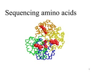
4. Sequence Determination
Frederick Sanger was the first (in 1953),
he sequenced the two chains of insulin
• Sanger's results established that all of the
molecules of a given protein have the same
sequence.
• Proteins can be sequenced in two ways:
- real amino acid sequencing
- sequencing the corresponding DNA in
the gene
4
5. Insulin consists of two polypeptide chains, A and B,
held together by two disulfide bonds.
The A chain has 21 residues and the B chain has 30
residues.
The primary structure of bovine insulin
5
6. Determining the Sequence
1. If there is more than one polypeptide chain, separate them
2. Cleave (reduce) any disulfide bridges
3. Determine amino acid composition of each chain
4. Determine N- and C-terminal residues
5. Cleave each chain into smaller fragments and determine the
sequence of each chain
6. Repeat step 5, using a different cleavage procedure to
generate a different set of fragments
7. Reconstruct the sequence of the protein from the sequences
of overlapping fragments
8. Determine the positions of the disulfide cross-links
6
8. Step 3: Determine Amino Acid
Composition
Complete hydrolysis in 6 N HCl
followed by quantitative analysis
Figure 7-6. Amino acid analysis.
Reverse-phase HPLC separation of
amino acids derivatized with a
fluorescent reagent.
8
9. Determining the Sequence
An Eight Step Strategy
1. If more than one polypeptide chain, separate
2. Cleave (reduce) any disulfide bridges
3. Determine amino acid composition of each chain
4. Determine N- and C-terminal residues
5. Cleave each chain into smaller fragments and determine the
sequence of each chain
6. Repeat step 5, using a different cleavage procedure to
generate a different set of fragments
7. Reconstruct the sequence of the protein from the sequences
of overlapping fragments
8. Determine the positions of the disulfide cross-links
9
10. Step 4: Identify N- and C-terminal residues of polypeptide chains
N-terminal analysis:
– Dansyl chloride method
– Edman's reagent
(phenylisothiocyanate)
If more than 1 end group
is discovered, this means
there is more than 1
polypeptide chain
10
11. The Edman degradation
Detect the N-terminal amino acid
by HPLC or GC-MS
left intact
By subjecting the polypeptide
chain through repeated cycles
of Edman degradation, we can
determine the AA sequence of
the entire polypeptide
Releases N-terminal
AA as
Edman's reagent
11
12. • C-terminal analysis
Enzymatic analysis (carboxypeptidase) is common
– Carboxypeptidase A cleaves any residue except Pro, Arg,
and Lys
– Carboxypeptidase B (hog pancreas) only works on Arg and
Lys
Carboxypeptidases cleave
AAs from the C-terminal end
in a successive fashion
Exhibit selectivity
towards side chains
12
13. Determining the Sequence
An Eight Step Strategy
1. If more than one polypeptide chain, separate
2. Cleave (reduce) any disulfide bridges
3. Determine amino acid composition of each chain
4. Determine N- and C-terminal residues
5. Cleave each chain into smaller fragments and determine the
sequence of each chain
6. Repeat step 5, using a different cleavage procedure to
generate a different set of fragments
7. Reconstruct the sequence of the protein from the sequences
of overlapping fragments
8. Determine the positions of the disulfide cross-links
13
14. Steps 5 and 6:
Fragmentation of the chains
1. Enzymatic fragmentation
– trypsin, chymotrypsin,
clostripain, staphylococcal
protease
2. Chemical fragmentation
– cyanogen bromide
– Trypsin: * Most important
* Cleaves peptide bond after
positively harged AAs
From Lehninger
Principles of Biochemistry
14
15. Step 1: Separation of chains
Subunit interactions depend on weak forces
Separation is achieved with:
- extreme pH
- 8 M urea
- 6 M guanidine HCl
- high salt concentration (usually ammonium sulfate)
Step 2: Cleavage of Disulfide bridges
1) Performic acid oxidation
2) Sulfhydryl reducing agents
- mercaptoethanol
- dithiothreitol or dithioerythritol (Cleland's reagent)
- to prevent recombination, follow with an alkylating agent like
iodoacetate
15
16. Step 7: Reconstructing the Sequence
• Use two or more fragmentation agents in
separate fragmentation experiments
• Sequence all the peptides produced (usually by
Edman degradation)
• Compare and align overlapping peptide
sequences to learn the sequence of the original
polypeptide chain
16
17. The amino acid sequence of a polypeptide chain is
determined by comparing the sequences of 2 sets of
mutually overlapping peptide fragments
1
2
3
4
By joining together 1, 2, 3, & 4 you can get the sequence
17
18. Reconstructing the Sequence
Compare cleavage by trypsin and staphylococcal protease on a
typical peptide:
• Trypsin cleavage:
A-E-F-S-G-I-T-P-K L-V-G-K
• Staphylococcal protease:
F-S-G-I-T-P-K L-V-G-K-A-E
• The correct overlap of fragments:
L-V-G-K A-E-F-S-G-I-T-P-K
L-V-G-K-A-E F-S-G-I-T-P-K
• Correct sequence:
L-V-G-K-A-E-F-S-G-I-T-P-K
18
19. Step 8: Assignment of disulfide bond positions
• Cleave the native protein with its disulfide
bonds intact so as to contain 2 peptide
fragments linked through Cys residues
19
21. Nature of Protein Sequences
• Sequences and composition reflect the function of proteins
• Membrane proteins have more hydrophobic residues, whereas
fibrous proteins may have atypical sequences
• Homologous proteins from different organisms have homologous
sequences
• For example, cytochrome c is highly conserved
21
25. What is DNA Microarray?
• Scientists used to be able to perform genetic analyses of a few genes
at once. DNA microarray allows us to analyze thousands of genes in
one experiment!
25
26. Purposes Of DNA microarray
• So why do we use DNA microarray?
– To measure changes in gene expression levels – two samples’ gene
expression can be compared from different samples, such as from
cells of different stages of mitosis.
– To observe genomic gains and losses. Microarray Comparative
Genomic Hybridization (CGH)
– To observe mutations in DNA.
26
27. The Plate DNA microarray
• Usually made commercially.
• Made of glass, silicon, or nylon.
• Each plate contains thousands of spots, and each spot contains a probe
for a different gene.
• A probe can be a cDNA fragment or a synthetic oligonucleotide, such
as BAC (bacterial artificial chromosome set).
• Probes can either be attached by robotic means, where a needle
applies the cDNA to the plate, or by a method similar to making
silicon chips for computers. The latter is called a Gene Chip.
27
31. STEP 1: Collect Samples.
This can be from a variety of organisms. We’ll use two
samples – cancerous human skin tissue & healthy human
skin tissue
31
32. STEP 2: Isolate mRNA.
• Extract the RNA from the samples. Using either a column, or a
solvent such as phenol-chloroform.
• After isolating the RNA, we need to isolate the mRNA from the rRNA
and tRNA. mRNA has a poly-A tail, so we can use a column
containing beads with poly-T tails to bind the mRNA.
• Rinse with buffer to release the mRNA from the beads. The buffer
disrupts the pH, disrupting the hybrid bonds.
32
33. STEP 3: Create Labelled DNA.
Add a labelling mix to the RNA.
The labelling mix contains poly-T
(oligo dT) primers, reverse
transcriptase (to make cDNA),
and fluorescently dyed
nucleotides.
We will add cyanine 3 (fluoresces
green) to the healthy cells and
cyanine 5 (fluoresces red) to the
cancerous cells.
The primer and RT bind to the
mRNA first, then add the
fluorescently dyed nucleotides,
creating a complementary strand
of DNA
33
34. STEP 4: Hybridization.
• Apply the cDNA we have
just created to a microarray
plate.
• When comparing two
samples, apply both samples
to the same plate.
• The ssDNA will bind to the
cDNA already present on the
plate.
34
35. STEP 5: Microarray Scanner.
The scanner has a laser, a computer,
and a camera.
The laser causes the hybrid bonds to
fluoresce.
The camera records the images
produced when the laser scans the
plate.
The computer allows us to immediately
view our results and it also stores our
data.
35
36. STEP 6: Analyze the Data.
GREEN – the healthy sample hybridized more
than the diseased sample.
RED – the diseased/cancerous sample
hybridized more than the nondiseased sample.
YELLOW - both samples hybridized equally
to the target DNA.
BLACK - areas where neither sample
hybridized to the target DNA.
By comparing the differences in gene
expression between the two samples, we can
understand more about the genomics of a
disease.
36
37. Benefits.
• about $60,000 for an arrayer and scanner setup.
• The plates are convenient to work with because they are small.
• Fast - Thousands of genes can be analyzed at once.
37
38. Problems.
• Oligonucleotide libraries – redundancy and
contamination.
• DNA Microarray only detects whether a gene is
turned on or off.
• Massive amounts of data.
http://www.stuffintheair.com/very-big-problem.html
38
41. The Future of DNA Microarray.
• Gene discovery.
• Disease diagnosis: classify the types of cancer on the basis of the
patterns of gene activity in the tumor cells.
• Pharmacogenomics = is the study of correlations between therapeutic
responses to drugs and the genetic profiles of the patients.
• Toxicogenomics – microarray technology allows us to research the
impact of toxins on cells. Some toxins can change the genetic profiles
of cells, which can be passed on to cell progeny.
41
43. 43
Mutagenesis
Mutagenesis -> change in DNA sequence
-> Point mutations or large modifications
Point mutations (directed mutagenesis):
- Substitution: change of one nucleotide (i.e. A-> C)
- Insertion: gaining one additional nucleotide
- Deletion: loss of one nucleotide
44. 44
Consequences of point mutations within a coding
sequence (gene) for the protein
Silent mutations:
-> change in nucleotide sequence
with no consequences for protein
sequence
-> Change of amino acid
-> truncation of protein
-> change of c-terminal part of protein
-> change of c-terminal part of protein
45. 45
Applications of directed mutagenesis
-> site-directed mutagenesis
-> point mutations in
particular known area
result mutated DNA
(site-specific)
46. 46
General strategy for
directed mutagenesis
Requirements:
- DNA of interest (gene or promoter) must be
cloned
- Expression system must be available -> for
testing phenotypic change
47. 47
Protein Engineering
-> Mutagenesis used for modifying proteins
Replacements on protein level -> mutations on DNA level
Assumption : Natural sequence can be modified to
improve a certain function of protein
This implies:
• Protein is NOT at an optimum for that function
• Sequence changes without disruption of the structure
• (otherwise it would not fold)
• New sequence is not TOO different from the native sequence
(otherwise loss in function of protein)
Objective: Obtain a protein with improved or new
properties
48. 48
Rational Protein Design
Site –directed mutagenesis !!!
Requirements:
-> Knowledge of sequence and preferable Structure
(active site,….)
-> Understanding of mechanism
(knowledge about structure – function relationship)
-> Identification of cofactors……..
53. Screening: Basis for all screening & selection methods
Expression Libraries
->link gene with encoded product which is responsible for enzymatic
activity
54. Low-medium throughput screens
-> Detection of enzymatic activity of
colonies on agar plates or ”crude cell
lysates” -> production of fluorophor
or chromophor or halos
-> Screen up to 104 colonies
-> effective for isolation of enzymes
with improved properties
-> not so effective for isolation of
variants with dramatic changes of
phenotype
Lipase: variants on Olive oil plates
With pH indicator (brilliant green)
55. 55
Protein Engineering - Applications
Site-directed mutagenesis -> used to alter a single property
Problem : changing one property -> disrupts another
characteristics
Directed Evolution (Molecular breeding) -> alteration of
multiple properties
57. Tools in BIOTECHNOLOGY
• One of the basic tools of modern biotechnology is
gene splicing.
• This is the process of removing a functional DNA
fragment ( a gene) from one organism and
combining it with the DNA of another organism
to study how the gene works.
• The desired result is to have the new organisms
carry out the expression of the gene that has
been inserted.
57
58. What are the Applications of Genetic Engineering?
Transgenic Organisms, GMO (Genetically Modified Organisms)
58
59. Genetic engineering is a technique that makes it possible to transfer DNA
sequences from one organism to another
1. What is genetic engineering?
2. What are transgenic organisms?
Organisms that contain genes from other species
Examples of Transgenic organisms
Transgenic
microorganisms
Transgenic
plants
Transgenic
animals
59
60. They reproduce rapidly and are easy to grow
3. Why to use transgenic bacteria?
Production of insulin, growth hormone, and clotting factor
4. How do humans benefit from transgenic microorganisms?
Transgenic microorganisms
Production of
•Substances to fight cancer
•Plastics
•Synthetic fibers
•Food production
5. What do we expect to achieve in the future?
60
61. How to Transform Bacteria?
4. The recombinant
plasmid replicates and
a large number of
identical bacteria are
cloned. They produce
human insulin.
2. Remove a plasmid from a
bacterium and treated with
3. Bind the plasmid with
the human gene to
form a recombinant
plasmid. Then the
recombinant plasmid
is re-inserted back into
the bacterium
1. Remove the DNA from a human body cell,
then isolate the human gene of insulin using
restriction enzymes.
61
62. 7. How do humans benefit from transgenic plants?
• Increase crop productivity
• Corps able to resist weed-killing chemicals
•Crops that produce a natural insecticide, not need to spray pesticides
6. What are transgenic plants?
Plants that contain genes from other species
Transgenic Plants
Transgenic Corn
62
63. • Golden rice is a variety of rice produce through genetic
engineering to include vitamin in the edible parts of rice.
• Golden rice was developed as a fortified food to be used in areas
where there is a shortage of dietary vitamin A.
• No variety is currently available for human consumption.
Although golden rice was developed as a humanitarian tool, it has
met with significant opposition from environmental and anti-
Genetically Modified Organism (GMO)
Golden Rice
63
64. 8. What are transgenic animals?
Animals that contain genes from other species
9. How do humans benefit?
• Increase meat productivity
•Livestock with extra copies of growth hormone
genes to grow faster and produce leaner meat
• Transgenic chickens resistant to bacterial
infections
Transgenic Animals
10. What do we expect to achieve in the future from
transgenic animals?
64