
Empfohlen
Weitere ähnliche Inhalte
Was ist angesagt?
Was ist angesagt? (20)
Andere mochten auch
Andere mochten auch (10)
Ähnlich wie Bone disorders
Ähnlich wie Bone disorders (20)
Kürzlich hochgeladen
Kürzlich hochgeladen (20)
Bone disorders
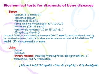
- 1. Biochemical tests for diagnosis of bone diseases Serum •Calcium (2 -2.6 mmol/l) •corrected calcium •Albumin (35-50 g/l ) •serum alkaline phosphatase (30 -120 IU/l) •Phosphate (0.8-1.5 mmol/l) •parathyroid hormone ( 10 to 55 pg/mL. ) •25-hydroxy vitamin D Serum 25-OHD concentrations between 50-75 nmol/L are considered healthy but optimal vitamin D status is when serum concentrations of 25-OHD are 75 nmol/L (30 micrograms/L) or more. Urine •Calcium •Phosphate •urinary markers, including hydroxyproline, deoxypyridinoline, C- telopeptide, and N -telopeptide Correct total Ca( mg/dL) =total Ca ( mg/dL) + 0.8( 4-alb[g/dL[ (
- 4. Arthritis is not a single disease; it is an informal way of referring to joint pain or joint disease. There are more than 100 different types of arthritis and related conditions. TYPES OF ARTHRITISTYPES OF ARTHRITIS • Degenerative Arthritis (Osteoarthritis) • Inflammatory Arthritis (rheumatoid ) • Infectious (Septic) Arthritis • Metabolic (Gouty) Arthritis symptoms Common arthritis joint symptoms include swelling, pain, stiffness and decreased range of motion. Symptoms may be mild, moderate or severe
- 6. Osteoporosis is a bone disease that occurs when the body loses too much bone, makes too little bone, or both. Osteoporosis means “porous bone”. Osteoporotic bones have lost density or mass and contain abnormal tissue structure. As bones become less dense, they weaken and are more likely to break
- 7. Postmenopausal osteoporosis occurs in 5% to 20% of women, with a peak incidence in the 60s.The incidence in women is eight times higher than that in men. Estrogen deficiency is thought to underlie this form of osteoporosis, rendering the skeleton more sensitive to parathyroid hormone (PTH), resulting in increased calcium resorption from bone. This in turn decreases PTH secretion, 1,25- dihydroxyvitamin D production, and calcium absorption and ultimately causes loss of trabecular bone, leading to vertebral crush fractures and Colles' fractures. Senile osteoporosis occurs in women or men more than 70 years of age and usually is associated with decreased bone formation along with decreased ability of the kidney to produce 1,25(OH)2D3. The vitamin D deficiency results in decreased calcium absorption, which increases the PTH level and therefore bone resorption. In type 2 osteoporosis, cortical and trabecular bone is lost, primarily leading to increased risk of hip, long bone, and vertebral fractures.
- 8. Treatment of symptomatic osteoporosis has had limited success. Prevention is preferable to treatment, since no therapy fully restores lost bone mass. •PHARMACOLOGIC TREATMENT:PHARMACOLOGIC TREATMENT: •Hormonal Replacement Therapy (HRT) such as Estrogen •Non-hormonal Replacement Therapy (NHRT) such as biphosphonates (alendronate), calcitonin, selective estrogen-receptor modulators (raloxifene), and fluoride •Others ( testosterone, human parathyroid hormone & analougs(Teriparatide), and growth hormone) (NON-PHARMACOLOGIC(NON-PHARMACOLOGIC PREVENTION)PREVENTION) • The combination of calcium (1.2 g/day) with vitamin D3 (800 IU/day) • Exercise regularly • Prevent falls • Avoid smoking & excessive alcohol • Maintain an appropriate body weight
- 9. This type of osteoporosis is associated with a variety of conditions, including: Hormonal imbalances (eg, cushing's syndrome); Cancer (notably multiple myeloma); Gastrointestinal disorders (especially IBD causing malabsorption); Drug use (eg, corticosteroids, cancer chemotherapy, anticonvulsants, heparin, barbiturates, valporic acid, gonadotropin-releasing hormone excessive use of aluminum- containing antacids); Pathological conditions: chronic renal failure; hyperthyroidism; hypogonadism in men; immobilization,rheumatoid arthritis); and Poor nutrition (including malnutrition due to eating disorders).
- 10. Osteomalacia is a disorder marked by inadequate or defective mineralization of the skeleton, resulting in soft or fragile bones. When the disease occurs in children before the growth plates have closed, it is known as rickets and tends to produce obvious skeletal deformities. It typically occurs either when there are insufficient amounts of vitamin D in the diet or, when the body is unable to properly absorb and metabolize vitamin D, which is essential for the absorption of the calcium needed to maintain strong, healthy bones. It can also occur with calcium and phosphorus deficiency plus other genetics disorders Vitamin D deficiency is most often caused •insufficient exposure to sunlight and nutritional deficiency •Gastrointestinal malabsorption •Liver and kidney disease * drugs
- 11. Hypocalcaemic seizures or tetany, particularly in the neonatal period. From the age of 6 months, children often present with bony deformities of the chest, pelvis and skull, delayed dentition, and bone pain. Children may be irritable and manifest impaired growth of all body organs. Increased susceptibility to infections and respiratory symptoms. Severe vitamin D deficiency can result in cardiomyopathy and potentially fatal heart failure. Widespread bone pain and tenderness (especially low back pain and in the hips), proximal muscle weakness & lethargy are the main features of vitamin D deficiency in adults. Skeletal deformity The patient may experience signs of hypocalcaemia & multiple fractures which are bilateral and symmetrical Low bone density on dual-energy X-ray rickets osteomalacia
- 12. • BIOCHEMICAL FINDINGS Blood biochemistry: renal function, electrolytes (including serum calcium and phosphate), LFTs, parathyroid hormone level: More than 80% of adults with osteomalacia have a high concentration of serum alkaline phosphatase. Hypocalcaemia (NV:9-11mg/dl), hypomagnesaemia and hypophosphataemia (NV: 5-7mg/dl) may be present, depending on the severity and chronicity of the disease and the patient's dietary calcium intake. Secondary hyperparathyroidism is typical in hypocalcemic rickets. Full Blood Count: Anaemia suggests possible malabsorption. Urine microscopy to help determine whether the patient has underlying chronic kidney disease. Vitamin D status is most reliably determined by assay of serum 25- hydroxyvitamin D (25-OHD): Vitamin D deficiency: individuals with symptomatic osteomalacia or rickets have serum 25-OHD of less than 25 nmol/L (10 micrograms/L)
- 13. TREATMENT General management Education: dietary advice (refer to a dietician). Encourage exposure to sunlight. Vitamin D supplementation. Calcium supplementation. Treatment of any underlying condition. Treatment of pain. Orthopaedic intervention may be required. Children Oral calciferol in the form of either ergocalciferol or colecalciferol is the treatment of choice for children with rickets (6000 IU( Calcium supplementation is advisable during the first weeks of therapy. Adult Calciferol in a daily dose of 10000 IU
- 14. CAUSES The cause of Paget disease is unknown. Both genetic and environmental factors have been implicated. SYMPTOMS •Most people who have Paget's disease of bone have no symptoms. When symptoms occur, the most common complaint is • bone pain. •Joint pain Nerve problems:
- 15. • PATHOGENESIS Three phases of Paget disease have been described: lytic, mixed lytic&blastic, and sclerotic. Paget disease begins with the lytic phase, in which normal bone is resorbed by osteoclasts that are more numerous and larger than normal osteoclasts. Bone turnover rates increase to as much as 20 times normal. The second phase, the mixed phase, is characterized by rapid increases in bone formation from numerous osteoblasts. Although increased in number, the osteoblasts remain morphologically normal. The newly made bone is abnormal, however, with collagen fibers deposited in a haphazard fashion rather than linearly, as with normal bone formation. In the final phase of Paget disease, the sclerotic phase, bone formation dominates and the bone that is formed has a disorganized pattern (woven bone) and is weaker than normal adult bone. This woven bone pattern allows the bone marrow to be infiltrated by excessive fibrous connective tissue and blood vessels, leading to a hypervascular bone state.
- 16. • BIOCHEMICAL DIAGNOSIS Measurement of serum alkaline phosphatase—in some cases, bone- specific alkaline phosphatase (BSAP)—can be useful in the diagnosis of Paget disease. Elevated levels of urinary markers, including hydroxyproline, deoxypyridinoline, C-telopeptide, and N -telopeptide, may help identify patients with Paget disease. Procollagen I N -terminal peptide (PINP) has emerged as a sensitive serum marker for bone formation. Hypercalcemia or hypercalciuria may develop with immobilization or coincident primary hyperparathyroidism. Hyperuricemia from Paget disease is more common in men than women and appears to be caused by the increased turnover of nucleic acids from high bone turnover. Serum total acid phosphatase is an osteoclastic enzyme that may be elevated in active Paget disease.