
Empfohlen
Weitere ähnliche Inhalte
Was ist angesagt?
Was ist angesagt? (20)
Ähnlich wie Urinalysis a comprehensive review
Ähnlich wie Urinalysis a comprehensive review (20)
Kürzlich hochgeladen
Kürzlich hochgeladen (20)
Urinalysis a comprehensive review
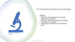
- 1. Outline: • Collection and Handling of urine sample • Types of urine specimen • Physical & chemical examination of urinalysis • Discrepancies in urinalysis • Microscopic examination of urine sediment • Quiz… Alyazeed hussein, BSc-SUST A Comprehensive Review of Urinalysis Medical Laboratory Science
- 2. Urinalysis A complete urinalysis is composed of multiple tests, including macroscopic, physical, chemical, and microscopic examination. Specimen Collection and Handling: Use clean, dry container, receive and analyze the sample within 2 hours!!? Types of urine specimen: 1. Random urine: Most common type, for routine tests. 2. First morning: Concentrated specimen used for routine screening, pregnancy test. 3. Fasting & 2-Hour postprandial: for DM(insulin monitoring), 2 hours after eating. 4. 24-Hour: Collected over a period of 24 hours for creatinine clearance, Glomerular Filtration Rate (GFR). 5. Midstream clean-catch (MSU): urine collected in the middle of urination; used for bacterial culture. 6. Catheterized: Collected from a tube placed through the urethra into the bladder; used for bacterial culture and routine screening. 7. Suprapubic aspiration: Needle inserted into the bladder through the abdominal wall; used for bacterial culture and cytologic testing. 8. Pediatric collection: Use small, clear plastic bags with adhesive to adhere to the genital area. Alyazeed hussein, BSc-SUST
- 3. URINE SPECIMEN STORAGE AND HANDLING • Most common form of preservation, refrigeration at 2°C to 8°C, is suitable for the majority of specimens. Any urine specimen for microbiological studies should be refrigerated immediately if it cannot be transported directly to the laboratory, the specimen remains suitable for culture for up to 24 hours. • Before testing, urine must be brought to room temperature. • Other preservatives are: Boric acid (acceptable for culture), Thymol (cells & casts), formalin (cellular preservative) Alyazeed hussein, BSc-SUST
- 4. Physical examination of urine (Color, Appearance and Specific gravity) A. color: 1. Pale yellow & yellow: normal color of urine(urochrome: urobilin). 2. Colorless: may due to dilution, or Diabetes Meletus. 3. Dark yellow: may due to dehydration, or First morning( concentrated), usually with high specific gravity. 4. Orange or dark yellow-amber: Bilirubinemia occurs from liver problems, such as hepatitis > bilirubinuria, yellow foam forms when urine is shaken due to the presence of conjugated bilirubin. Smith iodine test positive (green ring), hay's test (sulfur powder) positive. 5. Red/pink: (RBCs, (hemoglobin-brown and myoglobin-muscle) or menstrual contamination. 6. Green/blue: medication or pseudomonas. Note that! Uroerythrin adds a slight pink pigment, mostly apparent following refrigeration, when the pigment attaches to precipitated amorphous urates. Alyazeed hussein, BSc-SUST
- 6. B. Appearance and clarity: 1. Clear: normal. 2. Slightly cloudy: May be due to the presence of low numbers of formed elements. 3. Cloudy or milky: presence of amorphous, crystals, pus cells, epithelial cells, also due to Chyluria (W. bancrofti, lymphatic filariasis). Bence jones protein: light chain of immunoglobulins in urine(multiple myeloma) = (heat test). C. Specific Gravity: determines the kidney's reabsorption ability. • Normal range: 1.015 to 1.030 • Low specific gravity: loss of the kidney's ability to concentrate urine or presence of disease, It can also be found normally with large fluid intake. • High specific gravity may result from adrenal insufficiency, diabetes mellitus(glycosuria). Note that! If urine pH >8.0, add 0.005 to the reading. Alyazeed hussein, BSc-SUST
- 7. Chemical examination of urine multi-parameter reagent strip (Multistix) Procedure: MUST BE FOLLOWED EXACTLY TO ACHIEVE RELIABLE TEST RESULTS 1. Collect FRESH urine specimen in a clean, dry container. Mix well immediately before testing. 2. Remove one strip from bottle and replace cap. Completely immerse reagent areas of the strip in FRESH urine and remove immediately to avoid dissolving out reagents 3. While removing, run the edge of the strip against the rim of the urine container to remove excess urine. Hold the strip in a horizontal position to prevent possible mixing of chemicals from adjacent reagent areas/or contaminating the hands with urine. 4. Compare reagent areas to corresponding Color chart on the bottle label at the time specified. Hold strip close to color blocks and match carefully. Avoid laying the strip directly on the Color chart as this will result in the urine soiling the chart. For optimal results, read the ketone test at 15 seconds after dipping; read the bilirubin test at 20 seconds; glucose at 30 seconds; blood at 40 seconds; urobilinogen at 45 seconds; and specific gravity from 45 to 60 seconds after dipping. Alyazeed hussein, BSc-SUST
- 9. False-negativeFalse-positiveTest Not mixed will, high proteinuria, glucosuria, Boric acid Expired strip, formalinLeukocyte esterase Formalin, lack of nitrateImproper storage (bacterial proliferation) Nitrite Formalin, high vitamin CPeroxidasesBlood High ketones, high ascorbic acid Alkaline urine, oxidizing agent : bleach, Glucose Ascorbic acid, boric acid, sample exposed to light Colored substancesBilirubin Boric acid, formalin, hypochlorite, delay in examination, volatilization Highly pigmented urine, drugs Ketones Bence-jones protein, sperms Alkaline urine, drugsProtein Formalin, hypochlorite, antibiotics Sulfonamide, drugs, beetUrobilinogen Discrepancies Alyazeed hussein, BSc-SUST
- 10. Discrepancies • Renal glycosuria: presence of glucose in urine with normal blood glucose level! Due to defect in renal tubular dysfunction or by glucagon hormone. (>180mg/dl) • Strip positive for blood with absence of RBCs microscopically: hemoglobinuria(Hb from lysed RBCs) or diluted urine (Ghost RBCs) pH > 7, SG < 1.010, handling, old sample, high temperature, peroxidase positive bacteria (E. coli), myoglobin, too fast centrifugation, • Sterile pyuria, presence of pus in urine with no bacteria: using of antibiotics. • RBCs can be confused with yeast cells or oil droplets. Diluted acetic acid can be used to lyse RBCs, leaving only yeast, oil droplets, and WBCs. • Positive leukocyte esterase with Glitter cells or absence of WBCs microscopically: dilute alkaline urine. • Excessive shaking or taping of sediment against edges of table to mix it up> cause the casts to dissolve. • Note that!! Native urine: urine not centrifuged > counting chamber method > small volume of urine only. Alyazeed hussein, BSc-SUST
- 11. • 10 to 15 mL (12ml) Centrifuged at 400 – 450 g (RCF), 1500 – 2000 (1600) (RPM) for 5 mins hold the test tube upside down and count to 3, then turn the test tube again and stand it upright mix drop (20 μL) in a slide + glass cover slip (carefully to avoid air bubbles) examine. • Report RBCs/WBCs using high-power magnification (i.e., high-power field [hpf]), report casts and crystals using low-power magnification (i.e., low-power field [lpf]). • Normal Urines: Contain 0-4 RBCs (hpf), 0-3 WBCs (hpf), 0-2 hyaline casts (lpf), several epithelial cells (hpf), some types of crystals, and mucus. • Casts have a tendency to locate near the edges of the cover slip (LPF scanning around the cover slip). Microscopic examination Alyazeed hussein, BSc-SUST
- 13. Alyazeed hussein, BSc-SUST Sediment constituents such as bacteria, yeast cells, crystals, and spermatozoa are not counted, but instead given as crosses
- 14. Urine cells Alyazeed hussein, BSc-SUST
- 15. Isomorphic RBCs Dysmorphic RBCs, Have cellular blebs (mickey mouse ear), associated with glomerular bleeding (glomerulonephritis). Ghost RBC In dilute urine, absorb water, swell, and lyse rapidly, releasing hemoglobin. Examined under reduced light. RBCs, Seen in kidney or urinary tract diseases or menstrual blood contamination, <3 /HPF is normal Alyazeed hussein, BSc-SUST
- 16. Pus cells, neutrophils in acute infections, eosinophils in interstitial nephritis, lymphocytes in renal transplantation Alyazeed hussein, BSc-SUST Elongated WBCs
- 17. Macrophages, Contain digested material, lipids, seen in chronic inflammation and radiation therapy Alyazeed hussein, BSc-SUST
- 18. Renal tubular cells (RTEs) line the nephron, seen in renal tubular damage (acute tubular necrosis, viral infection, or renal transplant rejection) Alyazeed hussein, BSc-SUST
- 19. Sperms, found in men with retrograde ejaculation, post-prostatectomy, or in sample collected soon after ejaculation Alyazeed hussein, BSc-SUST
- 20. Squamous epithelial cells, from urethra, skin and vaginal mucosa Alyazeed hussein, BSc-SUST
- 21. Clue cell: Gardnerella vaginalis Alyazeed hussein, BSc-SUST
- 22. Tadpole cells or caudate cells Alyazeed hussein, BSc-SUST Transitional epithelial cells (Urothelial cells), from renal pelvis, ureter and bladder, increased in infections, renal stones, bladder cancer, and post-catheterization. Chronic urinary tract infection
- 24. Transitional cell carcinoma Alyazeed hussein, BSc-SUST
- 25. Decoy cells: viral infection (polyoma virus) or malignancy Alyazeed hussein, BSc-SUST
- 26. Oval fat bodies: with marked proteinuria, acute tubular necrosis or nephrotic syndrome Alyazeed hussein, BSc-SUST
- 27. Urine casts Alyazeed hussein, BSc-SUST
- 28. Fatty casts, with marked proteinuria, acute tubular necrosis or nephrotic syndrome Oval fat bodies Alyazeed hussein, BSc-SUST
- 29. Renal tubular epithelial cells (RTEs) casts: Found in tubular damage, acute tubular necrosis, or renal transplant rejection Alyazeed hussein, BSc-SUST
- 30. White blood cells (WBCs) casts: in pyelonephritis, lupus or allergic nephritis Alyazeed hussein, BSc-SUST
- 31. Granular casts: found in normal urine following stress, strenuous exercise, or in renal diseases Alyazeed hussein, BSc-SUST
- 32. Hyaline casts: seen in normal urine following strenuous exercise also seen in renal diseases Alyazeed hussein, BSc-SUST
- 34. Red blood cells (RBCs) casts: indicates serious renal disease, acute nephritis, glomerular injuries, or glomerulonephritis and malignant hypertension Alyazeed hussein, BSc-SUST
- 35. Hemoglobin casts Alyazeed hussein, BSc-SUST
- 36. Myoglobin cast Alyazeed hussein, BSc-SUST
- 37. Bilirubin/bile cast Alyazeed hussein, BSc-SUST
- 38. Waxy cast: found in sever renal disease or acute glomerulonephritis Alyazeed hussein, BSc-SUST
- 39. Broad cast: poor prognosis, renal failure Alyazeed hussein, BSc-SUST
- 40. Crystals cast: amorphus Alyazeed hussein, BSc-SUST
- 41. Myeloma cast Alyazeed hussein, BSc-SUST
- 42. Mixed cast (yeast, WBC, granular ,hyaline) Alyazeed hussein, BSc-SUST Bacterial cast
- 43. Urine crystals at acid pH Alyazeed hussein, BSc-SUST
- 44. Cystine crystals: in patients with cystinosis, a congenital condition, most common aminoaciduria Alyazeed hussein, BSc-SUST
- 45. Sulfonamide crystals: form renal calculi in dehydrated patients, infrequently seen today Alyazeed hussein, BSc-SUST
- 46. Ampicillin crystals Alyazeed hussein, BSc-SUST
- 47. Drug crystals Alyazeed hussein, BSc-SUST
- 48. Uric acid crystals: associated with hyperuricemia, uric acid stone, tumor lysis syndrome, gouty nephropathy Alyazeed hussein, BSc-SUST
- 52. Found in concentrated urine associated with fever and dehydration, may hide bacteria, casts and crystals Amorphus urate (acidic urine) Amorphus phosphate (alkaline urine) Alyazeed hussein, BSc-SUST
- 53. Bilirubin crystals: in urine contain high amounts of bilirubin Alyazeed hussein, BSc-SUST
- 54. Calcium oxalate crystals: patients consume tomatoes, apples, oranges (rich in oxalic acid) may have these crystals in urine. Found in renal calculi. Alyazeed hussein, BSc-SUST
- 55. Cholesterol crystals: associated with nephrotic syndrome Alyazeed hussein, BSc-SUST
- 56. Hippuric acid: in person who eat a diet rich in benzoic acid, may also seen in liver diseases Alyazeed hussein, BSc-SUST
- 57. Leucine crystals: in liver disease, appears in association with tyrosine crystals Alyazeed hussein, BSc-SUST
- 58. Tyrosine crystals: seen in hepatic failure Alyazeed hussein, BSc-SUST
- 59. Alyazeed hussein, BSc-SUST Acid urate: old sample not significant
- 60. Sodium urate: no clinical significance Alyazeed hussein, BSc-SUST
- 61. Urine crystals at neutral or alkaline pH Alyazeed hussein, BSc-SUST
- 62. Calcium carbonate: no clinical significance Alyazeed hussein, BSc-SUST
- 63. Calcium phosphate: in normal urine may cause renal calculi Alyazeed hussein, BSc-SUST
- 64. Ammonium biurate crystals (thorn apple): seen in old urine sample (teaching sample), have no clinical significance Alyazeed hussein, BSc-SUST
- 65. Ammonium magnesium phosphate (triple phosphate, struvite crystals, coffin lid), appear fern-like feathery (dissolved), normal in urine but may be associated with bacterial growth (Proteus). Calculi seen in chronic UTI Alyazeed hussein, BSc-SUST
- 67. Bacteria: in urinary tract infection, or may be a vaginal or fecal contaminant Alyazeed hussein, BSc-SUST
- 68. Yeast/fungi: candida albicans, appears as budding yeast or pseudohyphae, may be from a vaginal contaminant, bladder or kidney inaction Alyazeed hussein, BSc-SUST
- 69. Adult female of E. vermicularis Eggs of E. vermicularis Trophozoites of T. vaginalis Egg of S. haematobium Alyazeed hussein, BSc-SUST
- 71. Alyazeed hussein, BSc-SUST Oil droplets Corpora amylacea Glass artifacts
- 73. Mucus threads: infections of lower urinary and vaginal tract. Alyazeed hussein, BSc-SUST
- 74. Pollen grains: from contaminate urine and urine container Alyazeed hussein, BSc-SUST
- 75. Alyazeed hussein, BSc-SUST Air bubbles Starch globules
- 76. Alyazeed hussein, BSc-SUST Fecal contamination Rotifer Alternaria Pubic lice Dust mite
- 78. • This has been a presentation of Alyazeed Hussein. • Thanks for your attention and kind patience. • Any questions, additions, or comments? Alyazeed hussein, BSc-SUST
- 79. References • Urinalysis Benchtop Reference Guide, CAP. • Kjeldsberg's Body Fluid Analysis, ASCP. • A Handbook Of Routine Urinalysis, Sister Laurine Graff. • Textbook of urinalysis and body fluids, Landy J. McBride-Lippincott. • Graff's textbook of urinalysis and body fluids, Lillian A. Mundt, 3rd e • Success in clinical laboratory science, ANNA P. CIULLA, 4th e • www.researchgate.net • www.sciencedirect.com • www.diagnostics.roche.com Alyazeed hussein, BSc-SUST