Leukemia in Children
•Als PPTX, PDF herunterladen•
70 gefällt mir•33,297 views

Childhood Leukemias

Empfohlen
Weitere ähnliche Inhalte
Was ist angesagt?
Was ist angesagt? (20)
Ähnlich wie Leukemia in Children
Ähnlich wie Leukemia in Children (20)
Mehr von CSN Vittal
Mehr von CSN Vittal (20)
Kürzlich hochgeladen
Kürzlich hochgeladen (20)
Leukemia in Children
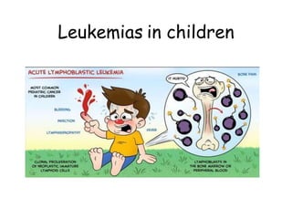
- 2. Leukemia in Children Leukemias are the most common cancers affecting children. Acute lymphoblastic leukemia (ALL) accounts for 73%, Acute myeloid leukemia (AML) accounts for approximately 18%. Chronic myeloid leukemia(CML) is rarely seen, accounting for less than 4%.
- 3. Epidemiology ALL: 3–4 cases per 100,000 white children Peak incidence between 2 and 5 years of age Accounts for 25–30% of all childhood cancers In ALL, boys more commonly affected than girls Incidence of AML is similar for all age groups
- 4. 10/30/2017 4 Factors predisposing ALL GENETIC ENVRONMENTAL Down’s Ionising radiation Diamond Blackfan syndrome Drugs NF Type1 - alkylating agents Ataxia telengiectasia - nitrosourea Turner - epipodophyllotoxin Klinefelter - benzene exposure Fanconi anemia Advanced maternal age Blooms syndrome Paternal smoking
- 5. Pathology • Acute leukemia –is characterized by clonal expansion of immature hematopoietic or lymphoid precursors. • Chronic leukemia –refers to conditions characterized by the expansion of mature marrow elements
- 6. CLINICAL FEATURES General Systemic Effects 1. Fever (60%). 2. Lassitude (50%) 3. Pallor (40%) Hematologic Effects Arising from Bone Marrow Invasion 1. Anaemia – pallor, fatigability, tachycardia, dyspnoea & CHF 2. Neutropenia – fever, ulceration of buccal mucosa and infection. 3. Thrombocytopenia – petechial, purpura, easy bruisability, bleeding from mucous membrane and internal bleeding.
- 7. CLINICAL FEATURES Clinical Manifestations Arising from Lymphoid System Infiltration 1. Lymphadenopathy 2. Splenomegaly. 3. Hepatomegaly
- 8. CLINICAL FEATURES Clinical Manifestations of Extramedullary Invasion –CNS‐ ICT symptoms, seizures –Genitourinary ‐ painless testicular swelling –Bone joints‐ bone pain –Skin ‐ bleeds –Git ‐ bleeds
- 9. CLINICAL FEATURES – Childhood Cancer CONTINUOUS FEVER, WEIGHT LOSS HEADACHES, EARLY MORNING VOMITION INCREASED SWELLING OR PERSISTENT PAIN IN BONES, JOINTS, BACK OR LEGS LUMP OR MASS – ABDO, NECK, CHEST, PELVIS, ARMPITS DEVELOPMENT OF RASH, BLEEDING, BRUISION CONSTANT / RECURENT INFECTIONS AWHITISH COLOR BEHIND PUPIL NAUSEA – PERSISTANT OR VOMITING WITHO OR W/O SEIZURE CONSTANT TIREDNESS EYE OR VISON CHANGES RECURRENT OR PERSISTENT FEVER
- 10. CLASSIFICATION • Light microscopy (FAB) – morphology L1, L2, L3 • Cytochemistry ‐ staining ‐ MPO, ESTERASE • Immunophenotyping (WHO) – CD numbering • Cytogenetics ‐ chromosome/gene rearrangement
- 11. Cytologic Features L1 L2 L3 Cell Size Small cells predominate Large, heterogenous in size Large and heterogenous Nuclear chromatin Homogenous Variable, heterogenous Finely stippled & homogenous Nuclear shape Regular, occasional clefting or indentation Irregular, clefting & indentation common Regular, oval to round Nucleoli Not visible, or small & inconspicuous One or more present, often large Prominent, one or more vesicular Amount of cytoplasm Scanty Variable, often moderately abundant Moderately abundant Basophilia of cytoplasm Slight or moderate, rarely intense Variable, deep in some Very deep Cytoplasmic vacuolation Variable Variable Often prominent
- 12. FAB types of acute lymphoblastic leukemia (ALL). A) L1 morphology with uniform‐sized blasts. B) L2 ALL with more blast cell variation. ( C) L3 blasts with more clumped nuclear chromatin, nucleoli, basophilic cytoplasm, and cytoplasmic vacuoles.
- 13. Imjunologic Subtype % of cases FAB Subtype Cytogentetic abnormalities Pre B ALL 75 L1, L2 T(9;22), t(4;11), t(1;19) T Cell ALL 20 L1, L2 14q11 or 7q34 Mature B Cell ALL (Burkitt Leukemia) 5 L3 T(8;14)
- 14. INVESTIGATIONS • Blood count • Haemoglobin: Moderate to marked reduction • Blood smear: Blasts are present on blood smear. Very few to none (in patients with leukopenia). • White blood cell count: Low, normal, or increased • Thrombocytopenia: 92% of patients have platelet counts below normal. Very few to none (in patients with leukopenia).
- 15. INVESTIGATIONS – Bone Marrow • Leukemia must be suspected when the bone marrow contains more than 5% blasts. • The hallmark of the diagnosis of acute leukemia is the blast cell, are relatively undifferentiated cell with diffusely distributed nuclear chromatin, one or more nucleoli and basophilic cytoplasm.
- 16. Bone marrow changes Normal marrow Entire marrow replaced by blast 10/30/2017 16
- 17. IAP UG Teaching slides 2015‐1618 A, Acute lymphoblastic leukemia with CD 22,19,10 positiv Acute lymphoblastic leukemia with CD 22,19,10 positivity
- 18. INVESTIGATIONS • Chest radiograph: Mediastinal mass in T‐cell leukemia. • Blood chemistry: Electrolytes, blood urea, uric acid, • Liver function tests, Immuno globulin levels. • Coagulation profile: Decreased coagulation factors that frequently occur with AML are: hypofibrinogenemia, factors V, IX and X.
- 19. INVESTIGATIONS - CSF – CNS1 , < 5 WBCs/mm3 with no blasts; – CNS2 , < 5 WBCs/mm3, a positive "cytospin" for blasts; – CNS3, > 5 WBCs/mm3, blasts on cytocentrifuge slide Cerebrospinal fluid: Chemistry and cells.
- 20. TREATMENT • Three phases: 1. remission induction, 2. consolidation (or intensification), and 3. continuation (or maintenance). • Protocol adopted depends on the institution • Modified BFM or COG protocol is often the choice
- 21. INDUCTION • Prednisolone 60 mg/m2/day • Inj.VCR 1.5mg2/day • Inj DNR 30 mg/m2 • L ASPARGINASE 10000u/m2 • MTX I/T
- 22. INTENSIFICATION & CNS PROPHYLAXIS • Inj CYCLOPHOSPHAMIDE 1gm/m2 • InJ CYTARABINE 75mg/m2/day • 6 MP 60 mg/m2/d • I/T MTX • CRANIAL IRRADIATION
- 23. MAINTENANCE • Inj. VCR 1.5 mg/m2 one in a month • Tab PREDNISOLONE 60 mg/m2 for one wk • T.6MP 50 mg/m2 p.o daily • T.MTX 20 mg/m2 p.o wkly The optimal duration of therapy remains unknown. Most investigators continue to treat patients for 2 to 3 years, based on results of older studies
- 24. FOLLOW UP If the patient completes chemotherapy for 2 years without relapse-stop chemo and follow up. No relapse within 5 years-can be declared as cured.
- 25. SUPPORTIVE CARE • A total of 10 mg/kg/day of allopurinol in divided doses is given in all cases before the commencement of antileukemic drugs. • When the blast cell count is more than 50,000/mm3 or there are large tumour masses, allopurinol is obligatory, together with a fluid intake of 2–3 L/m2/day
- 26. SUPPORTIVE CARE • use of packed red cells • When high fever and possible septicemia occur in the presence of neutropenia, antibiotic therapy should be started after taking appropriate blood cultures and a chest radiograph.(NEUTROPENIA REGIME) • Platelet transfusions should be administered to patients with overt bleeding or when the platelet count is below 10,000/mm3.
- 27. ALLOGENIC STEM CELL TRANSPLANTATION • Usually done in second remission. • Can be done in first remission in high risk patients - WBC > 25000, - philadelphia chromosome positive, - poor initial response to remission induction.
- 28. NEWER DRUGS Monoclonal antibodies rituximab (CD20), epratuzumab (CD22) Antimetabolites clofarabine, nelarabine Tyrosine kinase inhibitor imatinib, nilotinib,.
- 29. REMISSION • Patients with .0.01% leukemic cells after the end of induction have a worse prognosis and may require more intensive therapy.
- 30. RISK STRATIFICATION FACTOR FAVOURABLE UNFAVOURABLE Age (yrs) 1 – 9 < 1 OR > 10 WBC count < 10,000 > 2,00,000 Immunophenotype Precursor B Cell T Cell Genetics Hyoperploidy Hypoploidy CNS Status CNS 1 CNS 3 MRD (end of induction) < 0.01% 0.5 or 1% Testicular / CNS involvement Absent Present FAB Type L 1 L 3 Ethnicity White Black
- 31. DD • ITP‐ – isolated thrombocytopenia, – well child with – no lymph node enlargement or spenomegaly • Aplastic Anaemia – Pancytopenia with – no organ enlargement • Juvenile Rheumatoid Arthritis • Infectious mononucleosis – Atypical lymphocytes • Metastatic solid tumours
- 32. RELAPSE • Despite current intensive front‐line treatments, 20% of children with ALL experience bone marrow relapse. • Relapse may be an isolated event in the bone marrow or may be combined with relapse in other sites