Weitere ähnliche Inhalte
Ähnlich wie Isnocon 2017 Meningioma (20)
Kürzlich hochgeladen (20)
Isnocon 2017 Meningioma
- 1. RESEARCH POSTER PRESENTATION DESIGN © 2015
www.PosterPresentations.com
Meningioma's are the most common Primary intracranial neoplasms of the Central Nervous
System(1). Arising from the arachnoid cap cells of the arachnoid granulations, they are
exposed to venous blood from the sinuses. The induction of cell division in these cells is
thought to be important step in the neoplastic transformation of the cells. (2)
World Health Organization, in the year 2000 stratified them into three histological subtypes.
These subtypes have prognostic correlation with Grade 1 having good prognosis and Grade 3
having poor prognosis. Indication for radiotherapy also varies between the Grades of
Meningioma's. Grade 1 Meningioma's are the most common subtype accounting to 90-95
percent . Microsurgery with an intent to achieve gross total resection is the mainstay of
treatment. The indication for radiotherapy in Grade 1 Meningioma's are recurrence ,subtotal
resection and inoperability when critical vascular and neural structures are encased within or
in the path of access to the tumor
Introduction
Review of Literature
Results(Contd)
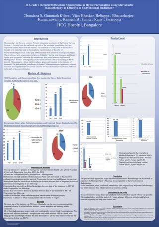
WHO grading and Recurrence Rate five years after Gross Total Resection
only(1), Subtotal Resection only (2) ,
Recurrence Rates after Subtotal resection and External Beam Radiotherapy(3),
Hypofractionated Radiotherapy (SRT) and Meningioma(4,5)
HCG Hospital, Bangalore
Chandana S, Gurunath Kilara , Vijay Bhaskar, Beliappa , Bhattacharjee ,
Kumaraswamy, Ramesh B , Sunita , Rajiv , Swaroopa
In Grade 1 Recurrent/Residual Meningioma, is Hypo fractionation using Stereotactic
Radiotherapy as Effective as Conventional Radiation?
Materials and Methods
This is a retrospective analysis of meningioma patients treated in Health Care Global Hospitals
, Cyber knife Department from Sept 2009- Jan 2016.
All cases are Histopathologically proven Grade 1 Meningioma's
Performa's were made and filled before analysis. Phone calls were made to the patient to
evaluate the meningioma specific survival, Progression free survival and Disease free survival.
Meningioma Specific survival was defined as the duration between date of diagnosis to death of
patient due to meningioma or last follow up.
Progression free survival was defined as duration between date of last treatment by SRT till
death /Progression /last follow up.
Disease free survival was defined as duration between date of last treatment by SRT till
Progression /last follow up.
Residue is defined as , when radiotherapy was started within 90 days of surgery.
Recurrence is defined as when treated anytime after 3 months of surgery.
Results
The mean age of the patients was 51.8years. Headache was the most common presenting
symptom followed by visual deficits, seizures, motor weakness and memory related
complaints.
All of them had undergone surgery and were histologically proven Grade 1 meningiomas. This
was the only adjuvant treatment , except in one case which received SRT as a boost after
Image guided radiotherapy. Median RT dose delivered was 25.7Gy The mean isodose line for
prescription of dose was 82.2%.
Meningioma Specific Survival after a
Median Follow up of 3.5 years was 84.6%
Progression Free Survival after a Median
Follow up of 3.5 years was 69.2%
Disease Free Survival after a Median
Follow up of 3.5 years was 84.6%
.
Conclusion
The present study argues that Hypo fractionated Stereotactic Radiotherapy can be offered to
patients with Meningioma of <30ccm as it is comparable to that of conventional
Radiotherapy.
It also shows that, when irradiated immediately after surgery(Early Adjuvant Radiotherapy )
has better response than when treated in a recurrence setting.
Limitations of the study
It is a retrospective study, hence no scheduled analysis of follow up side effects was possible.
The median follow up of this study is 3.5 years, a longer follow up period would help us
conclude regarding the long term control rate.
References
1. Ostrom QT, Gittleman H, Fulop J, et al. CBTRUS Statistical Report: Primary Brain and Central Nervous System Tumors Diagnosed in the United States in
2008-2012. Neuro Oncol 2015; 17 Suppl 4:iv1.
2. Drummond KJ, Zhu JJ, Black PM. Meningiomas: updating basic science, management, and outcome. Neurologist 2004; 10:113– 130
3. Jaaskelainen, J. Seemingly complete removal of histologically benign intracranial meningioma (Late recurrence rate and factors predicting recurrence in
657 patients) . Surg Neurol. 1986;26:461–469.
4. Mirimanoff RO, Dosoretz DE, Linggood RM, et al. Meningioma: analysis of recurrence and progression following neurosurgical resection. J Neurosurg
1985; 62:18–24
5. Glaholm J, Bloom HJ, Crow JH. The role of radiotherapy in the management of intracranial meningiomas: the Royal Marsden Hospital experience with
186 patients. Int J Radiat Oncol Biol Phys 1990; 18:755–761.(5)
10(100%)
5
0
3(60%)
0
2
4
6
8
10
12
<30ccm >30ccm
Number of cases
Number of
recurrences