
Empfohlen
Empfohlen
Weitere ähnliche Inhalte
Was ist angesagt?
Was ist angesagt? (19)
Ähnlich wie 3 D Grid Modeling With Brain Lab
Ähnlich wie 3 D Grid Modeling With Brain Lab (20)
3 D Grid Modeling With Brain Lab
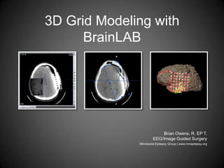
- 1. 3D Grid Modeling with BrainLAB Brian Owens, R. EP T.EEG/Image Guided Surgery Minnesota Epilepsy Group | www.mnepilepsy.org
- 2. Purpose 3D electrode grid modeling can be useful visualization electrode locale Can be combined with electrical stimulation data and/or MEG/fMRI data Can be helpful when used with stereotactic navigation
- 3. Method 3D grid creation is very easy using BrainLAB’siPlan application Before grid placement obtain a thin slice MR Post surgery obtain a thin slice volume CT scan Fuse CT to MR Electrodes are very high in contrast on CT compared to soft tissue Build grid from CT using ‘Object Creation’ and proper thresholding
- 5. Axial slices
- 8. Square image matrix (256x256 or 512x512)
- 10. Standard soft tissue algorithm
- 11. Circular or square FOV – the smallest FOV to encompass the head
- 12. Include the hard palate, tip of the nose, ears, top of the head & all fiducial markers if any
- 13. No gantry tilt
- 15. Thresholding Adjust the thresholding of the object abusively so that only the electrodes are created separately from soft tissue Use the image to the left as a guide
- 16. ROI In ‘Auto Segmentation’ you can adjust your region of intrest to encompass the electrode array You can do this in all planes
- 17. 3D Grid 3D object creation of grid complete Yellow voxels represent seizure dipoles obtained from MEG Blue voxel represents somatosensory dipole obtained from MEG
- 20. This slideshow has no official affiliation with BrainLAB. All images used were obtained using real patient data with no patient identifying information. The iPlan 3.x application is a product of BrainLAB, INC. www.mnepilepsy.org www.brainlab.com