stain & staining.pdf
•
2 gefällt mir•1,916 views

Staining is a method used to impart color to cells and tissues under a microscope. There are various staining techniques classified by chemical nature and method. Simple staining uses one stain like methylene blue. Differential staining distinguishes characteristics like Gram positive and negative bacteria. Special staining identifies structures like capsules, endospores, and flagella. Staining enhances contrast and highlights specific cellular components for examination and analysis in biological and biochemical research applications.

Empfohlen
Weitere ähnliche Inhalte
Was ist angesagt?
Was ist angesagt? (20)
Ähnlich wie stain & staining.pdf
Ähnlich wie stain & staining.pdf (20)
Mehr von benazeer fathima
Mehr von benazeer fathima (20)
Kürzlich hochgeladen
Kürzlich hochgeladen (20)
stain & staining.pdf
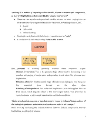
- 1. 1 *Staining is a method of imparting colour to cells, tissues or microscopic components, so they are highlighted and visualized better under a microscope.* There are a variety of staining methods used for various purposes ranging from the study of microscopic organisms to cellular structures, metabolic processes, etc., Simple Differential Special staining Staining is carried out with the help of a reagent termed as “stain“. It can be done in two ways, namely in-vitro and in-vivo. The protocol of staining generally involves three sequential stages: 1.Smear preparation: This is the primary stage, which involves the mixing of the inoculum with a drop of sterile water and spreading it until a thin film is formed over theglassslide. 2.Fixation of smear: It is the second stage, which involves drying and heat fixing the thin microbial layer formed on the glass slide. 3.Staining of the specimen: This is the final stage where the stain is applied onto the dried smear, which imparts colour to the microscopic matter. This procedure is carried out prior to microscopic examination and biochemical tests. *Stains are chemical reagents or dye that imparts colour to cells and tissue sections of the biological specimens and aids in its visualization under a microscope.* Stains work by increasing the contrast between different cellular components, thereby highlighting specific cell structures.
- 2. 2 Stains can be classified into the following types, depending upon its chemical nature and the type of staining methods. Based on chemical nature: There are three kinds of stain, acidic, basic and neutral, depending upon the chemical nature of the stain. Based on the staining method: There are four kinds of stain, viz. direct, indirect, differential and selective stains.
- 3. 3 Mechanism Stains are organic compound composed of a benzene ring, a chromophore group and an auxochrome group. Now benzene is a colourless solvent, and the chromophore group is a molecule that imparts colour to the benzene. As a result, the compound formed is called a ‘Chromogen’. Now, this chromogen is not a stain in itself, it is just a coloured compound. The second part of the stain, the auxochrome, is a chemical group that ionizes the chromogen i.e. it imparts a positive or negative charge to the chromogen group. As a result, the auxochrome enables the ionized chromogen to bind to cells or tissue fibres of opposite charge and thereby colour it. Types of Staining 1) Simple Staining It determines the cell shape, size and arrangement of the microorganisms. It is a very quick or simple method to perform and it makes the use of a single stain only. These are of two types, namely direct and indirect staining.
- 4. 4 Characteristic Differences between Direct and Indirect Staining: Characteristics Direct staining Indirect staining Stain used Basic stain Acidic stain Charge of stain Positive Negative Examples Methylene blue, crystal violet, carbol fuschin Nigrosine, india ink, congo red Outcome Stains the specimen Stains the background General view after staining Principle for discoloration Because of the positively charged stain, it gets attracted towards the negatively charged cell, hence it get fixed to the cell that retain the color of stain results in colorless background with colored cell. Because of the negatively charged stain, it gets repelled by the negatively charged cell, hence it does not fixed to the cell, results in colorless cell with colored background. 1) Differential Staining It differentiates between the physical and chemical properties of two different groups of an organism, depending on the cell-wall characteristics. It makes the use of multiple or more than one stains. It can be categorised into two types that are given below:
- 5. 5 A. Gram staining : It provides an important tool to differentiate the two major groups of bacteria, i.e. gram-positive and gram-negative. Dr Hans Christian Joachim Gram introduced this method in 1884. It is carried out by the use of differential stain known as Gram’s stain. Procedure: Gram staining Protocol Gram positive bacteria Gram negative bacteria Primary staining Heat fixed smear is flooded by crystal violet and allowed to stand for 1min. Mordanting After washing, iodine is then flooded and allowed to stand for 1min. Decolourization After washing, alcohol is added that is washed immediately Counter staining At last, safranin is flooded over the smear and allowed to stand for 30sec, then washed by water. Observation After air drying, place one drop of oil immersion over the smear and adjust the microscope to identify the specimen, whether it is gram negative or gram positive. Appear purple in colour because of teichoic acid that resist the primary stain. Appear pink in color due to lack of teichoic acid,alcohol creates pore in the cell which decolourizes the primary stain
- 6. 6 B. Acid fast Staining: It differentiates species of mycobacterium from the other groups of bacteria. Paul Ehrlich first developed it in 1882. And later, this technique was modified by a scientist named Ziehl Neelson. Procedure: Acid fast staining Protocol Acid fast bacteria Non acid fast bacteria Primary staining Heat fixed smear is flooded with carbol fuschin and allowed to stand for 1 min. Decolourization After washing, acid alcohol is added. Counter staining At last, methylene blue is flooded over the smear and allowed to stand for 30 sec, then wash it with water Observation After air drying, place one drop of oil immersion over the smear and adjust the microscope to identify the specimen, whether specimen is acid fast or not. Appears red in colour due to presence of mycolic acid that resist the color of primary stain and does not decolourize. Appears blue in colour, as they lack mycolic acid, alcohol creates pore in the cell that decolourizes the primary stain. 2) Special Staining It helps in the identification of particular internal and external structural components of the specimen. It includes capsule, endospore and flagella staining.
- 7. 7 A. Capsule staining: It differentiates the capsule from the rest of the cell body. This is carried out by the use of both positive and negative dyes. Procedure: Capsule staining Protocol Diagram Primary staining Drop of India ink is placed on a clean slide. Smearing Inoculum is then smeared in a dye. Dragging Use another slide to drag the mixture into thin film, and then air dried. Secondary staining Crystal violet is flooded over the thin film, and then air dried. Observation Examine the cells whether they are encapsulated or not. Interpretation of result Positive: Zone formation occurs against dark background Negative: Zone formation does not occur
- 8. 8 B. Endospore Staining: It differentiates the endospore from the vegetative cell and makes the use of both acidic and basic stains. Procedure: Endospore staining Protocol Diagram Primary staining Malachite green is flooded over the smear Heat fixing Then the mixture is heat fixed Decolourization Decolourized by water Counter staining Safranin is then flooded over the mixture and then air dried Observation Examine the slide under the microscope, whether endospore is present or not Interpretation of result: Positive: If Endospore present, it will appear green in color whereas vegetative cell appears as pink Negative: And if endospore is absent then only vegetative cells will appear pink in color
- 9. 9 C. Flagella Staining: It helps in the identification of the bacterial motility through the presence or absence of flagella. It makes the use of acidic and neutral stain. Procedure: Flagella staining Protocol Diagram Primary staining One drop of leifson’s stain is flooded over the smear Secondary staining After that methylene blue is added, and allowed to stand for one minute Observation Examine the appearance of flagella to know whether the bacteria is motile or not Interpretation of result: Positive: If flagella is present, then it will appear red in color while cell appears blue Negative: And if not present, only cell will appear blue in color
- 10. 10 Examples of Bacteria in different Staining Methods Simple staining Direct stain positive organism: Staphylococcus sp. , E.coli etc Indirect stain positive organism: Staphylococcus sp. ,Micrococcus luteus etc Differential staining Gram positive organisms: Streptococcus sp. , Enterococcus sp. , Listeria sp. , Bacillus sp. etc. Gram negative organisms: Pseudomonas sp. , Salmonella sp. , Klebsiella sp. , Yersinia sp. etc. Acid fast organisms: Mycobacterium sp. Non acid fast organisms: Enterobacter sp. Special staining Capsule stain positive bacteria: B.anthracis, K.pneumoniae etc. Capsule stain negative bacteria: Neisseria gonorrhoreae Endospore stain positive bacteria: Clostridium sp. , bacillus sp. etc. Endospore stain negative bacteria: E.coli , Salmonella sp. etc Flagella stain positive organisms: B.subtilis, pseudomonas sp. , E.coli etc Flagella stain negative organisms: shigella sp. , M.tuberculosis, C.diphtheriae Applications Staining methods have wide applicability in both biological and biochemical research. It is used in staining of metal. Used in staining of the wood. References: https://www.google.com/url?sa=i&url=https%3A%2F%2Fbiologyreader.com%2Fstaining .html&psig=AOvVaw2NElgkPBP4ZwHrdTjZbT0L&ust=1670133596045000&source=image s&cd=vfe&ved=0CBIQ3YkBahcKEwiI8rPO4tz7AhUAAAAAHQAAAAAQBA