
Empfohlen
Weitere ähnliche Inhalte
Was ist angesagt?
Was ist angesagt? (20)
Andere mochten auch
Andere mochten auch (20)
Ähnlich wie Leg ulcer
Ähnlich wie Leg ulcer (20)
Mehr von bbthapa
Kürzlich hochgeladen
Kürzlich hochgeladen (20)
Leg ulcer
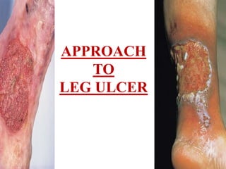
- 1. APPROACH TO LEG ULCER
- 2. INTRODUCTION • An ulcer can be defined as a break in the epithelial continuity. • Classified – Non- specific – Specific – Malignant • A leg ulcer is discontinuity of the squamous epithelium of the skin usually around the ankle or on the foot
- 3. • Leg ulcers are a common, chronic, recurring condition • Prevalence b/w 1.5 to 3 per 1000 and increases with age. • It’s estimated that up to 20 per 1000 people over 80 yrs will suffer from a leg ulcer • Following healing, re-ulceration rates at one year range from 26% - 69%
- 4. Classification of Chronic Leg Ulcers VASCULAR ARTERIAL • Atherosclerosis • Burger’s disease • Vasuclitis • Raynaud’s disease VENOUS • Chronic venous insufficiency • Varicose vein • Post sclerotherapy LYMPHATIC • Chronic lymphedema
- 5. • INFECTIVE – Pyogenic – Osteomyelitis – Tuberculosis – Syphillis – Tropical disease – Fungal disease • TRAUMATIC – Radiation – Thermal burns – Bites – Decubitus • NEOPLASTIC – Melanoma – SCC – BCC – Kapsoi’s sacroma
- 6. • SYSTEMIC METABOLIC – Ulcerative colitis – Diabetes – Martolle’s ulcer – Sickel cell disease – Avitminosis • NEUROPATHIC – Cord lesion – Pheripheral Neuropathies – Trauma – Diabetes – Tabes dorsalis – Alchoholism
- 7. Leg ulcer aetiology Grey J.E et al. ABC Wound Healing BMJ 2006; 332: 347-50 • Venous insufficiency 80 - 85% (60 – 70 %) • Arterial disease ( chronic lower limb ischemia) 20% • Mixed arterial and venous disease • Neuropathic Diabetic ulcer • Rheumatoid arthritis - 7% • Systemic vasculitis • Lymphoedema • Trauma • Malignant ulcer (BCC, SCC, Malignant melanoma) • Infective
- 8. CHRONIC VENOUS ULCER • CVI- chronic venous insufficiency who have developed irreversible skin damage as the result of sustained ambulatory venous hypertension • CVU- chronic venous ulceration defined as a break in the skin , present for more than 6 months , b/w the malleoli and tibial tuberosity that is presumed to be wholly or partially due to venous disease
- 9. CVI grade III is CVU • Basle Classification ( widmer) • CVI-I – Corona phlebectatica ( venous, or malleolar, Flare) • CVI-II – Hyper or dipigemented areas ( lipodermatosclerosis, atrophe blanche) • CVI-III – Open or healed ulceration
- 10. Others Classification • SVS/ISCVS classification – Clinical 0-3/ anatomical 0-7/ orgin 0- 2 /severity – Clinical Class 3- ulceration • CEAP classificaton – Clincal class 5- healed ulceration – Clinical class 6- active ulceration
- 11. Epidemiology of chronic venous ulcer • At any point – A third to a half will have trunk varicose veins – 7% will have CVI- I – 1-3%will have CVI- II – 1% will have CVI- III – Age – 3.6% over 65yrs – M:F – 1:1 • Common association are – Lower social class – Occupation like prolong standing predisposes – Inc BMI assoc with delayed CVU healing – Fibre deficent diet n constipation aggravate – Pregnancy n multiparity – Caucasians
- 12. PATHOPHYSIOLOGY CVU • CVI, culminating in CVU is the end result of sustained ambulatory venous hypertension acting upon a dermal microcirculation designed to operate in the presences of low venous pressure • Ambulatory venous pressure (AVP) – Normal - < 25Hg – >30 – inc incidence of CVU • 31-40 mmHg- 15% • >90mmHg- 100%
- 13. Pathophysiology contd… Macrovascular • Muscle pump dysfunction – Ageing General debility/ MSK n neurological LL pathologies ( fixed ankle, flat foot) • Valvular reflux (90% in CVI) – Primary alvular incompetence ( elastin/collagen) – Secondary valvular incompetence (post phlebitic) • Venous obstruction • Valvular hypoplasia/ agenesis
- 14. Microvascular • Venous stasis n hypoxia • Arteriovnous fistula ( stealing blood from skin) • Peri-capillary fibrin cuff( Browse n Burnand) • Leucocyte Migration (coleridge –smith) • Capillary Proliferation
- 15. CVU as a failing wound • Ulcer exduate inhibit keratinocyte migration and fibroblast activity • Increased proteolysis • Upregulation of metaloproteinases- breakdown important growth factor necessary for epithelialization • Epithelial cells in CVI refractory to the normal signalling process
- 16. ARTERIAL LEG ULCER Chronic limb ischemia n Ulcer Definition of CLI: • Persistent rest pain that requires regular adequate analgesia for at least 2 weeks, with resting ankle pressure of < 50mmHg or a toe pressure of < 30 mmHg • Presence of gangrene/ Ulcer of the foot/ toes with the same resting pressure
- 17. • Arterial ulcers account for 10% - 15% of leg ulcers • Major risk factors for Peripheral occlusive disease – Men > 45 yrs – Women >55 yrs or premature menopause – Family history of premature atherosclerosis – Smoking – HDL < 35 mg/dl – Hypertension • Other risk factors – DM – Def antioxidant vitamins – Elevated plasma fibrinogen level n homocysteine level – Sedentary life style
- 18. Clinical categories of chronic limb ischemia Grade Category Clinical description 0 0 Asymptomatic 1 Mild claudication I 2 Moderated claudication 3 Severe claudication II 4 Ischemic rest pain III 5 Minor tissue loss- non healing ulcer, focal gangrene with diffuse pedal edema 6 Major tissue loss- extending above TM level, Functional foot no longer salvagable
- 19. Fontaine classification- severity of CLI • Stage I- Asymptomatic • Stage II- intermittent claudciation limiting lifestyle • Stage III- Rest pain due to ischemia • Stage IV- Ulcer/ Gangrene due to ischemia
- 20. Patho - physiology of Arterial ulcer • Macrovascular – Fatty streak – Fibrous plaque – Complicated lesion • Ulcer • Hemorrahage • Calcification • Necrosis • Microvascular – High blood viscosity – Endothelial swelling – Platelet/ RBC/ PMN plugging – Inc permiability n tissue edema – Reduced n unevenly distributed flow
- 21. Natural History of Atherosclesrosis Macro n Microvascular changes Chronic limb ischemia Intermittent claudication ( 60% stabalise, 20 %improve) (20%)Decrease claudication distance Nocturnal ischemic pain Rest pain Progressive ischemia
- 22. Contd.. Tissue loss Ulceration n gangrene Minor trauma Secondary infection Ischemia n edema Decrease perfusion Thrombosis in microcirculation Spread proximally to small arteries
- 23. DIABETIC FOOT ULCER • 2% of whole population • 10- 25% of all DM develop some foot problems • 1 in 10 to 1 in 5 diabetic inpatient are due to foot ulceration, infection or gangrene • 5- 10% of all diabetic will require a lower extrimity amputation (1% in UK) • 45 % of all amputation are on diabetics • Amputation on diabetics are 20 times higher than non-diabetic
- 24. Pathophysiology Diabetic foot ulcer • Diabetic foot diseases is truly multifactorial – Neuropathy – Macrovascular ds – Microvascular ds – Infection – Connective tissue abnormalities – Haematological disturbances
- 25. Neuropathy Ischemia Ulcer Infection Trauma SOMATIC SENSORY SOMATIC MOTOR VISCERAL SENSORY AUTONOMIC
- 26. Classification of Diabetic foot ulcers • 1981 Wagner FW: Wagner Classification • 1996 Lavery & Armstrong: University of Texas Diabetic Wound Classification System • 1999 Macfarlane & Jeffcoate: S(AD)SAD system
- 27. Wagner FW Classification Gr O no obvious ulcer, but deformity, hyperkeratosis, or bony abnormality Gr 1 superficial ulcer, no infection sign Gr 2 deep ulcer with infection, but no bony involvement Gr 3 deep ulcer with abscess & bony involvement Gr 4 local gangrene (e.g., toe, forefoot) Gr 5 whole foot gangrene
- 28. LEG ULCERATION IN RA • Leg ulceration is often poorly defined in research • Surprisingly little is known about the prevalence and aetiology • The validated overall prevalence of leg ulceration in 324 in-patients was over 8% (Wilkinson & Kirk, 1965) • A postal survey of 250 RA patients taken from a diagnostic register found a validated overall prevalence of 9% (Thurtle & Cawley, 1983)
- 29. Factors affecting tissue viability in RA • Articular(joint) disease • Extra-articular (systemic) disease – anaemia, nodules, vasculitis, peripheral neuropathy • Peripheral vascular disease • Side effects of medication • Reduced mobility and self care capacity • Poor nutrition
- 31. Figure 2
- 32. LEG ULCER IN VASCULITIS • Inflammation with destruction of the vascular wall. • Cutaneous vasculitis is more common than systemic • Clinical presentation ranges from reticulated erythema to widespread purpura and finally necrosis leading to ulceration. Common caueses of vasculitis are • Infection, – Meningococcus/mycobacterium leprae/ rickettsia/ spirochetes • Immunoloigcal – Connective tissue disorder – Cryoglobuinemia – Goodpasture syndrome – Drugs ( antibiotics n NSAID) • Idiopathic – PAN/ giant cell arteritis/ Takayasu diseases
- 33. Pathophysiology • Immune complexes- deposited in wall of blood vessles • This often starts in the legs because of the high hydrostatic pressure in the post-capillary venules due to gravity. • activates the complement system, causing the chemotaxis of neutrophil • Activated neutrophil release proteolytic enzymes • Inflammation n vessel wall destruction
- 34. LEG ULCER IN LYMPHEDEMA Chronic oedema – defined as oedema that has been present for more than 3 months and does not go down overnight (Moffatt et al 2003, Williams 2003)
- 35. Aeitology • Primary lymphoedema – aplasia/hypoplasia/valvular incompetence • Secondary lymphoedema – Cancer treatment – Recurrent infection – Trauma – Parasitic infection – Obstruction by tumor – Immobility • Lymphovenous oedema due to CVI or traditional venous ulcer bandaging systems
- 36. Pathophysiology • Although chronic oedema is not directly responsible for ulcer development, it will affect wound healing (Mortimer and Browse, 2003). • This is due to the reduced oxygenation of the tissues resulting from the presence of edema (Casley- Smith, 1997). • As such, skin damage to an edematous limb may lead to ulceration. • • Ulceration can also follow superficial infection
- 37. EVALUATION OF LEG ULCER • HISTORY • EXAMINATION OF ULCER N SURROUNDING • EXAMINATION OF VASCULAR INSUFFICENCY • EXAMINATON OF NERVE LESION
- 38. The history of an ulcer History – Mode of onset /Precipitating event – Duration – Symptoms (pain/ discharge/ – Systemic features – Risk factors – Peripheral ischemic symptoms – Previous interventions – Treatment
- 39. The history of an ulcer Venous – Varicose veins – DVT/thrombophlebitis – Previous ulceration – Previous treatments – Sclerotic changes – Oedema – Painless – Precipating factors (age, occupation, BMI, low socioeconomic) – Coexsistence • PVD (20%), DM (5%), RA (8%)
- 40. The history of an ulcer Arterial • Intermittent Claudication • rest pain Risk factors • HTN, smoking, hyperlipidemia, sedentary • Previous arterial intervention Vasculitis – History of autoimmune disease – Painful – Lack chronic arterial occlusive symptoms – Systemic symptoms of autoimmune disease
- 41. The history of an ulcer • Infective – Exposures – Associated cellulitis – Systemic symptoms – Exudate – Spreading – Foul smell • Neoplastic – Chronicity – Previous malignancy – Risks • Exposures • UV radiation • Ionising radiation
- 42. CLINICAL ASSESSMENT OF LEG ULCER FEATURES VENOUS ARTERIAL NEUROTROPHIC TRAUMATIC MALIGNANT SITE gaiter area 70%, lateral 20%, circumferential 5% malleoli, heel, metatarsal heads, 5 MT base pressure area site of trauma face, lips, toungue SIZE/SHAPE large shallow ,vertically oval, small deep variable variable variable MARGIN irregular regular regular EDGE sloping punched out sloping everted/roll ed FLOOR granulation slough/ necrotic slough variable black mass
- 43. FEATURES VENOUS ARTERIAL NEURO TRAUMA MALIGNANT BASE fix /no induration no no no indurated deep up to DEPTH shallow deep bone variable shallow DISCh serous/ pureulent/ sero-sanguineous/ gree nish/bloody TENDER painless painful painless painful painless SKIN edema/eczema/ pigmentation/ atrophie blanche/ varicose vein Thin skin/ dec hair growth/ loss of sc fat/ loss of shininess/ brittle nails callus/ loss of sensation cellulitis fix to underlying str LYPMH NODE absent absent absent present except BCC
- 44. Uncommon variants of leg ulcer • Marjolin’s ulcer • Gummatous ulcer • Meleney’s ulcer • Erythrocyanoid ulcer • Martorell’s ulcer
- 45. DISTINGUISHING FEATURES OF ARTERIAL AND VENOUS ULCERS C/F ARTERIAL ULCER VENOUS ULCER Gender M>F F>M Age >60yrs 40 -60 yrs Risk Factors Smkn, HTN, DM, Hyperlipidemia DVT,Thromphophilia, Varicose vein Past History peripheral, coronary, Cerebrovascular DVT,Long bone # Symptoms severe pain except DM neuropathy 3rd have slight pain Site malleoli, heel, metatarsal heads, 5 th MT base Medial (70%), Lateral (20%) or both and gaiter area Edge Regular,Punched out, Indolent irrregular wit neo epithelium Base deep , green (soughy) or black (necrotic) with no granulation tissue, may comprise major tendon, bone n jt pink n granulating but may be covered in yellow green slough Surrounding skin features of severe limb ishemia LDS, Varicose eczema, atrophie blanche Veins empty full Swelling usually absent often present
- 46. EVALUATION OF VASCULAR INSUFFICENCY • Signs of ischemia – Thinning of skin/diminished hair growth/loss of subcutaneous fat/ loss of shininess/ brittle nails/ ulcer in pressure areas/ coldness • Burger’s postural test/ Burger’s angle – angle of 30 degree indicates severe ishemia • Capillary filling time- 20- 30 sec • Crossed leg test (Fuschsig’s test)- detect popliteal pulsation • Palpation of Blood vessles • ABPI • Bordie-Terendelenburg test – To determine the incompetency of the saphenofemoral valve and other communicating systems • Tourniquet test • Perthes’s test- – Cramping pain in DVT • Schwartz test- • Pratt’s test- position of leg perforators • Fegan method- • Homan’s sign • Moses’s sign
- 47. ANKLE BRACHIAL PRESSURE INDEX (ABPI) ABPI < 0.9 ABPI 0.9 – 1.1 ABPI >1.1 Palpable foot pulse PVD possible , assess furtther PVD likely Calcification • An ABPI of < 0.8 at rest indicates haemodynamically significant lower limb arterial disease • 0.7- claudication • 0.4- rest pain • < 0.3 suggests imminent necrosis present- PVF possible, assess further No palpable foot pulse PVD probable, assess further PVD likely assess further Calcification present- PVD probable, assess further
- 48. PULSES OF LOWER LIMB • The four pulses are: – femoral – popliteal – posterior tibial – dosalis pedis
- 49. Locate the superior border of the pubis in the mid line of the body; this is the uppermost part of the pubic symphysis. Feel the anterior limit of the iliac crest. The femoral pulse can be found midway between these two bony points (the mid-inguinal point)
- 50. Popliteal pulse Ask the subject to bend the knee so that it is flexed to about 90 degrees. Sit on the right hand edge of the bed close to the subject right foot. As before gently clasp the sides of the knee (of either limb) and press the pulps of your fingers into the popliteal fossa.
- 51. Locate the medial malleolus. 2-3cm below and behind it you should find the posterior tibial pulse. when taking the pulse on your own foot it is easier to use the thumb. In clinical practice the pulse is palpated using the pulps of the index and middle fingers. The artery is slightly deeper placed than the dorsalis pedis and therefore more concentration is often required to feel its pulsations Place your fingers half way down the dorsum of the foot on the bony area in the line between the first and second toes. The bones you can feel are the dorsal aspect of the navicular and the intermediate cuneiform bones. The pulse is palpated where the artery passes over this area.
- 52. MANAGEMENT PRINCIPLE: • Determine etiology • Accurate assessment of ulcer • Identify n correct co morbid factors • Treat underlying causes • Adequate drainage and desloughing • Avoid adherent dressing
- 53. The Investigation of a Leg Ulcer General Investigations Haematological FBC U&E, LFT’s Blood Glucose Lipid profile CRP Other ECG X Ray Swab C&S (remember TB)
- 54. The Investigation of a Leg Ulcer Arterial ABI’s Doppler USG Duplex USG Arteropraphy Digital substraction angiogram (DSA) CTA MRA Plethysmography Transcutaneous oxygen tension Isotope blood flow Venous Doppler USG Duplex USG Ambulatory Venous Pressure (AVP) measurement Phlebography (Ascending/ Descending) Photoplethysmography Vacricography CT/MR Venography Other wound swab c/s Biopsy RF, ANA DNA binding Compliment Levels
- 55. Doppler USG • A standard Doppler probe emits a sound when blood flows past the transmitting and receiving crystals. • It may be uniphasic or biphasic • Minimum level of investigation • Quick/ Easy/ Non-invasive/ Inexpensive • Useful in – Patency of vein – Venous reflux (SFJ > SPJ) – Exclude arterial diseases • ‘Doppler signal’ indicates moving blood, not viablility
- 56. Duplex Ultasonography • Generates 3 types of image information based on • Grey scale B mode ultrasound/ Color flow (triplex)/ Pulsed Doppler spectral analysis • Uses – blood velocity n valve closure time – Detailed analysis of anatomy n physiology – Valvular reflux / incompetnecy • Red in color map flow – Infrainguinal DVT – Origin of varicose vein n venous ulceration – Preoperative marking of LSV for FP bypas – Post operative surveillence of vein grafts – Arterial stenosis • 100% peak systolic volume rise compared with a normal area
- 57. Limitation of duplex • Cannot reliable distinguish deep venous incompetence due to primary from secondary • Cannot reliably deterimine the presence of functional severity of venous outflow obstruction • Unreliable in suprainguinal venous system • Time consuming and operator dependent • Oversensetive to clinically insignificant degrees of reflux
- 58. Phlebography • Replaced by combination of duplex and plethysmograpy • Anatomy of deep venous system • Presence of residual thrombus • Extent of recanalisation • Distriution of collaterals • Functional phlebography enable to detect the site of phlebography Venogram showing post-thrombotic venous occlusions with collateral pathways.
- 59. Varicography
- 60. Plethysmography • Amount of blood in dermal circulation is measured by the amount of light absorbed and reflected • Screening test • Mesures the refilling time(RT) in dermal circulation • RT > 20 sec is normal • Incompetency is indicated by short RT • Little practical use in arterial disease
- 61. CT n MR venography • Ideal for imaging for large central vein • Not widely used for peripheral venous disease
- 62. Ambulatory Venous Pressure • Direct method has little practical use • Measurement – Stand motionless- 90 mmHg – Walking (10 tip toe mov)- 15-30 mmHg – Again stand still- RT 90 is 18- 40 sec • Close relationship b/w AVP and ulcer – <30 mmHg- none – 41-50 mmHg- 22% – 61- 70 mmHg- 57% – >80mmHg- 73%
- 63. Transcutaneous oxygen tension measurement • Metabolic state of tissue • Dorsum of the foot/ calf/ thigh • Testing tcpo2 is 60 mmHg • Useful in determining the site of amputation
- 64. Arteriography • Gold standard for assessment of LL • DSA is more advance method and provide the image of contrast within the lumen of the artery without the surrounding tissues • Site- Femoral/ Brachial/ Common femoral vein • RF n INR should be normal • Stop oral-hypoglycemic and anticoagulant
- 65. lymphangiogram
- 66. Overveiw of TREATMENT of Leg Ulcer • MEDICAL MANAGEMENT n LIFESTYLE • DRESSING n TOPICAL AGENTS • PHARMACOTHERAPY • PHYSICAL THERAPIES • SURGICAL MANAGEMENT
- 67. SELF CARE GUIDELINES • Keep mobile with regular walking if possible • Elevate legs when immobile • Use emollient and examine legs regularly for broken skin, blisters, swelling or redness • Lose weight if appropriate • Stop smoking
- 68. DRESSING n TOPICAL AGENT IDEAL DRESSING • Control (absorb) odour, exudates and or bleeding • Exclude pathogenic bacteria and minimize colonization • Relieve pain • Ehance the would environment to speed up healing • Protect the wound from further environmental injury • Main the wound at body temperature • Reduce excessive scarring and or recurrence • Hide the wound form sight
- 69. Venous leg Ulcer – treatment • There is no proven role of antiseptics • Irrigate the wound with warm tap water or saline, then dry. • • Remove slough or necrotic tissue by gentle washing • potassium permanganate 0.01% soak if the ulcer is malodorous • For uncomplicated, non-infected ulcers apply a simple low-adherent dressing like paraffin emollients n gauge and Zinc & replace weekly. • Other dressings may be used if needed - pain (hydrocolloid), heavy exudate (alginate) or slough (hydrogel) • Biological dressing provide growth factors n scaffold for the ulcer healing n is more promising
- 70. Venous leg ulcer - treating infection • All chronic wounds are colonised with bacteria • Antibiotics should be used only if there is evidence of cellulitis or active infection (e.g. pyrexia, increasing pain, enlarging ulcer) • If there are clinical signs of infection present, clean ulcer with warm tap water or saline before taking a swab • Start immediate empiric treatment with an anti-staphylococcal antibiotic i.e. flucloxacillin or erythromycin 500mg qds for seven days • Change dressing daily or alternate days to assess if infection is improving • Do not use antimicrobial dressings • Do not start compression therapy if ulcer is infected
- 71. Physical therapies • Elevation n Bed rest will eventually heal virtually all CVU • Regular exercise will improve AVP • Compression therapy has been the main stay of treatment for CVI • Elastic bandage has better healing rates • For moderate to sever CVI class III (34-45 mmHg) n IV( >45mmHg) • Commence at metatarsal head terminate at tibial tuberosity • Multilayer has advantages over single layer – Charing cross four-layered bandage ( 42 at ankle n 17 below knee) • In mixed ulcer 3 layered compression bandage is used • Compression is contraindicated if the ABPI < 0.6 • Replace every 6 months • Graduated compression stockings should be used for at least 5 years after ulcer healing
- 72. Surgery- varicose ulcer • Varicose vein – SFJ / SPJ ligation , GSV stripping , Avulsion of varicosities . • Open perforator surgery • Sub fascial perforator surgery • Deep vein reconstruction – Vein valve repair – Vein valve transplantation – Vein diameter reduction – Veno-venous bypass ( palma porcedure) • Skin Graft – SSG – Pinch graft • Amputation
- 73. TREATMENT OF ARTERIAL ULCER • Infection can cause rapid deterioration of an arterial ulcer • It is not appropriate to debride arterial ulcers as this may produce further ischaemia and formation of a larger ulcer • Compression bandaging should not be applied • Choice of dressing is dictated by the nature of the wound • Treatment options include – Risk factor modification – Pharmacotherapy – Lumbar sympathectomy – Angioplasty – Graft – Amputation • To relieve pain n remove a life threatining ischemic • Below knee have great advantage of rehabilitation • Above knee have better wound healing
- 74. TREATMENT OF NEUROPATHIC ULCER • GENERAL ISSUES – Good well fitting shoes with sufficient depth – Look n feel before putting shoe – Careful toe nail cutting – Never to attempt trimming calluses • Chiropody (nail, callus, padding) • orthotics • Surgery- excision of prominent bone • Eradication of infection • Dressing • Surgical debridement and or amputation for neuroischemic or ishcemic ulcer
- 75. Treatment option for Rheumatoid leg ulcer • Treat the etiology • Relieve the pain • Local support n dressing • Bed rest n Elevation • Anti inflammatory agent – Colchicine (1.5mg daily) – Dapsone (100mg daily) – Prednisolone 20-40mg daily – Triamcinolone acetate (Kenalog 10mg/ml) 0.75-1ml for ulcer of 5cm diameter • Topical growth factor • Surgical option- SSG
- 76. Approach to vasculitic leg ulcer
- 77. Approach to lymphedema n ulcer
- 78. THANK YOU