
Empfohlen
Weitere ähnliche Inhalte
Was ist angesagt?
Was ist angesagt? (20)
Andere mochten auch
Andere mochten auch (20)
Ähnlich wie Deep Vein Thrombosis
Ähnlich wie Deep Vein Thrombosis (20)
Mehr von bbthapa
Kürzlich hochgeladen
Kürzlich hochgeladen (20)
Deep Vein Thrombosis
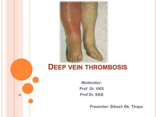
- 1. DEEP VEIN THROMBOSIS Moderator: Prof Dr. VKS Prof Dr. SKB Presenter: Bikash Bk. Thapa
- 2. DEEP VEIN THROMBOSIS A thrombus is a mass formed from blood constituents within a vessel or the heart during life. A venous thrombus is the formation of a semi-solid coagulum within flowing blood in the venous system.
- 3. The incidence of DVT ranges between 5 and 9 per 10,000 person-years in the general population, and the incidence of VTE (DVT and PE combined) is approximately 14 per 10,000 person-years. Major surgery have a significant incidence of perioperative DVT (15 to 40%). Major trauma : 40- 80% Hip or knee surgery : 40- 60% Spinal cord injury : 60-80%
- 4. EPIDEMIOLOGY OF DVT occurs first time in about 1 to 2 persons per 1,000 20 – 30% of general surgical patients Male :Female = 1:1 This incidence increases with increasing age, doubling with each decade of life after 40 More than two thirds of these patients have DVT alone, and the 30% will have symptomatic pulmonary embolism with mortality of 17%
- 5. 50 to 60% of DVT are asymptomatic The recurrence rate with anticoagulation has been noted to be 6% to 7% in 6 months DVT in upper extremity is less common- 5% of all documented DVT but one third result in PE Upper limb DVT may be primary or secondary
- 6. Stasis Activation of Coagulation Vessel Damage Virchow's Triad PATHOPHYSIOLOGY
- 7. PATHOPHYSIOLOGY Starts as a platelet nidus, usually in the venous valves of the calf. Platelet adhere to the endothelium and to each other forming a projecting mass The liberation of thromboplastin initiates a chemical cascade leading to coagulation If the rate of flow is slow red cells are entangled so that the lumen is occluded In front and behind platelet mass, the blood stagnates further formation of fibrin takes place, resulting in a large solid coagulum
- 8. SEQUELS OF THROMBOSIS Fibrinolysis Organisation Calcification Incorporation Embolism
- 9. A thrombus often develops in the soleal veins of the calf, Propagate and extend up to next venous branch and break down to embolize Thrombus localise to calf have less tendency to embolise than thromboi that extend to the thigh vens Aproximately 20% of cases of calf DVT propagate to the thigh, and 50% of cases of thigh or proximal DVT embolize
- 10. SEQUELE OF DVT DVT- Phlegmaisa alba dolens- Phlegmasia cerulea dolens in 1 to 2 days- Secondary arterial insufficiency venous gangrene in 1 to 2 days Inc capillary hydrostatic pressure leading to massive intersital edema and hypovolemic shock- Venous gangrene leading to amputation PCD is assoc with amputation of 50% 12-40% PE Mortality rate of 20%
- 11. RISK FACTORS A. Malignancy Tumor cell activation of clotting cascade Indirect clotting activation Reactive thrombocytosis B.Endothelial injury Adhesion of tumor cells Chemotherapeutic drugs C. Venous stasis Immobility, venous obstruction, inc pressure, inc blood viscosity Reduced clerance of activated clotting factors Endothelial hypoxia- inc expression of TF D .OCP Odd ratio of 3-5 for risk of DVT
- 12. E. Hypercoagulable states Primary Dec activation of protein C Impaired heparin binding of antithrombin III Downregulation of membrane associated plasimin production Inc serum prothrombin lefels Decrease thrombogenic inhibitors (antithrombinIII, protein C, Protein S Secondary Antiphospholipid syndrome Venous trauma Surgery Cancer Chemotherapy Myeloproliferative syndromes Heparin induced thrombopathy hyperhomocysteinemia
- 13. RISK FACTORS FOR DVT
- 14. In pregnant women, it has an incidence of 0.5 to 7 per 1,000 pregnancies, and is the second most common cause of maternal death in developed countries after bleeding general anesthesia and prior episode of DVT have a 5 times increased risk Patients with acute MI who are not receiving anticoagulation have a 26-38% rate of DVT 40% of postoperative neurosurgical patients develop DVT. Type A blood is associated with lower levels of antithrombin III and higher levels of factor VIII than type O blood. Women of reproductive age with type A blood are 4 times as likely to develop DVT.
- 15. PRESENTATION Pain n swelling of unilateral calf Bilateral is uncommon-30% Asymptomatic n may present as features of pulmonary embolism Pleuritic chest pain, haemoptysis n SOB Phlegmasia alba dolens and phlegmasia cerulea dolen
- 16. Phlegmasia alba dolens : Deep venous channels of the extremity are affected while sparing collateral veins and therefore maintaining some degree of venous return. Present with blanching of the extremity, edema and discomfort Phlegmasia cerulea dolens: Occurs with extension of thrombus into the collateral venous system, resulting in limb pain and swelling with cyanosis a sign of arterial ischemia
- 17. PHYSICAL SIGN Pitting edema Dilated superficial veins stiff calf and tenderness over the course of the deep veins Erythema Bluish discoloration Absent or decreased pulse Homans’ sign – resistance (not pain) of the calf muscles to forcible dorsiflexion – Pratt's sign: Squeezing of posterior calf elicits pain Low grade fever PE- cyanosis, dyspnoea, raised neck veins, a fixed split second heart sound and a pleural rub
- 18. DIFFERENTIAL DIAGNOSIS Cellulitis, lymphangitis Lymphedema Ruptured Baker cyst Varicose veins Superfical thrombophlibitis
- 19. Score (wells score) Clinical Parameter Score +1 Active cancer (treatment ongoing, or within 6 mo or palliative) +1 Paralysis or recent plaster immobilization of the lower extremities +1 Recently bedridden for >3 d or major surgery <4 wk +1 Localized tenderness along the distribution of the deep venous system +1 Calf swelling >3 cm compared with the asymptomatic leg +1 Pitting edema (greater in the symptomatic leg) +1 Previous DVT documented +1 Collateral superficial veins (non varicose) -2 Alternative diagnosis (as likely or greater than that of DVT) WELLS SCORE FOR DVT
- 20. Total of Above Score >3 High probability 1 or 2 Moderate probability < 0 Low probability WELLS SCORE
- 21. LAB CBC PT ,PTT , INR ESR D-dimer ABG Protein C , S Antithrombin III LFT RFT
- 22. IMAGING DUPLEX ULTRASOUND combines real-time B-mode ultrasound with pulsed Doppler capability. HELICAL CT VENOGRAPHY CONTRAST VENOGRAPHY MRI OTHERS CHEST X-RAY ECHO PULMONARY ARTERIOGRAPHY V/Q SCAN
- 23. DUPLEX ULTRASONOGRAPHY 73% Sensitivity in calf vein 95-97% Sensitivity in proximal vein 95-98% Specificity lack of spontaneous flow inability to compress the vein absence of color filling of the lumen by color flow DUS, loss of respiratory flow variation, Venous distension
- 24. DUPLEX ULTRASONOGRAPHY Advantage helpful to differentiate venous thrombosis from hematoma, Baker cyst, abscess, and other causes of leg pain and edema. Non-invasive inexpensive Disadvantage Venous thrombi in iliac and tibial vein are difficult to visualize early n fresh thrombi not be able to differentiate between old and new clots
- 25. VENOGRCTAPHY/ CT VENOGRAPHY (GOLD STANDERD) o The gold standard is intravenous venography, o A positive study result is failure to fill the deep system with passage of the contrast medium into the superficial system or demonstration of discrete filling defects o Similar sensitivity as Ultrasound
- 26. MRI – In the second and third trimester of pregnancy, MRI is more accurate than duplex ultrasonography because the gravid uterus alters Doppler venous flow characteristics. – In suspected calf vein thrombosis, MRI is more sensitive than any other noninvasive study.
- 27. D-dimer testing D-dimer antibodies account for their high sensitivity for venous thrombo embolism. D-dimer level may be elevated in any medical condition where clots form. D-dimer level is elevated in trauma, recent surgery, hemorrhage, cancer, and sepsis. The D-dimer assays have low specificity for DVT; therefore, they should only be used to rule out DVT, not to confirm the diagnosis of DVT.
- 28. D-dimer results should be used as follows: A negative D-dimer assay result rules out DVT in patients with low-to-moderate risk and a Wells DVT score less than 2. All patients with a positive D-dimer assay result and all patients with a moderate-to-high risk of DVT (Wells DVT score >2) require a diagnostic study (duplex ultrasonography).
- 29. TREATMENT Principles are: To prevent clot propa
- 30. Major general , urological. Gyne. Cardiothoracic,vascular or neruological surgery Age > 40 yrs Mahor medical illness Major trauma or burns Minor surgery or trauma in pat with previous DVT, PE Orthopedic surgery or amputation of LL Lower limb paralsis Major pelvic or abdominal surgery for cancer Major surgery, trauma or illness in pat with previous DVT, PE, Thrombophillia Full limb paralysis Maojor limb amputation Moderate risk High risk
- 31. Frequency of Fatal PE Frequency of Proximal Vein Thrombosis Frequency of Calf Vein Thrombosis Category >1% 10-30% 40-80% High-risk 0.1-1% 1-10% 10-40% Moderate- risk <0.1% <1% <10% Low-risk
- 32. PROPHYLAXSIS All patient admitted should be assessed for risk for DVT Overall, low-dose UFH and LMWH reduce the risk for symptomatic and non symptomatic VTE by 60 to 70%. Low risk Graduated compression stockings, Early mobilisation and leg elevation Moderate risk Leg elevation, early mobilization, and either graduated compression stockings or s/c UFH or LMWH High risk Leg elevation, early mobilisation, TEDs, UFH or LMWH
- 33. Heparin 5,000 unit ,S/C 2 hours before surgery and every 8 or 12 hourly Graduated elastic compression (GEC) stockings reduce the incidence of asymptomatic DVT by approximately 50-60% Intermittent pneumatic compression (IPC) reduces the incidence of asymptomatic DVT by approximately 69% Aspirin reduces DVT by 30% and PE by 50% Prophylactic insertion of IVC filters Fondaparinux has similar results like LMWH
- 34. • In the Women's Health Study, supplementation with vitamin E (alpha-tocopherol, 600 IU every other day) reduced the risk of venous thrombo embolism in women, especially those with a prior history or genetic predisposition. • High-risk patients should also be prescribed a single prophylactic subcutaneous 40 mg dose of enoxaparin prior to a long plane trip (>6 h).
- 35. TREATMENT Principles are: To prevent clot propagation Reduce the risk of PE and Enhance the resolution of the clot to minimize the post – thrombotic syndrome Options are Anticoagulants/ Pharmacotherapy Catheter directed intra-thrombus thrombolysis Inferior vena caval filters Venous thrombectomy
- 36. ANTICOAGULATION THERAPY HEPARIN Increases the binding of coagulatoin factors IXa, Xa, XIa, XII and thrombin to antithrombin-III Discourage further clot formation and facilitate endogenous clot lysis onset of action 10-18 mins IV 20-60 min S/C ( peak at 4-6 hrs n last up to 12 hrs) Half life – 90 mins Safe in pregnancy Monitoring done with APTT 6 hrly (1.5 to 2.5 times) Platelet count should be done every 3 days APTT >1.5 should be achieved with in 24 hrs
- 37. Heparinization Continous IV infusion Loading dose- 5000 to 10000 unit( 80 U/kg) Continuous infusion- 1300 units /hour (18 U/Kg) Intermittent intravenous adminstration 70-100 units /kg every 4 hours Subcutaneous adminstration 5000 unit every 8 0r 12 hourly Should be continued for 4-6 days Oral anticoagulant after 24 hours and overlapped for 4 days Heparin should be discontinued when INR is >2 for 2 consecutive days Warfarin is continued for minimum 3 months to maintain INR between 2.0 to 4.5
- 38. Advantages of Low Molecular Weight Heparin Greater activity against factor Xa Lower risk of bleeding Longer half life(4-6 hrs) Produce little effect on test APTT or PT so no need of monitoring Can be started simultaneously with warfarin Weight-based once- or twice-daily SC LMWH injections Incidence of HIT is<2% Can be used as outpatient therapy Less recurrence (8.6% vs 6.9%)
- 39. Adverse affects are Harmorrhage- 5% Thrombocytopenia( HAAbs) 1-5% Repeated exposure 21% HIT type I HIT type II Hypersensitivity hyperkalemia
- 40. WARFARIN Inhibits the enzyme vitamin K epodxide Thus indirectly inhibit the the synthesis of vitamin K dependent clotting factors 5mg orally Onset: 36 -48hrs Duration: 2-6 days Half life: 36-40hrs Bioavailability: 100% Can cross placenta/ appears in milk/ The target INR 2 to 3 Adverse affects Haemorrhage (0.5 to 1% per month) Skin necrosis Teratogenecity Liver damage
- 41. Duration First attack- 3 mth Second attack- 1 year Third attack- life long Life time warfarin is aslo indicated for Thrombophilia Patients with first VTE and malignancy Recurrence rate is 2.6% for life long therapy
- 42. NEWER AGENTS Fondaparinux synthetic pentasaccharide Its five-polysaccharide sequence binds and activates antithrombin, causing specific inhibition of factor Xa The drug is administered SC once daily with a weight- based dosing protocol: 5 mg, 7.5 mg, or 10 mg for patients weighing <50 kg, 50 to 100 kg, or >100 kg, respectively Half life is 17 hrs The rates of recurrent VTE ranged from 3.8 to 5%, with rates of major bleeding of 2 to 2.6%, for all treatment arms
- 43. Direct Thrombin inhibitors recombinant hirudin, argatroban, and bivalirudin bind to thrombin, inhibiting the conversion of fibrinogen to fibrin as well as thrombin-induced platelet activation. reserved for (a) patients in whom there is a high clinical suspicion or confirmation of HIT, and (b) patients who have a history of HIT or test positive for heparin- associated antibodies administered for at least 7 days, or until the platelet count normalizes. Warfarin may then be introduced slowly, overlapping therapy with a DTI for at least 5 days
- 44. LONG TERM ANTI THROMBOTIC THERAPY FOR DVT
- 45. INFERIOR VENACAVAL FILTER Kimray-Greenfield filter in 1973 Traps emboli as small as 3mm Made of titanium or nickel titanium alloy Placed percutaneously under LA via the common femoral, internal juglar or antecubital vein Positioned in the infra-renal cava 20 years follow up – 96 % patency Complications associated with IVC filter placement include insertion site thrombosis, filter migration, erosion of the filter into the IVC wall, and IVC thrombosis. The rate of fatal complications is <0.12%
- 46. Absolute indication are DVT or PE with contraindication to anticoagulation therapy prior hemorrhagic stroke Recent neurosurgical procedure or major surgery Multiple trauma Active internal bleeding Intracranial neoplasm Bleeding diathesis Pregnancy DVT or PE having complication with anticoagulation therapy Failure of anticoagulation therapy Free-floating iliofemoral or caval thrombus Relative indication Prior history of chronic pulmonary HTN Morbidly obese with BMI >55
- 47. Catheter Directed Intra-thrombus thrombolysis Placement of thrombolysis catheter within the clot Rapid clearance of the DVT SITE: IJV, contralateral CFV, popliteal, tibial vein Thrombolytic drugs: streptokinase, alteplase , reteplase, and tenecteplase, urokinase convert plasminogen to plasmin, which leads to the degradation of fibrin. Higher success in acute DVT n new onset DVT and upper limb < 10 days with 34% and > 10 days 19% 1 year patency 79% for complete lysis and 32% for partial lysis Overall one year patency is 60% Major bleeding -11% Mortality 0.4% Contraindicated in:Recent surgery, active or recent bleeding, bleeding diathesis, pregnancy and cerebral disease
- 48. VENOUS THROMBECTOMY Inidcation Phlegmasia alba dolens Phlegmasia cerulea dolens with impending venous gangrene threatening to the patient’s life and limb Failed or contraindicated anticoagulation and thrombolysis If the patient has phlegmasia cerulea dolens, a fasciotomy of the calf compartments is first performed Useful in relatively young patient who has cause other than malignant dis for the DVT
- 49. An intraoperative completion venogram is obtained to determine if any residual thrombus or stenosis is present. Post op managed with limb elevation, calf exercise, compression stockings, antibioitc therapy, heparin for 5 days and warfarin for 6 months or life long Good result if performed with in 7-10 days of event Mortality rate is 1% Intraoperative PE occurs in 20% 10 yrs follow up patency 77% Severe PCD and venous gangrene have poor outcome
- 50. TREATMENT IN PREGNANCY The treatment of DVT in pregnancy is similar to the treatment of non pregnant. Heparin SC or small pump infusion Avoid warfarin in pregnancy If warfarin therapy is essential, it should be avoided at least during the first trimester (because of teratogenicity) and from about 2 to 4 weeks before delivery to reduce risk of hemorrhagic complications Compression stockings
- 51. COMPLICATIONS OF DVT • pulmonary embolism • post-thrombotic syndrome • occurs in 15% of patients with deep vein thrombosis (DVT). It presents with leg oedema, pain, nocturnal cramping, venous claudication, skin pigmentation, dermatitis and ulceratiaion (usually on the medial aspect of the lower leg).
- 52. High clinical pretest probability- DVT likely Doppler ultrasound Ultrasound positive for DVT Diagnoses of DVT confirmed Begin treatment Ultrasound negative for DVT D-Dmer test (if available and reliable) Otherwise skip to repeat ultrasound D-Dimer positive Repeat ultrasound in 1 week D-Dimer negative DVT ruled out Repeat ultrasound positive for DVT Diagnoses of DVT confirmed Begin treatment Suspect DVT Low clinical pretest probability- DVT likely Consider starting with D-dimer test first (if available and reliable ) Or skip to ultrasound D-dimer positive D-Dimer negative DVT ruled out Doppler ultrasound Ultrasound positive for DVT Diagnose of DVT confirmed Begin treatment Ultrasound negative for DVT DVT ruled out (consider repeat ultrasound if D-dimer not available)