2. Contents:
1) Why electrophoresis??
2) What is electrophoresis??
3) Principle
4) Agarose gel electrophoresis
5) Sds-PAGE (Polyacrylamide gel
electrophoresis)
6) Factors effecting electrophoresis
7) applications
3. Why electrophoresis?
To separate DNA fragments
from each other
To determine the sizes of
DNA fragments
To determine the presence
or amount of DNA
To analyze restriction
digestion products.
Determination of
molecular weight of
proteins.
4. What is electrophoresis?
It describes migration of charged
particles or molecules under the
influence of electric field.
It is standard method for separation,
identification, analysis and purification of:
–DNA molecules
–protein molecules
5. PRINCIPLE
separates molecules from each other on
the basis of
–size
–charge
–shape
basis of separation depends on how the
sample and gel are prepared?
6. Gel electrophoresis
What is a gel?
Gel is a cross linked polymer whose
composition and porosity is chosen based on the
specific weight and porosity of the target molecules.
Types of Gel:
Agarose gel
Polyacrylamide gel
7. Agarose gel electrophoresis
Agarose gel:
Agarose is extracted in the form of agar from several species
of red marine algae, or seaweed, found in California and
eastern Asia, dissolves in near-boiling water, and forms a gel
when cools.
Has gelling temperature 35-38˚C and melts at 90-95 ˚C.
Used to separate macromolecules such as nucleic acids, large
proteins and protein complexes.
Composition:
It is prepared by dissolving 0.5% agarose in boiling water and
allowing it to cool to 40°C.
It is fragile because of the formation of weak hydrogen bonds
and hydrophobic bonds.
8. Material required for gel
electrophoresis
Electrophoresis chamber
Agarose gel
Gel casting tray
Buffer
Staining agent (dye)
A comb
DNA ladder
Sample to be separate
10. Gel Casting Trays
available in a variety of
sizes and composed of
UV-transparent plastic.
The open ends of the
trays are closed with
tape while the gel is
being cast, then
removed prior to
electrophoresis.
11. Applied voltage
• voltage, rate of migration
• The higher the voltage, the more quickly the
gel runs
• But if voltage is too high, gel melts
• The best separation will apply voltage at no
more than 5V/cm of gel length.
12. Buffers
During electrophoresis water undergoes
hydrolysis : H2O H + OH-
Buffers prevent the pH from changing by
reacting with the H+ or OH- products
Most common buffer used is called TRIS
– [tris (hydroxymethyl) aminomethane]
13. Buffers (cont.)
Another compound is added to make Tris an
effective buffer — either boric or acetic acid
Another compound is added to bind metals
EDTA
The buffer is either TBE or TAE
TBE is made with Tris/Boric Acid/EDTA
TAE is made with Tris/Acetic Acid/ EDTA
14. Staining of DNA
To make DNA fragments visible after electrophoresis, the DNA
must be stained
The favorite—ethidium bromide
When bound to DNA it fluoresces under ultraviolet light
(reddish –orange color)
Convenient because it can be added directly to the gel
Sensitive—detects 0.1ug of DNA.
ethidium bromide is mutagenic so care must be taken while
handling the dye.
Other alternatives for ethidium bromide :
Methylene blue
Syber safe
xylene cyanol
bromphenol blue
15. A Comb
A comb is placed in the
liquid gel after it has
been poured
Removing the comb
from the hardened gel
produces a series of
wells used to load the
DNA
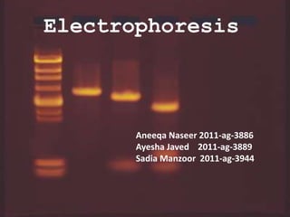
16. DNA ladder
It is a solution of DNA
molecules of different length
DNA Ladder consists of known
DNA sizes used to determine
the size of an unknown DNA
sample.
The DNA ladder usually
contains regularly spaced sized
samples which when run on an
agarose gel looks like a
"ladder".
17. Method For Electrophoresis
Add running buffer, load samples and marker
Run gel at constant voltage until band separation occurs
Pour into casting tray with comb and allow to solidify
View DNA on UV light box and show results
Prepare agarose gel
Melt, cool and add Ethidium Bromide. Mix thoroughly.
18. Get your sample
obtained from previous
purifying technique
(i.e. PCR)
Set up gel, remove
comb
Load Buffer
Load Sample
Run Gel
Stain and look at with UV
light
19. DNA is negatively charged.
+-
Power
DNA
When placed in an electrical field, DNA will migrate toward the
positive pole (anode).
H
O2
An agarose gel is used to slow the movement of DNA and separate
by size.
20.
21. SDS
Since we are trying to separate many different
protein molecules of a variety of shapes and sizes,
we first want to get them to be linear so that the
proteins no longer have any secondary, tertiary or
quaternary structure.
22. SDS (cont.)
To have proteins with linear structures we use sodium
dodecyl sulfate (SDS).
SDS is a detergent that can dissolve hydrophobic
molecules, also have negative charge (sulphate)
attached to it allowing it to run properly through the gel
(from negative to positive).
If cell is incubated with it
o membranes will be dissolved
o Proteins will be solubilized by it
o All proteins covered with negative charges
23. End result has two important features:
1. Proteins contain only primary structures.
2. In electric field migrate towards positive
pole.
24. Why PAGE?
Proteins separate only on the basis of charges
in electric field.
We also need to separate it on basis of size.
To separate it on basis of size we use PAGE.
25. It is polymer of acrylamide monomers
when its polymer is formed it turns into gel
and we use electricity to pull proteins
through the gel, process is called
polyacrylamide gel electrophoresis.
Polyacrylamide gel
Tunnels of different diameters
What is PAGE?
26. Polyacrylamide gel
Polyacrylamide is a polymer of acrylamide
monomers.
Like Agarose Gels, Polyacrylamide gels are used
to separate protein molecules by shape, size and
charge.
Polyacrylamide is specifically used for proteins
because it provides the protein with an
environment where it will not become
denatured.
Allowing different sized proteins to move at
different rates.
27. Polyacrylamide gel
When electricity passed proteins tend to move
through gel in bunches, or bands, since there are so
many copies of each protein and they are all same
size.
Controlled time is given for run so that proteins not
reached to other side of the gel.
After it stain proteins.
Bands
29. Gel has 5 number lanes where 5 different
samples of proteins are applies to gel.
Lane 1: molecular weight standards of known
sizes.
Lane 2: mixture of 3 proteins of different sizes
with a being biggest and c being smallest.
Lane 3: protein a by itself
Lane 4: protein b by itself
Lane 5: protein c by itself
31. The Sample:
Charge/mass ratio of the sample dictates its electrophoretic mobili
ty. The mass consists of not
only the size (molecular weight) but also the shape of the molecule.
• Charge: The higher the charge, greater is the
• electrophoretic mobility. The charge is dependent on pH of the
medium.
b) Size: The bigger molecules have a small electrophoretic mobility
compared to the smaller particles.
c) Shape: The globular protein will migrate faster than the fibrous
protein
32. DNA or RNA Molecular Weight
The length of the DNA molecule is
the most important factor, smaller molecules travel
farther.
voltage
The higher the voltage, the faster the DNA moves.
But voltage islimited by the fact that it heats and
ultimately causes the gel to melt. High voltages also
decrease the resolution (above about 5 to 8 V/cm)
33. Agarose
• Agarose gel electrophoresis can be used for the separation of DNA
fragments ranging from 50 base pair to several megabases (millions
of bases) using specialized apparatus. Increasing the agarose
concentration of a gel reduces the migration speed and enables
separation of smaller DNA molecules.
• The distance between DNA bands of a given length is determined
by the percent agarose in the gel. In general lower concentrations
of agarose are better for larger molecules because the result in
greater separation between bands that are close in size. The
disadvantage of higher concentrations is the long run times
(sometimes days). Instead high percentage agarose gels should be
run with a pulsed field electrophoresis (PFE), or field inversion
electrophoresis.
34. Buffer
• The most common buffers for agarose gel:
TAE: Tris acetate EDTA
TBE: Tris/Borate/EDTA
SB: Sodium borate.
• TAE has the lowest buffering capacity but provides the best resolution for
larger DNA. This means a lower voltage and more time, but a better
product.
visualization
Ethidium bromide dye which is used as staining dye, in staining is carcinogen.
36. Vaccine analysis
• Electrophoresis is widely used in vaccine
analysis method.
• Several vaccines have been purified,
processed and analysed.
e.g. influenza vaccine, polio vaccine,
hepatitis vaccine.
37. DNA analysis
• Through electrophoresis specific DNA
sequences can be analysed, isolated and
cloned.
• Analysed DNA may be used in forensic
investigations and paternity tests.
38. Protein analysis
• Electrophoresis has advanced our
understanding on the structure and function
of protein.
• Amount of protein in your urine and blood is
measured and compared to established
normal values- lower or higher than the
normal levels usually indicates a disease.
39. Antibiotic analysis
• . With electrophoresis, experts are not only
able to synthesize new antibiotics but are also
able to analyze which types of bacteria are
antibiotic-resistant.
• These drugs, such as penicillin, are among the
widely prescribed drugs against bacterial
infections