Ultra Sound Imaging
•Als PPTX, PDF herunterladen•
17 gefällt mir•2,041 views

This ppt describes basic principles of Ultra Sound Imaging. Advantages, Applications, limitations and modes of operations are also included.

Empfohlen
Weitere ähnliche Inhalte
Was ist angesagt?
Was ist angesagt? (20)
Andere mochten auch
Andere mochten auch (20)
Ähnlich wie Ultra Sound Imaging
Ähnlich wie Ultra Sound Imaging (20)
Kürzlich hochgeladen
Kürzlich hochgeladen (20)
Ultra Sound Imaging
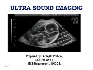
- 1. ULTRA SOUND IMAGING Prepared by : Abhijith Prabha , L6A ,roll no : 6 , ECE Department , SNGCE. 1
- 2. INTRODUCTION An ultrasound imaging device is a machine that is commonly used to examine underlying features of the human body. Its advantages are that no surgery is needed in order to obtain a clear picture of entities such as kidney stones. The main parts of an ultrasound equipment are the ultrasound transducer or probe, the electrical control of the probe (including "beam former") and the visualization system. This section will focus particularly on the visualization system. 2 Abhijith Prabha
- 3. Principle The ultrasound machine transmits high-frequency (1 to 5 megahertz) sound pulses into your body using a probe. The sound waves travel into your body and hit a boundary between tissues (e.g. between fluid and soft tissue, soft tissue and bone). Some of the sound waves get reflect back to the probe, while some travel on further until they reach another boundary and get reflected. 3 Abhijith Prabha
- 4. Fig : schematic diagram of principle of Ultra Sound imaging 4 Abhijith Prabha
- 5. Principle The reflected waves are picked up by the probe and relayed to the machine. The machine calculates the distance from the probe to the tissue or organ (boundaries) using the speed of sound in tissue (5,005 ft/s or1,540 m/s) and the time of the each echo's return (usually on the order of millionths of a second). The machine displays the distances and intensities of the echoes on the screen, forming a two dimensional image 5 Abhijith Prabha
- 6. Relation between attenuation and frequency in body. 6 Abhijith Prabha
- 7. Attenuation coefficient of different body tissues 7 Abhijith Prabha
- 8. Ultra Sound Machine 8 Abhijith Prabha
- 9. The Ultrasound Machine A basic ultrasound machine has the following parts: transducer probe - probe that sends and receives the sound waves central processing unit (CPU) - computer that does all of the calculations and contains the electrical power supplies for itself and the transducer probe transducer pulse controls - changes the amplitude, frequency and duration of the pulses emitted from the transducer probe 9 Abhijith Prabha
- 10. display - displays the image from the ultrasound data processed by the CPU keyboard/cursor - inputs data and takes measurements from the display disk storage device (hard, floppy, CD) - stores the acquired images printer - prints the image from the displayed data 10 Abhijith Prabha
- 11. Fig : working of Ultrasonic Machine 11 Abhijith Prabha
- 12. Ultrasound generator . 12 Abhijith Prabha
- 13. Fig: block diagram of Ultra Sound generator 13 Abhijith Prabha
- 14. Ultrasound receiver 14 Abhijith Prabha
- 15. Fig : ckt diagram of a typical Ultrasound Receiver 15 Abhijith Prabha
- 16. Modes of operation A MODE A stands for Amplitude. Information of the reflected signal in a single ultrasound beam is continually displayed distance from the transducer and intensity are shown by position and amplitude in a line on an oscilloscope. This mode is mainly of historical interest, may be rarely used in gynaecology or ophthalmology. 16 Abhijith Prabha
- 17. Modes of operation B MODE B stands for Brightness. In this case A-mode information from many beams, typically forming a sector in a plane of the body, is shown as pixel intensity on a monitor. B mode is often referred to as 2D, and is the most important modality for anatomic assessment and orientation in the body, also for localising and as a background for display of other information such as Doppler signals. 17 Abhijith Prabha
- 19. Modes of operation M MODE M stands for motion. This approach is used for the analysis of moving organs. It is based on A-mode data from a single ultrasound beam that are represented as function of time. This does not require a sweep through many ultrasound beams which allows for high temporal resolution. 19 Abhijith Prabha
- 20. Fig : illustration of Ultrasound image in Mode M 20 Abhijith Prabha
- 21. Modes of operation Doppler mode Doppler mode exploits the frequency shift due to relative motion between two objects. With this approach information regarding blood velocity and cardiac valves can be obtained. Doppler mode can be obtained by continuous or pulsed wave (PW); in addition, velocity data can be shown as overlaying colour on B-mode images (colour Doppler, power Doppler and Tissue Doppler). 21 Abhijith Prabha
- 22. Transducers Mechanical Probe: seldom used now. Electronic Probe: – Linear array transducers • piezoelectric elements linearly arranged • sequentially activated to produce an image – Phased array transducers • smaller scanning surface (foot print) • good for echocardiography • more expensive • elements are activated with phase differences to allow steering of the ultrasound signal 22 Abhijith Prabha
- 23. Applications Obstetrics and Gynaecology measuring the size of the foetus to determine the due date determining the position of the foetus to see if it is in the normal head down position or breech checking the position of the placenta to see if it is improperly developing over the opening to the uterus (cervix) seeing the number of foetuses in the uterus checking the sex of the baby (if the genital area can be clearly seen) checking the foetus's growth rate by making many measurements over time 23 Abhijith Prabha
- 24. Applications Cardiology seeing the inside of the heart to identify abnormal structures or functions measuring blood flow through the heart and major blood vessels Urology measuring blood flow through the kidney seeing kidney stones detecting prostate cancer early 24 Abhijith Prabha
- 25. Advantages Ultrasonic can be easily focused, i.e., they are directional. They are inaudible. It is possible to investigate the properties of very small structures. Information obtained by US , particularly in dynamic studies, cannot be acquired by other more convenient technique. 25 Abhijith Prabha
- 26. Limitations development of heat - tissues or water absorb the ultrasound energy which increases their temperature locally formation of bubbles (cavitations) - when dissolved gases come out of solution due to local heat caused by ultrasound 26 Abhijith Prabha