Weitere ähnliche Inhalte Ähnlich wie Dental anomalies in axenfeld rieger syndrome (1) (20) Kürzlich hochgeladen (20) 1. International Journal of Paediatric Dentistry 2005; 15: 459–463
© 2005 BSPD and IAPD 459
Blackwell Publishing, Ltd.
Dental anomalies in Axenfeld–Rieger syndrome
E. M. O’DWYER & D. C. JONES
Department of Oral and Maxillofacial Surgery, Arrowe Park Hospital, Upton, UK
Summary. The authors describe the case of a 10-year-old girl presenting with Axenfeld–
Rieger syndrome (ARS), a rare autosomal dominant condition. The patient showed
severe hypodontia, microdontia and short roots. Early diagnosis of the syndrome from
its dento-facial and systemic features is important so that subsequent ocular compli-
cations may be prevented.
Introduction
Axenfeld–Rieger syndrome (ARS) is a rare autosomal
dominant condition. It incorporates features of the
Axenfeld and Rieger syndromes, indicating that
both these conditions are variable expressions of the
same gene abnormality. The syndrome is characterized
by deranged or arrested development of neural crest
cells in the anterior chamber of the eye, facial
bones, teeth, periumbilical skin and cardiovascular
system. Early diagnosis of the syndrome from its
dento-facial and systemic features is important since
subsequent ocular complications may be prevented.
A case presenting in a 10-year-old girl is described.
Case report
A 10-year-old female with missing teeth was referred
by her dentist to the Department of Oral and Maxil-
lofacial Surgery, Arrowe Park Hospital, Upton, UK.
She was the second child of noncosanguinous parents.
Axenfeld–Rieger syndrome had been diagnosed
by comprehensive ophthalmic examination when the
subject was 3 years old. This was performed because
of the strong family history of glaucoma. Her mother
had undergone a trabeculectomy, surgery to reduce
intraocular pressure by creating an alternative drainage
channel for aqueous humour. Clinical examination re-
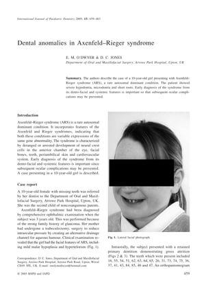
vealed that the girl had the facial features of ARS, includ-
ing mild malar hypoplasia and hypertelorism (Fig. 1). Intraorally, the subject presented with a retained
primary dentition demonstrating gross attrition
(Figs 2 & 3). The teeth which were present included
16, 55, 54, 51, 62, 63, 64, 65, 26, 31, 73, 74, 75, 36,
37, 41, 43, 84, 85, 46 and 47. An orthopantomogram
Correspondence: D. C. Jones, Department of Oral and Maxillofacial
Surgery, Arrowe Park Hospital, Arrowe Park Road, Upton, Wirral
CH49 5PE, UK. E-mail: emilymodwyer@hotmail.com
Fig. 1. Lateral facial photograph.
2. 460 E. M. O’Dwyer & D. C. Jones
© 2005 BSPD and IAPD, International Journal of Paediatric Dentistry 15: 459–463
revealed severe hypodontia. (Fig. 4). Unerupted teeth
included 17, 14, 24, 25, 33, 34 and 44, with an
unerupted, rudimentary upper right second premolar
(15). In total, 15 permanent teeth (18, 13, 12, 11,
21, 22, 23, 27, 28, 32, 35, 38, 42, 45 and 48) were
absent (Table 1). All first permanent molars and
lower second molars were in situ, with the upper left
canine and lower left first premolar partially erupted.
No other permanent teeth were identified. This
severe hypodontia with missing central and lateral
incisors and second premolars is characteristic of
ARS. All teeth were microdont and had short roots.
The patient remains under review in a specialist
paedodontic clinic and may require extensive oral
rehabilitation when growth of the cranio-facial
skeleton is complete. This will comprise of a multi-
disciplinary team approach involving the general dental
practitioner, the restorative dentist, the orthodontist,
and the oral and maxillofacial surgeon.
Discussion
History
Vossius first documented bilateral anterior cham-
ber defects of the eyes associated with hypodontia
Fig. 2. Frontal occlusion photograph showing a retained primary
dentition, diastema and a bilateral maxillary posterior cross-bite.
Fig. 3. (a & b) Lateral occlusion photographs.
Fig. 4. Orthopantomogram displaying severe hypodontia with
absent central and lateral incisors, and lower second and upper
right premolar. NB Tooth 15 is rudimentary.
Table 1. Chart of the subject’s teeth which were recorded as present,
unerupted or missing using the Zsigmondy–Palmer and FDI systems.
Variable Teeth
Zsigmondy–Palmer system
Present 6 E D A | B C D E 6
7 6 E D 3 1 | 1 C D E 6 7
Unerupted 7 5 4 | 4 5
4 | 3 4
Permanent teeth missing 8 3 2 1 | 1 2 3 7 8
8 5 2 | 2 5 8
F.D.I. system
Teeth Present 16, 55, 54, 51 | 62, 63, 64, 65, 26
47, 46, 85, 84, 43, 41 | 31, 73, 74, 75, 36, 37
Teeth
Unerupted
17, 15, 14 | 24, 25
44 | 33, 34
Permanent teeth missing 18, 13, 12, 11 | 21, 22, 23, 27, 28
48, 45, 42 | 32, 35, 38
3. Axenfeld–Rieger syndrome 461
© 2005 BSPD and IAPD, International Journal of Paediatric Dentistry 15: 459–463
in 1883. In 1820, Karl Axenfeld, an ophthalmolo-
gist, described the syndrome as ‘a white line in the
posterior aspect of the cornea and tissue strands
from the periphery of the iris to this line’ [1].
Herwigh Rieger described Axenfeld’s findings plus
changes in the iris, stromal atrophy, pupillary abnor-
malities and nonocular development defects as an
inherited condition in 1935 [2,3].
The incidence of ARS is estimated at one per
200 000 of the population [4]. It is an inheritable
developmental disorder with complete penetrance
and variable expression [1,2]. Inheritance is auto-
somal dominant in 70% of cases with 30% of cases
arising de novo [4,5]. Grosso et al. 2002 described
cases with overlapping phenotypes which demon-
strate high ‘intrafamilial variability’. When diagnos-
ing a syndrome, this variability can cause confusion
since the classic features may not be expressed.
Clinical features of the Rieger ocular anomaly and
the Axenfeld anomaly
According to Jorgenson, an anomaly is ‘a specific
defect’ [7]. The Rieger ocular anomaly and the Axenfeld
anomaly are both disorders of embryological develop-
ment of the anterior chamber of the eye alone. They are
known as anterior chamber cleavage syndromes [1].
Glaucoma and visual loss occurs in 60% of all
children who are diagnosed with the Rieger or
Axenfeld anomalies [2]. The anterior chamber angle
abnormalities in the Rieger anomaly, Axenfeld
anomaly and ARS are parts of the spectrum of a
single disorder with a broad overlapping phenotype [8].
Rieger syndrome
A syndrome refers to ‘commonly associated defects’
or ‘a recognized pattern of malformations’ [7].
Rieger syndrome includes ocular features, such as
goniodysgenesis (a developmental aberration of the
anterior ocular segment), with dental, craniofacial
and skeletal defects. The extraocular features which
are consistent include hypodontia and failure of the
periumbilical skin to involute [7,9]. The dental mani-
festations of Rieger syndrome differentiate it from
other ocular syndromes, such as the Peter anomaly,
the Rieger anomaly, juvenile glaucoma or the Axen-
feld anomaly [10]. Associated conditions are learn-
ing difficulties, cerebellar hypoplasia, conduction
deafness, limb malformations, hypospadias in males
and congenital heart defects [9,11].
Shields et al. discussed the difficulty differentiat-
ing the Axenfeld and Rieger anomalies clinically,
and proposed the collective term Axenfeld Riegers
Syndrome (ARS) for all variations within this spectrum
of disorders [1]. Axenfeld–Rieger syndrome illustrates
three clinical expressions of the same gene [7]. Patients
have ‘a bilateral developmental disorder of the eyes,
autosomal dominant inheritance, no sex predilection,
frequently nonocular developmental defects and a
high incidence of secondary glaucoma’ [1].
Aetiology of Axenfeld–Rieger syndrome
It has been suggested that ARS is a result of
damaged or abnormally migrated neural crest cells,
the ectodermal mesostroma, late in gestation [12].
Neural crest cells are responsible for initiating cranio-
facial, dental and ocular development. This results
in underdeveloped associated neural crest tissues,
i.e. the dental and facial bones.
Clinical features of Axenfeld–Rieger syndrome
Table 2 describes the clinical features of ARS.
These patients often have maxillary hypoplasia (not
observed in this case, see Fig. 1), prominent supra-
orbital ridges, telecanthus, a broad nasal bridge, and
a protrusive lower and recessive upper lip [13].
An empty or enlarged sella turcica has also been
described. Hypodontia may result in an underdevel-
oped maxillary alveolus at the site of the missing
teeth, resulting in a reduced occlusal face height
[14]. In a study by Ozekis in 1999, 43% of patients
with ARS had dental anomalies, while 25% had
facial anomalies [15].
Mathias first reported the dental anomalies in 1936
[1]. The oral manifestations of ARS vary: These
patients exhibit microdontia, hypodontia, enamel
hypoplasia and taurodontia. Central incisors, lateral
incisors and second premolars are frequently micro-
dont or missing [4,13]. The case described here had
some absences in the permanent dentition.
Genetic features
The ARS chromosomal abnormality has been
linked to loci at chromosomes 4q25, 6p25, 13q14,
16q24 and 11. Two genes, FOX-C1 and P1TX2, on
chromosomes 4q25 and 6p25 have been identified
[16,17]. Mutations in the P1TX2 coding sequence
lead to various phenotypes of ARS and other anterior
4. 462 E. M. O’Dwyer & D. C. Jones
© 2005 BSPD and IAPD, International Journal of Paediatric Dentistry 15: 459–463
ocular segment malformations [17]. It has been
suggested that the Dlx2 gene expression is a reg-
ulator of branchial arch development and plays a
role in tooth morphogenesis in patients with ARS
[18].
Management
Patients with ARS require regular ophthalmic
appointments to monitor intraocular pressure and
optic nerve head changes throughout their lives so
that glaucoma can be diagnosed. Since the syndrome
is frequently familial, blood relatives should also
be investigated. Over 50% of patients with ARS
develop secondary glaucoma, which poses the threat
of visual loss and blindness.
The initial therapy for glaucoma is medical, and
involves the topical application of alpha-agonists,
beta-blockers or carbonic anhydrase inhibitors in an
attempt to decrease intraocular pressure [1]. Surgical
intervention is required in the majority of patients,
and this includes goniotomy (a procedure whereby
an opening is made in the skin blocking the drainage
gap of the eye, thus providing a passage for aqueous
fluid to flow out of the eye and reduce intraocular
pressure), trabeculectomy (a passageway/drainage
tube in the sclera, hidden under the upper eyelid,
by which the aqueous fluid inside the eye can escape
to lower the intraocular pressure) and trabeculotomy
(a piece of tissue in the drainage angle of the eye
is removed to create an opening that allows aqueous
humour to drain from the eye) [1]. Trabeculectomy
with antimetabolite therapy is the treatment of
choice for patients suffering from ARS.
The aim of conservative dental rehabilitation is
twofold, to improve both aesthetics and function. In
the long term, dental implants remain the most
likely treatment option for these patients. Close
liaison between dental professionals is essential to
monitor facial growth and dental development,
and to coordinate appropriate timing for dental
treatment.
Conclusion
In clinical practice, it is necessary to differentiate
de novo hypodontia from that which is syndrome-related.
This involves close interdisciplinary management
between dentists, paediatricians and geneticists.
Early diagnosis of the dental, cranio-facial and
systemic presentation of ARS could prevent the
devastating ocular effects of infantile glaucoma.
Table 2. Clinical manifestations of Axenfeld–Rieger syndrome.
Condition Clinical manifestation
Ocular Axenfeld anomaly
Rieger ocular abnormality (as described)
goniodysgenesis
Glaucoma
Cranio-facial Maxillary hypoplasia/prognathic profile
Mandibular hypoplasia
Hypertelorisim
Prominent supraorbital ridges
Telecanthus
Broad nasal bridge
Protrusive lower lip/recessive upper lip
Enlarged sella turcica
Dental Hypodontia
Hypoplasia
Taurodontia
Microdontia
Hyperplastic frena
Associated
systemic
conditions
Failure of periumbilical skin to involute
Cerebellar hypoplasia
Conduction deafness
Vertebral/limb malformations
Hypospadias
Congenital heart defects
Myotonic dystrophy
Scoliosis
Kyphosis
Lipodystrophy
Inguinal hernia
Sternum abnormalities
Kidney malformations
Retarded bone growth
What this case report adds
• A 10-year old girl with Axenfeld-Riger syndrome presented
with a retained primary dentition demonstrating gross attrition.
• In total 15 permanent teeth were absent (including third
molars).
• This severe hypodontia with missing central and lateral
incisors and second premolars is characteristic of the
syndrome.
Why this case report is important to paediatric dentists
• Early diagnosis of the Axenfeld-Riger syndrome from its
dento-facial features is important since subsequent ocular
complications may be prevented.
• Children with this syndrome exhibit a wide range of
craniofacial and dental disturbances such as maxillary and
mandibular hypoplasia, hypodontia, enamel hypoplasia and
microdontia.
• The aim of dental rehabilitation is to improve both
aesthetics and function. Dental implants is the most likely
treatment option for these patients.
5. Axenfeld–Rieger syndrome 463
© 2005 BSPD and IAPD, International Journal of Paediatric Dentistry 15: 459–463
Acknowledgements
The authors wish to thank Dr J. Cooper, Specialist
Registrar in Orthodontics at Arrowe Park Hospital,
for his clinical photographs.
Résumé. Le syndrome de Axenfeld-Rieger (ARS)
est une maladie autosomique dominante rare. Elle
comprend des éléments des syndromes de Axenfelds
et Riegers, indiquant que ces deux maladies sont des
expressions variables d’une même anomalie de gène.
Le syndrome est caractérisé par un développement
arrêté ou perturbé des cellules de la crête neurale
dans la chambre antérieure de l’œil, les os de la face,
les dents, l peau péri-ombilicale et le système cardio-
vasculaire. Un diagnostic précoce de ce syndrome à
partir des caractéristiques dento-faciales et systémiques
est important car les complications oculaires sub-
séquentes peuvent être prévenues. Le cas d’une
jeune fille de 10 ans est décrit.
Zusammenfassung. Axenfeld-Rieger Syndrom (ARS)
ist eine seltenen Erkrankung mit autosomal dominanter
Vererbung. Diese beeinhaltet Symptome von Axenfeld
Syndrom und Rieger Syndrom, was darauf hindeutet,
dass beide Krankheitsbilder auf Veränderungen des
gleichen Gens zurückzuführen sind. Charakteristisch
für dieses Syndrom ist veränderte oder unterbliebene
Entwicklung von Zellen aus der Neuralrinne mit
Auswirkungen auf Auge, Gesichtsknochen, Zähne,
periumbilikale Haut und das kardiovaskuläre System.
Die frühzeitige Diagnose anhand der dentofazialen
und systemischen Symptome ist wichtig, da dann
Komplikationen am Auge verhindert werden
können. Der Fall einer 10 jährigen Patientin wird
vorgestellt.
Resumen. El síndrome de Axenfeld-Riegers (SAR)
es una rara alteración autosómica dominante. Incorpora
rasgos de los síndromes de Axenfelds y Riegers e
indica que ambas alteraciones son expresiones variables
de la misma anormalidad genética. El síndrome se
caracteriza por desarrollo retrasado o detenido de las
células de la cresta neural en la cámara anterior del
ojo, huesos faciales, dientes, piel periumbilical y
sistema cardiovascular. Es importante el diagnóstico
precoz del síndrome por sus rasgos dento-faciales y
sistémicos para que puedan prevenirse las compli-
caciones oculares posteriores. Se describe la presentación
de un caso en una niña de 10 años.
References
1 Shields MB, Buckley E, Klintworth GK, Thresher R. Axenfeld–
Rieger syndrome. A spectrum of developmental disorders.
Survey of Ophthalmology 1985; 29: 387.
2 Wesley RK, Golnick AL. Rieger’s Syndrome. Journal of Pedi-
atric Ophthalmology and Strabismus 1978; 15 (2): 67–70.
3 Tabbara KF, Khouri FP, Der Kaloustian V. Rieger’s Syn-
drome with chromosomal anomaly. Canadian Journal of
Ophthalmology 1973; 8: 488–490.
4 Gorlin R, Pindborg J, Cohen M. Rieger’s syndrome. In: Gorlin
R, Pindborg J, Cohen M. (eds). Syndromes of the Head and
Neck, 2nd edn. New York, NY: McGraw-Hill, 1976: 649–651.
5 Riekman G. Rieger’s syndrome: case report. Canadian Jour-
nal of Dentistry for Children 1981; 48 (1): 55–56.
6 Grosso S, Farnetani MA, Berardi R, et al. Familial Axenfeld–
Rieger Anomaly, cardiac malformations and sensorineural
hearing loss: a provisionally unique genetic syndrome? Amer-
ican Journal of Medical Genetics 2002; 111: 182–186.
7 Jorgenson RJ, Stefan Levin L, Cross HE, Yoder F, Kelly TE.
The Rieger Syndrome. American Journal of Medical Genetics
1978; 2: 307–318.
8 Fitch N, Kabach M. The Axenfeld Syndrome and the Rieger
Syndrome. The Journal of Medical Genetics 1978; 15: 30–34.
9 Lapeer G. Rieger’s Syndrome, severe dental anomalies with
mild ophthalmic changes – a case report. Journal of the
Canadian Dental Association 1986; 11: 935–937.
10 Dimitrakopoulos J, Voyatzis N, Katopodi T. Rieger syndrome:
a case report. Journal of Oral and Maxillofacial Surgery
1997; 55: 517–521.
11 Heckenlively JR, Istenburg SJ, Fox LE. The Rieger Syn-
drome: a heritable disorder associated with glaucoma. The
Johns Hopkins Medical Journal 1982; 151 (6): 351–355.
12 Childers NK, Wright JT. Dental and craniofacial anomalies
of Axenfeld–Rieger syndrome. Journal of Oral Pathology
1986; 15: 534–539.
13 Prabhu NT, John R, Munshi AK. Rieger’s syndrome: a case
report. Quintessence International 1997; 28: 749–752.
14 Scarparo Caldo-Teixera A, Puppin-Rontani RM. Management
of severe partial hypodontia: case report. Journal of Clinical
Pediatric Dentistry 2003; 27 (2): 133–136.
15 Ozekis H, Shirai S, Ikeda K, Ogura Y. Anomalies associated
with Axenfeld–Rieger syndrome. Graese’s Archive for Clinical
and Experimental Ophthalmology 1999; 237 (9): 730–734.
16 Vaux C, Sheffield L, Gregory Keith C, Voullaire L. Evidence
that Rieger syndrome maps to 4q25 or 4q27. Journal of
Medical Genetics 1992; 29: 256–258.
17 Priston M, Kozlowski K, Gill D, et al. Functional analyses
of two newly identified PITX2 mutants reveal a novel molecular
mechanism for Axenfeld–Rieger syndrome. Human Molecular
Genetics 2001; 10 (16): 1631–1638.
18 Epinoza HM, Cox CJ, Semina EV, Amendit BA. A molecular
basis for differential developmental deformities in Axenfeld–
Rieger’s syndrome. Human Molecular Genetics 2002; 11 (7):
743–753.