The cell cycle consists of four main phases - G1, S, G2, and M. In G1, cells grow and undergo protein synthesis in preparation for DNA replication. The S phase is when DNA replication occurs. In G2, the cell prepares for mitosis by producing necessary proteins. During mitosis (M phase), the nucleus and cell contents divide to form two daughter cells each with identical DNA. The progression through the cell cycle phases is regulated by cyclins and cyclin-dependent kinases (CDKs). Different cyclins activate specific CDKs to promote the transition between phases, with checkpoints ensuring errors are corrected before progression.
2. 2
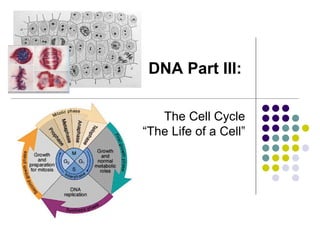
Cells must reproduce else they die. The "life of a cell" is termed the cell cycle.
The cell cycle has distinct phases, which are called G1, S, G2, and M.
Cells that have temporarily or reversibly stopped dividing are said to have
entered a state of quiescence called G0 phase.
Cells & Cell Reproduction
3. 3
• During this time organelles
are reproducing, protein
synthesis is occurring for
growth and differentiation.
• Because, transcription is
occurring, the DNA is
uncoiled.
• This phase is the most
variable, ranging from
almost nothing to years.
The G1 Phase of the Cell Cycle
4. The G1 Phase of the Cell Cycle
Most cells that differentiate will do
so during this phase. Cells arrested
in G1 may no longer have the
capability of reproducing and are
said to be in G0.
Certain cells in G0, however, when
given some external or internal
cues may revert back to G1 and
enter the cell cycle again.
Nerve and muscle cells are usually
arrested in G0.
5. 5
The S or synthesis phase is the second
phase of the cell cycle.
•DNA uncoils
•DNA replication occurs
•Additional organelle replication
occurs
•This phase ensures that each emerging
daughter cell will have the same
genetic content as the mother cell.
S Phase of the Cell Cycle
6. 6
The G2 or Gap 2 phase occupies
the time from the end of S until
the onset of mitosis.
•During this time, the cell
prepares for mitosis by making
and organizing necessary
proteins such as the tubulin
needed to construct
microtubules which used to
make spindle fibers.
•On the average this phase may
take four hours.
G2 Phase of the Cell Cycle
7. • During mitosis the nucleus is
replicated and the cytoplasm
divides to produce two
genetically identical daughter
cells.
• Remember that the DNA is
replicated in S prior to mitosis.
• The phases are triggered by the
accumulation of cell signals.
M Phase or Mitosis
8. 8
This graph represents the amount of DNA found in the
cell during the cell cycle. What caused the changes?
What happens at the end of Mitosis?
The Amount of DNA Varies During the Cell Cycle
9. 9
What Controls the Cell Cycle?
Pay attention to the 3 points in the
cycle that serve as “checkpoints”
10. 10
• The control of the cell cycle
is dependent on an
accumulation of “signal
molecules”.
• Quite often these signal
molecules must be
phosphorylated in order to
be functional. This are
simple illustrations.
Internal Controls of the Cell Cycle
11. Cyclins are a family of proteins that control the
progression of cells through the cell cycle by
activating cyclin-dependent kinase (Cdk) enzymes.
Only with the cyclin is the Cdk active. Cyclins were
originally named because their concentration varies
in a cyclical fashion during the cell cycle.
A kinase is a type of enzyme that transfers
phosphate groups from high-energy donor
molecules, such as ATP, to specific substrates, a
process referred to as phosphorylation.
.
11
Cyclins vs. Kinases
12. 12
Cyclins vs. Kinases
• Certain cyclins are made at certain times during the cell cycle, and their
concentration will rise and fall. Cyclins are also destroyed after they are no
longer needed by the cell.
• CDKs are not destroyed as they are only activated or deactivated.
• Which cyclin affects which phase of the cycle? (you don’t have to memorize
it but be able to read it in the graph!)
13. 13
•Certain kinases may have
two forms (active and
inactive).
•Kinases are enzymes
(proteins) that
phosphorylate certain
molecules or other enzymes.
•Most cell cycle signals are
phosphorylated by kinases.
Kinases Phosphorylate Cell Signal Molecules
Note: Kinases are a type of enzyme that transfers phosphate groups from high-energy donor
molecules, such as ATP, to specific substrates, a process referred to as phosphorylation.
They are not to be confused with phosphorylases, which carry out phosphorolysis, the
breaking of a bond using an inorganic phosphate group; or with phosphatases, which remove
phosphate groups. (They all start to sound alike, don’t they?)
14. 14
Cyclins Activate Kinases
Most cell cycle kinases are activated by molecules called cyclins. A kinase that
requires a cyclin for activation is called a cyclin-dependent kinase or Cdk.
15. The cyclin attaches to the Cdk. It is now called a cyclin-Cdk complex.
The complex that regulates the M (mitosis) portion of the cell cycle has 3
names (ugh!): the maturation-promoting factor, mitosis-promoting factor or
M-Phase promoting factor. Luckily they are all referred to as “MPF”.
MPF is activated at the end of G2 by a phosphatase, which removes an
inhibitory phosphate group added earlier.
The MPF is also called the M phase kinase because of its ability to
phosphorylate target proteins at a specific point in the cell cycle and thus
control their ability to function.
Cyclins Activate Kinases
16. An example of how MPF initiates mitosis ….
MPF promotes the entrance into mitosis (the M phase) from the G2 phase
by phosphorylating multiple proteins needed during mitosis. The steps
follow:
The nuclear lamina depolymerizes causing it to disassemble which in
turn causes the nuclear membrane to disassemble
Histone H1 binds to the DNA in chromosomes, causing the
chromosomes to condense
Cytoskeletal proteins allow cytoskeletal filaments to assemble which
leads to:
Formation of the mitotic spindle which separates the daughter chromosomes
formation of the cleavage furrow by microfilaments which allows cytokinesis
(constricting the cell at the center) to occur resulting in the formation of two
new cells
No – you don’t need to memorize the details – but a
basic understanding about how one thing can lead
to another is good!
17. 17
Cyclins Activate Kinases
Once the CDK phosphory-
lates certain signals, the
cyclin is destroyed.
In the cell, the concentration
of cyclins will rise and fall
depending on the phase of
the cell cycle.
When the cyclin is
destroyed the Cdk returns to
an inactive form (it is NOT
destroyed!).
19. 19
Cyclic Nature of Cyclins in the
Cell Cycle
This graph displays the cyclic nature of various cyclins in a given cell cycle.
Notice again that a number of cyclins are involved in the cell cycle and that they
activate a number of different kinases.
20. 20
Random info ….
The term cyclin was coined by R. Tim Hunt who discovered
them while studying the cell cycle of sea urchins cells. He
said he named it after his hobby of cycling and at the time he
did not realize the role of these molecules in the cell cycle.
However due the cyclic nature of the concentration of these
compounds and their role in the cell cycle, the name stuck.
Cyclins (D, E, A, B) are named based on the their protein
structure and conserved parts. Older classification of cyclins is
based on their role in the cell cycle. Most introductory books
use the terms like S cyclin and M cyclin.
R. Tim Hunt along with Leland Hartwell and Sir Paul Nurse
received the Nobel Prize in Medicine in 2001 for their
discovery and research in the role of cyclins and CDKs in the
cell cycle.
21. 21
Different Types of Cyclins
These are some known cyclin/CDK complexes and their
role in the cell cycle. (Don’t memorize – just understand
the general functions)
Cyclin/CDK
Complex
Cyclin Function of Cyclin/CDK Complex
G1-CDK Cyclin D Drives the transition G1 S transition
G1/S-CDK Cyclin E Cyclins bind to CDK at the end of G1 and commits the
cell to DNA replication.
S-CDK Cyclin A Cyclins bind the CDK during S and are necessary for the
initiation of DNA replication
M-CDK Cyclin B Cyclins promote the events of Mitosis
22. 22
Cyclins/CDKs Control the Cell Cycle
G1/S (R point) checkpoint is the primary determining factor for cell division to
take place. Growth factors are affecting the cell cycle, and cells are
growing. Once the R point is passed the DNA is going to be replicated. If a
cell receives a go-ahead signal at this check-point, it will complete the cell
cycle and divide. However, if the cell does not receive the go-ahead signal
in G1, the switches to a nondividing state called G0. If the cell passes the
G1 checkpoint, it is usually committed to cell reproduction.
The cell cycle has a number
of several external and
internal checkpoints
much like a timer or
clock. Often, moving
past these check points
involves chemical signals
that have been
phosphorylated by
cyclin-CDK complexes.
23. 23
Cyclins/CDKs Control the Cell Cycle
The M/spindle checkpoint ensures that all the chromosomes are attached to the
spindle in preparation of mitosis. The separation of the chromatids is
irreversible.
Once chromatids are replicated they are held together by a protein substance called
cohesin protein. Another protein called seperase can destroy this protein but has
two forms active and inactive.
The G2 checkpoint represents the
commitment for starting the
process of mitosis. This
checkpoint also ensures that
the DNA has been replicated
correctly. If the DNA has been
damaged, then the cell does not
continue to mitosis. Once the
CDK and cyclin combine, it is
called “maturation promoting
factor" or “mitosis promoting
factor” or MPF.
25. 25
External Signals also affect cell division
External Signals also affect cell
division. Mammalian cells
need certain nutrients and
regulatory proteins. In addition
external growth factors are can
determine cell division in
mammals. For example, when
the skin has been damage
(wound), platelets release a
substance called platelet-
derived growth factor (PDGF).
This growth factor stimulate
fibroblasts cells to start to
reproduce and make scar
tissue.
26. 26
External Factors Also Influence the Cell Cycle
Cell reproduction and the healing
of a wound.
I. Hemostasis - Bleeding is
contained via blood clotting.
II. Inflammation - Bacteria and debris
removed. Damaged platelets release
platelet derived growth factors. These
external growth factors signal fibroblast
to start to reproduce and make new cells.
III. Proliferation - Fibroblast reproduce
forming an extra cellular matrix
IV. Remodeling - Extra collagen and
cells that are not needed are removed.
Revisit this graph.
The term cyclin was coined by R. Tim Hunt who discovered them while studying the cell cycle of sea urchins cells. He said he named after his hobby of cycling and at the time he did not realize the role of these molecules in the cell cycle. However due the cyclic nature of the concentration of these compounds and their role in the cell cycle, the name stuck. Cyclins (D, E, A, B) are named based on the their protein structure and conserved parts. Older classification of cyclins is based on their role in the cell cycle. Most introductory books use the terms like S cyclin and M cyclin.
R. Tim Hunt along with Leland Hartwell and Sir Paul Nurse received the Nobel Prize in Medicine in 2001 for their discovery and research in the role of cyclins and CDKs in the cell cycle.
Revisit this graph.
.
Scroll over the bottom of the image in presentation mode and press play (the “Play” button on the screen is simply an image, thus inactive).
http://highered.mcgraw-hill.com/sites/0072495855/student_view0/chapter2/animation__how_the_cell_cycle_works.html
Scroll over the bottom of the image in presentation mode and press play (the “Play” button on the screen is simply an image, thus inactive).
This phase ensures that each emerging daughter cell will have the same genetic content as the mother cell.
The average time for S is around 9 hours, if the cell cycle lasts 24 hours.
Be sure to connect this information to the previous PowerPoint on DNA replication.
Scroll over the bottom of the image in presentation mode and press play (the “Play” button on the screen is simply an image, thus inactive).
Scroll over the bottom of the image in presentation mode and press play (the “Play” button on the screen is simply an image, thus inactive).
This animation ends with a summary of the entire process.
This serves as an introduction to the controls of the cell cycle. Emphasize the 3 points in the cycle that serve as “checkpoints”.