2. 12 2 Congenital Malformations and Abnormalities of Spleen Location and Histology
2.1 Accessory Spleen
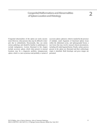
Fig. 2.1 Accessory spleen. A section of spleen with attached pancreas
and omentum. In the mass of tissue arising from the hilum, there is a
small nodule (approximately 2 cm) of dark red purple tissue. This is an
accessory spleen. They are seen in approximately 10 % of the popula-
tion and are most commonly seen in the splenic hilar region. (Image
courtesy of D. Farhi, Atlanta, GA, USA.)
a
b
Fig. 2.2 Accessory spleen. This pathologic specimen was submitted
as an “enlarged lymph node” with a suspicion of metastatic carcinoma.
It was seen adjacent to the pancreas and, as illustrated by histology (a)
and immunohistochemical stains for CD8 (b), is an accessory spleen.
CD8 staining highlights the distinctive splenic sinus architecture of the
spleen and can be useful in identifying spleen tissue at unusual sites
Fig. 2.3 Gross image of a sectioned accessory spleen. In this case, the
accessory spleen was approximately 5 cm in diameter. Note that the cut
surface has is a deep red color, similar to the spleen. (Image courtesy of
L. Morgenstern, Los Angeles, CA, USA.)
3. 132.3 Polysplenia
Fig. 2.4 Intrapancreatic spleen is a rare finding associated with embry-
ologic abnormality. Small fragments of spleen tissue are entrapped
within a portion, usually distal, of the pancreas (normal pancreatic tis-
sue; right). Although these can vary in size, they are usually quite small.
In this case, the elements are predominant red pulp components, sug-
gesting that this was a relatively early embryologic event. (Image cour-
tesy of R. Mills, Salt Lake City, UT, USA.)
2.2 Intrapancreatic Spleen
Fig. 2.5 Polysplenia. Gross image of polysplenia. In this case, the pri-
mordial separate lobes of splenic tissue do not fuse to make a single
unified organ. Rather, they are partly fused or remain separated by
fibrous bands. Although this anatomic abnormality can be seen associ-
ated with normal function, it is often associated with other, severe con-
genital abnormalities. (Image courtesy of D. Farhi, Atlanta, GA, USA.)
2.3 Polysplenia
Fig. 2.6 Polysplenia. Another example of a small gross specimen from
a fetus with polysplenia. Separated nodules of spleen tissue are present.
Each nodule would have its own artery and vein, which eventually
merge into the common splenic artery or vein. (Image courtesy of D.
Farhi, Atlanta, GA, USA.)
Fig. 2.7 Polysplenia. Still another example of polysplenia. Note that
the lobes of the spleen have not completely fused. (Image courtesy of
D. Farhi, Atlanta, GA, USA.)
4. 14 2 Congenital Malformations and Abnormalities of Spleen Location and Histology
Fig. 2.9 Splenosis. Microscopic image of splenosis. The tissue present
is composed of fibroadipose tissue from the peritoneum. At the intrap-
eritoneal surface (right and upper) are slightly fibrous lymphoid areas.
These represent fragments of spleen tissue that were likely “trans-
planted” to this site following abdominal trauma
Fig. 2.8 Splenosis. Gross image of splenosis. In this case, there are
nodular, dark red tissue fragments attached to segments of bowel. This
most often occurs after trauma, when the spleen is ruptured. Small frag-
ments of splenic tissue are seeded throughout the abdominal cavity.
Each establishes its own blood supply and functions as a tiny but com-
plete spleen. In cases of splenectomy and pathologic processes, such as
hemolytic anemias, these small displaced splenic fragments may
enlarge greatly. (Image courtesy of D. Farhi, Atlanta, GA, USA.)
2.4 Splenosis
Fig. 2.11 Spleno-gonadal fusion. Ectopic splenic tissue in a sectioned
testicle (lower). The splenic tissue has the usual deep red color, with
normal, somewhat compressed testicular tissue in the upper portion of
the sample
Fig. 2.10 Spleno-ovarian fusion. Gross image of spleno-ovarian (eg,
splenogonadal) fusion. In this case, there is a physical attachment
between the stretched and elongated splenic fragment, which has teth-
ered a portion of the left ovary. In some cases, the fusion may only
consist of a thin fibrous band, which may have small entrapped frag-
ments of splenic parenchyma
2.5 Splenogonadal Fusion
5. 152.6 Other Findings
a
b
Fig. 2.13 Tissue taken from “left inguinal hernia.” In this case, the
histology (a) and presence of a red pulp architecture staining by CD8
(b) show that the “inguinal hernia” was in fact caused by ectopic spleen
tissue. In the descent of the testes from the mesonephros, small portions
of spleen tissue may be transported. When these fragments are attached,
it is splenogonadal fusion; when unattached, these fragments may be
present as inguinal lumps, within the inguinal canal, simulating a
hernia
Fig. 2.12 Surface grooves. This spleen is enlarged by a low-grade
B-cell lymphoma. However, deep grooves are seen in the medial sur-
face (upper left), the upper pole (far right), and in the lateral/dorsal
surface (lower, middle). These grooves are a normal variant, and are
residual separations from the original embryologic splenic lobes that
are incompletely fused in the adult. They have no specific physiologic
consequences. (Image courtesy of W. Greaves, Houston, TX, USA.)
2.6 Other Findings a
b
Fig. 2.14 Needle biopsy, lung. A biopsy of the left lower lobe of lung
revealed this tissue. In this case, the lung was missed and a sample of
normal spleen was taken (a). Normal splenic architecture is highlighted
by CD8 staining (b), which is a useful stain for highlighting splenic red
pulp architecture