Periosteum: The Bone-Covering Membrane
•Als PPTX, PDF herunterladen•
6 gefällt mir•1,350 views

The periosteum is a membrane that covers the outer surface of bones and plays an important role in bone growth and repair. It has two layers - an outer fibrous layer and inner cambium layer. The cambium layer contains stem cells that can differentiate into bone-forming cells and is responsible for bone growth. The periosteum gets thinner with age and has decreasing ability to form new bone. It receives blood supply from surrounding muscles and fascia. The periosteum is used in procedures like bone grafting and repairing cartilage defects due to its osteogenic and chondrogenic potential.

Empfohlen
Weitere ähnliche Inhalte
Was ist angesagt?
Was ist angesagt? (20)
Ähnlich wie Periosteum: The Bone-Covering Membrane
Ähnlich wie Periosteum: The Bone-Covering Membrane (20)
Kürzlich hochgeladen
Kürzlich hochgeladen (20)
Periosteum: The Bone-Covering Membrane
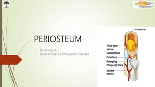
- 1. PERIOSTEUM Dr Sandip,PG1 Department of Orthopaedics, SMS&R
- 2. Definition Periosteum is derived from the Greek word Peri-, meaning "surrounding“, and -osteon, meaning "bone” The periosteum is a membrane that covers the outer surface of all bones, except at the articular surfaces of long bones and sesamoid bones. Has a major role in bone growth and bone repair. Also has an impact on the blood supply of bone as well as skeletal muscle.
- 3. Histology 2 distinct layers - Outer Fibrous Layer & Inner Cambium Layer (or Osteogenic Layer ) Outer Fibrous Layer – dense irregular connective tissue. Inner Cambium Layer – osteoblasts (bone forming cells) & osteoclasts (bone remodelling cells) The periosteum is attached to the bone by strong collagenous fibres called Sharpey's fibres.
- 6. Outer Fibrous Layer Subdivided into 2 parts – superficial and deep Superficial Portion - • generally inelastic. • highly vascularized and is a significant contributor to the blood supply of bone and even skeletal muscle. • It also includes a rich neural network. Deep Portion – • Fibroelastic layer , not highly vascularized • Periosteal tendon attachments usually terminate here.
- 7. Inner cambium layer highly cellular. composed of mesenchymal progenitor cells, differentiated osteogenic progenitor cells, osteoblasts and fibroblasts. responsible for increasing the width of a long bone and the overall size of the other bone types. After a bone fracture, the progenitor cells develop into osteoblasts and chondroblasts, which are essential to the healing process.
- 8. Inner Cambium Layer cont.… Thickest in the foetus Becomes progressively thinner with age. In the adult it becomes so thin that it cannot be distinguished from the overlying fibrous layer. With this atrophy comes a decrease in the osteoblastic potential as well.
- 10. Changes seen with Age Periosteum changes with age. The thick cellular vascular periosteum of infants and children readily forms new bone This capacity is evident when osteomyelitis or trauma destroys the diaphysis of a young individual’s bone and the periosteum regenerates a new diaphysis.
- 11. With increasing age periosteum becomes thinner and less vascular and its ability to form new bone decreases. Cells of the deeper layer become flattened and quiescent Yet they continue to form new bone that increases bone diameter They still have the potential to form bone and cartilage in response to injury.
- 12. Blood Supply A Plexus of small vessels lies on the outer fibrous layer of the periosteum. These periosteal blood vessels anastomose with vessels of the overlying muscle Branches of the vessels on the surface of the periosteum penetrate the fibrous layer These contribute to the vascular system of the deeper layer of the periosteum and to the blood vessels that penetrate bone And finally joins the Medullary Vascular System
- 14. The blood supply of periosteum is derived from 4 sets of vessels: (1) Intrinsic periosteal system. (2) Musculoperiosteal system. (3) Fascioperiosteal system. (4) Cortical capillary anastomoses.
- 15. The Intrinsic system of vessels lay within the fibrous layer of the periosteum. According to the pattern of these vessels they could be divided into: (a) a short vessel pattern, where there were many small vessels with no predominant direction; (b) a circular pattern, where the vessels encircled the bone; (c) a longitudinal pattern, where the vessels ran parallel to the long axis of the bone.
- 16. The Musculoperiosteal system consist of connections between the muscle circulation and the periosteal vessels at the sites of muscle origin. The Fascioperiosteal system consist of branches from a limb artery that run in a fascial plane between muscles to supply the periosteum. The Cortical capillary anastomosis consist of capillaries that run in the bone cortex between the intramedullary circulation and the periosteal vessels.
- 19. Longitudinal periosteal vessel arising from the nutrient artery of the radius. Arrow shows nutrient artery entering the nutrient foramen of the radius.
- 20. Nerve Supply Nerve Cell processes lie on the external periosteal surface. They accompany periosteal blood vessels. They help regulate periosteal blood flow.
- 21. Clinical Significance Cartilage Repair: • Cambium layer contains multipotent stem cells that can differentiate in both Osteoblasts as well as Chondroblasts. • The Periosteal Graft can help to repair the articular cartilage defect. Bone Repair: • Periosteum is also used to repair a bone defect • Fibula is one of the common bone used for bone grafting • Periosteal grafts and periosteal stripping can be used in limb equalizing procedures in patients with limb length discrepancies
- 22. Reference: Turek’s Orthopaedics: Principles and Their Application, 6th Edition Gray's Anatomy – The Anatomical Basis of Clinical Practice, 41st Ed https://www.ncbi.nlm.nih.gov/pmc/articles/PMC2826636/