1. Research article
492 The Journal of Clinical Investigation http://www.jci.org Volume 119 Number 3 March 2009
MT1-MMP and RECK are involved
in human CD34+ progenitor cell
retention, egress, and mobilization
Yaron Vagima,1 Abraham Avigdor,1,2 Polina Goichberg,1 Shoham Shivtiel,1
Melania Tesio,1 Alexander Kalinkovich,1 Karin Golan,1 Ayelet Dar,1 Orit Kollet,1
Isabelle Petit,1 Orly Perl,2 Ester Rosenthal,2 Igor Resnick,3 Izhar Hardan,2
Yechiel N. Gellman,4 David Naor,4 Arnon Nagler,2 and Tsvee Lapidot1
1Department of Immunology, Weizmann Institute of Science, Rehovot, Israel. 2Department of Hematology and Bone Marrow Transplantation,
Chaim Sheba Medical Center, Tel Hashomer, Israel. 3Department of Bone Marrow Transplantation and Cancer Immunotherapy and
4Lautenberg Center for General and Tumor Immunology, The Hebrew University — Hadassah Medical School, Jerusalem, Israel.
The mechanisms governing hematopoietic progenitor cell mobilization are not fully understood. We report
higher membrane type 1–MMP (MT1-MMP) and lower expression of the MT1-MMP inhibitor, reversion-
inducing cysteine-rich protein with Kazal motifs (RECK), on isolated circulating human CD34+ progenitor
cells compared with immature BM cells. The expression of MT1-MMP correlated with clinical mobilization
of CD34+ cells in healthy donors and patients with lymphoid malignancies. Treatment with G-CSF further
increased MT1-MMP and decreased RECK expression in human and murine hematopoietic cells in a PI3K/
Akt-dependent manner, resulting in elevated MT1-MMP activity. Blocking MT1-MMP function by Abs or
siRNAs impaired chemotaxis and homing of G-CSF–mobilized human CD34+ progenitors. The mobiliza-
tion of immature and maturing human progenitors in chimeric NOD/SCID mice by G-CSF was inhibited by
anti–MT1-MMP treatment, while RECK neutralization promoted motility and egress of BM CD34+ cells. BM
c-kit+ cells from MT1-MMP–deficient mice also exhibited inferior chemotaxis, reduced homing and engraft-
ment capacities, and impaired G-CSF–induced mobilization in murine chimeras. Membranal CD44 cleavage
by MT1-MMP was enhanced following G-CSF treatment, reducing CD34+ cell adhesion. Accordingly, CD44-
deficient mice had a higher frequency of circulating progenitors. Our results reveal that the motility, adhesion,
homing, and mobilization of human hematopoietic progenitor cells are regulated in a cell-autonomous man-
ner by dynamic and opposite changes in MT1-MMP and RECK expression.
Introduction
Release of hematopoietic progenitor cells (HPCs) to the circula-
tion is the outcome of signals provided by cytokines, chemokines,
adhesion molecules, proteolytic enzymes, and their inhibitors,
resulting in prevention of HPC retention in the BM and induction
of egress and recruitment of immature cells (1). Clinical mobiliza-
tion of immature human CD34+ cells to the peripheral blood (PB)
mimics enhancement of their physiological egress from the BM in
response to stress signals during injury and inflammation and is
achieved by chemotherapy and/or repeated G-CSF stimulations.
Understanding the molecular pathways governing egress of pro-
genitor cells from the BM to the circulation has major relevance
for clinical stem cell mobilization and transplantation protocols.
Accumulating data indicate that G-CSF–induced leukocyte pro-
liferation and recruitment from the BM are mediated by secreted
MMP-9 as well as several serine proteases, including elastase and
cathepsins G and K (1–4). For instance, cell membrane peptidase
CD26/DPPIV has also been implicated in G-CSF mobilization of
murine progenitor cells (5). In the course of mobilization, these
proteolytic enzymes cleave cell membrane– and ECM-anchored
molecules such as VCAM-1, c-kit, CXCR4, and SDF-1, affecting
cell adhesion and chemotaxis, though mice that lack some of these
proteases display unimpaired responses to G-CSF (reviewed in ref.
6). These findings indicate that distinct, nonoverlapping processes
and multiple levels of regulation are involved in HPC egress and
mobilization from the BM.
MMPs are members of the diverse family of proteases, which
mediate changes in tissue structure and cellular behavior both in
normal and disease conditions, and are key regulators of cell motil-
ity (7). Function of membrane type 1–MMP (MT1-MMP) is essential
for angiogenesis, wound healing, and connective tissue remodeling
(8), tumor growth and metastasis (9–11), and monocyte migra-
tion in vitro (12). MT1-MMP promotes cell invasion and motility
by pericellular ECM degradation as well as shedding of cell adhe-
sion molecules, such as CD44 (8). We have formerly reported that
adhesion and homing of immature human CD34+ cells to the BM
of immunodeficient mice is CD44 dependent (13). Of interest, clini-
cal data reveal that membranal levels of several adhesion receptors,
including CD44, are diminished on human G-CSF–mobilized HPCs
relative to BM-resident CD34+ HPCs (14, 15).
Among numerous regulatory mechanisms, MT1-MMP activity
is tightly controlled under normal and pathophysiological con-
ditions by endogenous inhibitors (16). Reversion-inducing cys-
teine-rich protein with Kazal motifs (RECK) is a transformation
Authorship note: Yaron Vagima, Abraham Avigdor, and Polina Goichberg contrib-
uted equally to this work.
Conflict of interest: The authors have declared that no conflict of interest exists.
Nonstandard abbreviations used: HPC, hematopoietic progenitor cell; MT1-MMP,
membrane type 1–MMP; MPB, mobilized PB; PB, peripheral blood; RECK, reversion-
inducing cysteine-rich protein with Kazal motifs; rh-, recombinant human.
Citation for this article: J. Clin. Invest. 119:492–503 (2009). doi:10.1172/JCI36541.
2. research article
The Journal of Clinical Investigation http://www.jci.org Volume 119 Number 3 March 2009 493
suppressor protein whose expression is inversely associated with
invasion of various tumor cell types (17). RECK is suggested to
act as a membrane-anchored inhibitor of MT1-MMP, MMP-2, and
MMP-9 expression and activation (18), though the mechanism is
not fully resolved. The role of MT1-MMP and RECK in the regula-
tion of HPC trafficking in vivo, stress-induced recruitment, and
clinical mobilization was not evaluated. In the present study, we
found that BM retention and egress of human CD34+ HPCs and
murine progenitors are dependent on MT1-MMP and RECK func-
tional levels and MT1-MMP–mediated CD44 membranal cleavage,
implicating MT1-MMP activity in G-CSF–induced mobilization.
Results
MT1-MMP expression is positively correlated with human CD34+ HPC
egress and G-CSF mobilization. First we examined MT1-MMP and
RECK expression on human HPCs. We identified membranal
MT1-MMP expression on immature human CD34+ progenitors
by flow cytometry (Figure 1A). We detected higher MT1-MMP sur-
face protein amounts on CD34+ cells enriched from steady-state
PB of healthy human donors (termed “steady state”), as compared
with their BM counterparts. Membranal MT1-MMP expression
was even more prominent on CD34+ cells found in mobilized PB
(MPB) of G-CSF–treated healthy donors for clinical BM transplan-
tation. Further, BM CD34+ HPCs from untreated healthy donors
displayed RECK expression (Figure 1A). Of note, BM CD34+ cells
expressed both MT1-MMP and RECK (Figure 1A, right), whereas
RECK labeling was not detected on CD34+ cells obtained from the
PB of steady-state and G-CSF–treated healthy donors (Figure 1A).
Only ex vivo G-CSF treatment elevated MT1-MMP levels on isolat-
ed CD34+ cells enriched from the BM of untreated healthy donors,
while other cytokines tested had no such effect (Figure 1B). These
data suggest that steady-state egress of human HPCs and, to a
higher extent, G-CSF–induced mobilization, are accompanied by
an increase in MT1-MMP and a parallel reduction in RECK expres-
sion. To evaluate the clinical relevance of the dynamic changes in
MT1-MMP expression during mobilization, we examined correla-
tions between MT1-MMP cell surface levels and the numbers of
immature CD34+ cells mobilized by G-CSF. RECK expression on
CD34+ cells enriched from PB of G-CSF–treated donors was rela-
tively low (Figure 1A) and was therefore not examined. Analysis of
samples obtained from the MPB of 21 consecutive G-CSF–treated
healthy donors harvested for allogeneic transplantations revealed
a significant correlation between MT1-MMP expression levels and
the numbers of mobilized CD34+ cells (Figure 1C; P < 0.01). We
also found that MT1-MMP expression was positively correlated
with CD34+ cell mobilization to the PB of 29 consecutive patients
with lymphoid malignancies treated with G-CSF in the recovery
phase of chemotherapy (Figure 1D; P < 0.001).
Opposite MT1-MMP and RECK expression upon steady-state release
and G-CSF–induced mobilization in chimeric NOD/SCID mice. Next
we studied changes in human MT1-MMP and RECK levels in the
functional in vivo model of immunodeficient NOD/SCID chi-
meric mice (19, 20). Inverse expression of MT1-MMP and RECK
mRNA by G-CSF treatment was detected in human cells repopu-
lating the BM of chimeric mice (Figure 2A). These findings were
also verified in a murine model of G-CSF–induced mobilization
in Balb/c mice (see Figure 3D) and further confirmed by immu-
nolabeling of BM sections from highly engrafted chimeric mice
(Figure 2B). G-CSF–induced changes in MT1-MMP and RECK
levels resulted in an increase in MT1-MMP activity in the BM cells
of chimeric mice (Figure 2C), indicative of functional MT1-MMP
involvement in the mobilization process. We then determined
that in control sham-treated chimeric mice, MT1-MMP surface
expression on human CD34+ HPCs and total CD45+ hematopoi-
etic cells was significantly higher in PB (P < 0.05) as compared
with human cells repopulating the BM of the same mice (Figure
2D, BM versus PB). Following G-CSF treatment, we observed a
3- to 4-fold increase in MT1-MMP surface labeling on total human
CD45+ leukocytes and, specifically, immature CD34+ cells in the
BM and PB (Figure 2D). In parallel, G-CSF treatment decreased
membranal RECK expression by 30%–40% on both immature
human CD34+ and maturing CD45+ cells in the BM and PB of
chimeric mice (Figure 2E). Importantly, the opposite changes by
G-CSF in MT1-MMP and RECK levels were also detected in the
rare population of primitive BM CD34+/CD38-/low human HPCs,
which contained repopulating stem cells (Figure 2, D and E).
Taken together, our data show that MT1-MMP and RECK expres-
sion are inversely regulated upon G-CSF mobilization, implying a
role for these dynamic changes in HPC egress.
G-CSF–induced changes in MT1-MMP and RECK expression are
dependent on PI3K/Akt activity. We further found that the effect
of ex vivo G-CSF treatment on MT1-MMP and RECK membran-
al expression on human CD34+ cells obtained from the BM of
NOD/SCID chimeric mice was reversed upon PI3K inhibition
(Figure 3A). In accordance, we detected increased phosphoryla-
tion of Akt, a substrate of activated PI3K, in the BM of G-CSF–
treated chimeric mice (Figure 3B), specifically, in human CD45+
leukocytes and immature CD34+ cells (Figure 3C). Based on these
results, we suggest that MT1-MMP and RECK regulation by
G-CSF depends on PI3K/Akt-mediated signaling. Next we found
that treatments with the drug rapamycin, an inhibitor of mTOR,
which is a downstream effector of PI3K/Akt activation (reviewed
in ref. 21), antagonized the inverse effects of G-CSF on MT1-MMP
and RECK expression in Balb/c mice (Figure 3D). These data indi-
cate that PI3K/Akt signaling is involved in the regulation of func-
tional MT1-MMP levels. Accordingly, we observed a decrease in
G-CSF–induced mobilization of immature murine CFU cells in
Balb/c mice co-injected with rapamycin (Figure 3E), indicating that
interference with G-CSF–induced PI3K signaling and MT1-MMP
activation antagonizes HPC mobilization.
MT1-MMP and RECK affect migration, homing, and engraftment
of human and murine HPCs. A major hallmark of hematopoietic
stem cells is their motility, which enables bidirectional traffick-
ing across the endothelium and ECM barriers during BM egress
and homing processes (22). The chemokine SDF-1 is the major
attractant implicated in chemotaxis in vitro of murine (23) and in
vivo repopulation of NOD/SCID mice by human stem and pro-
genitor cells (24). SDF-1–induced motility of MPB CD34+ cells in
vitro correlates with hematopoietic recovery after clinical autolo-
gous transplantation (25). To study MT1-MMP involvement in
SDF-1–induced chemotaxis, we administered human-specific
anti–MT1-MMP Abs, which inhibit its enzymatic function (26).
We found that treatment with anti–MT1-MMP Abs, but not with
control IgG, reduced the in vitro chemotactic response to SDF-1
via Matrigel of enriched human G-CSF–mobilized PB (MPB)
CD34+ cells (Figure 4A). TIMP-2, which hinders the enzymatic
activity of MT1-MMP, MMP-2, as well as several other MMPs, had
a similar inhibitory effect on SDF-1–induced motility of human
G-CSF–mobilized HPCs (Figure 4A, MPB CD34+). In accordance,
a 30%–50% reduction in MT1-MMP expression on enriched MPB
3. research article
494 The Journal of Clinical Investigation http://www.jci.org Volume 119 Number 3 March 2009
CD34+ cells by siRNA resulted in a 2-fold decrease in trans-
Matrigel migration of the immature cells, both spontaneous and
toward a gradient of SDF-1 (Figure 4B), whereas blocking RECK
function by neutralizing Abs enhanced migration of steady-state
human BM CD34+ cells via Matrigel compared with control IgG
or no treatment (Figure 4A, BM CD34+). Next, we found that
G-CSF mobilization increased SDF-1–induced trans-Matrigel
migration of primitive human CD34+/CD38-/low cells obtained
from the BM of chimeric mice (Figure 4C) as well as BM pro-
genitors from G-CSF–treated Balb/c mice (data not shown). This
increased motility was partially dependent on MMP activity, as it
was diminished in the presence of anti–MT1-MMP Abs or TIMP-2
(Figure 4C). We examined the SDF-1–induced chemotaxis of
MT1-MMP–deficient cells isolated from the BM following adop-
Figure 1
MT1-MMP expression positively correlates with egress and G-CSF mobilization of human CD34+ progenitors. (A) Left: Representative flow
cytometry analysis of membranal MT1-MMP and RECK expression on CD34+ cells enriched from the untreated (steady state) BM or PB, or PB of
G-CSF–mobilized healthy human donors (MPB). Dotted lines indicate background labeling with secondary IgG. Numbers denote MFI values of
MT1-MMP and RECK immunolabeling (mean ± SD of 3–6 independent experiments). Right: Representative flow cytometry analysis of MT1-MMP
and RECK co-expression on human steady-state BM CD34+ progenitors. (B) G-CSF treatment ex vivo increases MT1-MMP expression on steady-
state BM CD34+ cells. Flow cytometry analysis of MT1-MMP levels on CD34+ cells enriched from the BM of healthy donors (n = 6) and cultured
for 48 hours in the presence of 100 ng/ml G-CSF, IL-6, SDF-1, or SCF or left untreated (–). Results are shown as fold change in MFI relative to
untreated (mean ± SD; *P < 0.05). (C and D) Membranal MT1-MMP expression on MPB human CD34+ HPCs. Linear regression analysis (solid
lines) and 95% confidence interval (dotted lines) are shown. (C) CD34+ cells were enriched from the PB of 21 consecutive G-CSF–treated healthy
donors and immediately immunolabeled for MT1-MMP. (D) 29 consecutive patients were treated with chemotherapy and G-CSF, as described in
Methods. PB cells were isolated on the first collection day and co-immunolabeled with anti–MT1-MMP, anti-CD34, and anti-CD45 Abs.
4. research article
The Journal of Clinical Investigation http://www.jci.org Volume 119 Number 3 March 2009 495
tive transfer into congenic host. We detected impaired in vitro
trans-Matrigel migration of MT1-MMP–deficient cells compared
with WT counterparts (Figure 4D).
We further found that pretreatment with MT1-MMP neutral-
izing Abs interfered with homing of human G-CSF–MPB CD34+
cells to the BM of transplanted NOD/SCID mice compared with
cells treated with anti-human VLA-6 Abs (Figure 5A). Targeting
MT1-MMP expression by siRNA but not control transfections
decreased to a similar extent the homing of MPB CD34+ progeni-
tors (Figure 5B). BM cells obtained from MT1-MMP KO mice also
exhibited reduced homing capacity to the BM of transplanted
NOD/SCID mice compared with WT littermates (Figure 5C).
Finally, we applied a chimeric mouse model, in which BM cells
(CD45.2+) obtained from either MT1-MMP KO or WT 14-day-old
littermates were transplanted into sub-lethally irradiated B6.SJL
(CD45.1+) recipients. In line with an impaired homing capacity
of MT1-MMP–deficient cells into NOD/SCID mice (Figure 5C),
we found that engraftment levels of repopulating BM CD45.2+/
c-kit+ donor-derived progenitors from MT1-MMP KO mice were
significantly lower than the levels of CD45.2+/c-kit+ cells obtained
from their WT littermates in the BM of the recipient mice (Figure
5D; P < 0.01). Collectively, these results indicate a functional role
for MT1-MMP in directional motility and in vivo trafficking of
human and murine HPCs.
MT1-MMP activity is required for mobilization of human and murine
HPCs. To examine further the direct involvement of MT1-MMP in
HPC mobilization, we administered function-blocking MT1-MMP
Abs during the course of G-CSF treatment of NOD/SCID chi-
meric mice. MT1-MMP neutralization abrogated the increase in
numbers of maturing human PB CD45+ leukocytes, immature
CD34+ cells, and importantly, the more primitive CD34+/CD38–/low
HPCsfollowingG-CSFadministrations(Figure6A).Treatmentswith
anti-human VLA-6 Abs as a control did not interfere with G-CSF–
induced mobilization (Figure 6A). To substantiate these find-
ings, we examined G-CSF–induced mobilization of MT1-MMP–
deficient cells. MT1-MMP KO mice die at a very early age due to
Figure 2
MT1-MMP and RECK expression are inversely regulated by G-CSF. (A) Real-time PCR analysis of human MT1-MMP and RECK expression in
the BM cells of sham-injected (–) or G-CSF–treated (+) chimeric mice. mRNA levels upon G-CSF treatment are shown as fold change relative
to sham-injected mice (mean ± SD of 4 independent experiments, as normalized to human GAPDH control). **P < 0.01. (B) Representative
immunohistochemical analysis of MT1-MMP and RECK expression (brown staining) in BM section of highly engrafted G-CSF–treated or sham-
injected control chimeric mice. Scale bar: 50 μm. (C) Relative MT1-MMP activity determined in crude plasma membrane extracts from the BM
cells of sham-injected (control) or G-CSF–treated mice. Measurements of fluorogenic substrate cleavage (in arbitrary units) as a function of
incubation time with MT1-MMP–containing extracts are shown as mean ± SD of 3 independent experiments with 2 mice per treatment. *P < 0.05
between the treatments. (D and E) MT1-MMP and RECK expression on CD45+, CD34+, and CD34+/CD38-/low human cells detected in the BM
and PB of sham-injected or G-CSF–treated chimeric mice. Flow cytometry analysis data are represented as fold change in MFI relative to human
BM cells from sham-injected mice (mean ± SD of 6 independent experiments, 2 mice per treatment). **P < 0.01.
5. research article
496 The Journal of Clinical Investigation http://www.jci.org Volume 119 Number 3 March 2009
severe developmental defects (27) and can not sustain G-CSF injec-
tions. Therefore, we utilized the chimeric mouse model of sub-
lethally irradiated B6.SJL (CD45.1+) mice engrafted with either
MT1-MMP–deficient or WT CD45.2+ BM cells. In this system, we
observed an inferior G-CSF–induced mobilization of MT1-MMP–
deficient CD45.2+/c-kit+ cells compared with the WT counterparts
(Figure 6B). These results indicate that functional MT1-MMP is
important for an optimal HPC mobilization by G-CSF. Next we
found that under steady-state conditions, compromising human
RECK activity by the injection of neutralizing Abs resulted in a
4-fold increase in the numbers of maturing human CD45+ and
immature CD34+ cells in the PB of chimeric NOD/SCID mice
compared with control IgG-treated mice (Figure 6C). RECK down-
regulates MMP-2 activation and MMP-9 expression in vivo (18,
28). Accordingly, in the BM fluids of anti-RECK–injected mice, we
have detected increased levels of secreted MMP-2 and MMP-9 (Fig-
ure 6D), indicative of RECK inhibition by the Abs.
MT1-MMP is involved in membranal CD44 cleavage during G-CSF–
induced mobilization. To investigate potential targets of MT1-
MMP in progenitor cell mobilization, we examined proteolysis
of the CD44 adhesion molecule, as previous studies have impli-
cated shedding of the CD44 extracellular portion by MT1-MMP
in malignant cell motility (29). Following G-CSF–induced
mobilization in highly engrafted chimeric mice, we observed
reductions of human CD44 labeling in BM sections (Figure
7A), on human BM and PB CD45+ cells (data not shown), and
specifically, on the immature human CD34+ progenitors, as
determined by flow cytometry (Figure 7B). Moreover, G-CSF
treatment was accompanied by accumulation of CD44 cleav-
age products expected for MT1-MMP activity (30) in the BM
supernatants of G-CSF–treated mice (Figure 7C). In chimeric
mice co-injected with G-CSF and MT1-MMP–neutralizing Abs,
CD44 membranal expression on human CD34+ cells was simi-
lar to the levels in control mice (Figure 7B). Accordingly, fewer
cleavage products of CD44 were detected in the BM fluids of
G-CSF and anti–MT1-MMP cotreated mice compared with G-CSF
only counterparts (Figure 7C). Further, in the absence of G-CSF
stimulation, increasing functional MT1-MMP by anti-human
RECK Abs injection facilitated CD44 cleavage on BM cells (Fig-
ure 7C, αRECK). To demonstrate a role for MT1-MMP in G-CSF–
induced CD44 cleavage, we utilized the human U937 cell line as
a model for myeloid cells. Reducing MT1-MMP expression by
Figure 3
G-CSF–induced changes in MT1-MMP and RECK expression are dependent on PI3K/Akt signaling. (A) MT1-MMP and RECK membranal levels
on CD34+ progenitors detected in BM cells obtained from untreated chimeric mice following 48-hour treatment in vitro with G-CSF in the presence
of the PI3K inhibitor LY294002 (PI3Ki) or DMSO vehicle (–). MFI was determined by flow cytometry and expressed as fold change compared with
samples treated only with DMSO (mean ± SD of 3 independent experiments). *P < 0.05. (B) Representative immunohistochemical analysis of
phospho-Akt levels (brown staining) in the BM sections of chimeric NOD/SCID mice treated (+) with G-CSF or untreated (–). Scale bar: 50 μm.
(C) Relative changes in percentage of phospho-Akt–positive human hematopoietic cells repopulating the BM of chimeric mice treated with
G-CSF or untreated, as determined by flow cytometry. Data are shown as fold change relative to PBS-treated (–) counterparts (mean ± SD of at
least 3 independent experiments in duplicates). **P < 0.01. (D) Real-time PCR analysis of Mt1-mmp and Reck expression in the BM of Balb/c
mice treated with G-CSF and with or without rapamycin (RAPA) or left untreated. Data are represented as fold change (mean ± SD for 10 and
5 independent experiments for G-CSF and G-CSF + RAPA, respectively, normalized to HPRT control). *P < 0.05, **P < 0.01. (E) Number of
CFU cells (CFU-Cs; indicated as black circles) detected in 1 ml PB of Balb/c mice treated with G-CSF with or without RAPA or left untreated, as
described in D. n = 6 mice for each treatment from 3 independent experiments. *P < 0.05 (left asterisk compares left and middle columns; right
asterisk compares middle and right columns). Mean values for each group are indicated by horizontal lines.
6. research article
The Journal of Clinical Investigation http://www.jci.org Volume 119 Number 3 March 2009 497
siRNA attenuated the G-CSF–induced decrease in CD44 protein
on the membrane (Figure 7D). These results indicate that activa-
tion of MT1-MMP directly affects membranal CD44. Further,
CD44-mediated adhesion to hyaluronan of human CD34+ cells
obtained from the BM of chimeric mice was reduced 5-fold fol-
lowing G-CSF mobilization and was largely restored following
coadministration of anti–MT1-MMP Abs to G-CSF–treated mice
(Figure 7E). MT1-MMP activation by G-CSF or anti-RECK Abs
treatment ex vivo was also accompanied by a decrease in CD44-
dependent adhesion to hyaluronan of human CD34+ HPCs pres-
ent in the BM of control chimeric mice (Figure 7F). This effect
of G-CSF was partially diminished upon MT1-MMP neutraliza-
tion (Figure 7F, αMT1-MMP). We further detected significantly
increased frequencies of circulating immature colony forming
cells (Figure 7G; P < 0.01) and primitive lineage–c-kit+Sca-1+
HPCs (Figure 7H) in CD44-deficient (CD44–/–) mice as com-
pared with WT counterparts (CD44+/+). Based on our findings,
we propose that one of the mechanisms by which MT1-MMP
and RECK oppositely affect egress of HPCs to the circulation is
by regulating CD44 cell-surface levels.
Discussion
In the present study we found that the balance between MT1-MMP
and RECK expression is involved in the regulation of homing,
retention, egress, and mobilization of immature human CD34+
cells and maturing leukocytes.
We have detected that upon steady-state release and, more sig-
nificantly, following mobilizing stimuli by G-CSF, MT1-MMP
expression and activity were increased on human progenitor
cells. We have further established that MT1-MMP expression
was positively correlated with G-CSF–induced mobilization of
human CD34+ cells in healthy donors for allogeneic transplan-
tation and in mobilized patients with hematological malignan-
cies. These data point to the involvement of MT1-MMP in clinical
G-CSF–induced mobilization of human CD34+ HPCs. Analo-
gous mechanisms have been implicated in human and murine
HPC mobilization. In accordance, MT1-MMP and RECK expres-
sion were similarly affected by G-CSF in BM cells from humans
and Balb/c mice. The effect of G-CSF could be both direct and
indirect, mediated by cytokine secretion and microenvironmental
changes, as previously shown for mobilization of murine progen-
Figure 4
MT1-MMP and RECK oppositely affect in vitro motility of human and murine HPCs. (A) Enriched human MPB CD34+ cells were treated with
anti–MT1-MMP Abs, TIMP-2, or rabbit serum (control IgG). Enriched human BM CD34+ cells were treated with anti-RECK Abs (αRECK) or IgG1
(control IgG). Cells were allowed to migrate via Matrigel. Data are shown as percentage of migrating cells from the input cell number (mean ± SD
of 3 independent experiments in triplicates). *P < 0.05, **P < 0.01. (B) Enriched human MPB CD34+ cells were transfected with control (siCTRL)
or MT1-MMP (siMT1) siRNA. Data are shown as described in A (mean ± SD of 3 independent experiments in duplicates). *P < 0.05. Representa-
tive flow cytometry analysis of MT1-MMP expression in siCTRL and siMT1 transfected cells is shown. (C) BM mononuclear cells were obtained
form G-CSF– or sham-injected chimeric mice (control) and treated with αMT1-MMP or TIMP-2. Following trans-Matrigel migration, numbers of
human CD34+/CD38–/low were determined by flow cytometry. Data are expressed as percentage change compared with migration of untreated
cells from G-CSF–mobilized chimeric mice (set at 100%) (mean ± SD, 4 independent experiments performed in triplicate). *P < 0.05, **P < 0.01.
(D) BM mononuclear cells were obtained from chimeric B6.SJL (CD45.1+) mice treated for 3 days with G-CSF and previously transplanted with
CD45.2+ BM cells from MT1-MMP KO (Mt1-mmp–/–) mice or WT littermates (Mt1-mmp+/+) and allowed to migrate to SDF-1 via Matrigel-coated fil-
ters. Data are expressed as percentage of migrating CD45.2+c-kit+ cells from the input cell number as determined by flow cytometry (mean ± SD,
3 independent experiments performed in quadruplicate). **P < 0.01.
7. research article
498 The Journal of Clinical Investigation http://www.jci.org Volume 119 Number 3 March 2009
itor cells (31). We detected elevated MT1-MMP levels following
G-CSF treatment of isolated BM CD34+ cells from healthy
donors. These results point to a cell-autonomous mechanism of
MT1-MMP regulation by HPCs, which also takes place in the
absence of BM stroma components.
We next demonstrated that inverse expression levels of MT1-
MMP and RECK in human and murine progenitor cells were
dependent on the PI3K/Akt pathway. Inhibition of the PI3K-medi-
ated signaling by rapamycin treatment antagonized the G-CSF–
induced HPC mobilization in Balb/c mice. In support of our
results, a substantially greater release of HPCs to the circulation,
impaired BM retention (32), and self-renewal (33) were reported in
mice with abnormal PI3K activation due to PTEN deficiency.
MT1-MMP and RECK are established regulators of endothelial
and malignant cell motility (17, 34). Our data show that optimal
SDF-1–induced directional migration via a reconstituted ECM
barrier is dependent on MT1-MMP activity. Similarly, MMP-2 and
MMP-9 expression and secretion were demonstrated for G-CSF–
mobilized and steady-state circulating but not BM-residing
human CD34+ progenitors, which exhibited inferior spontane-
ous trans-Matrigel migration in vitro (35). Our results indicate
that in vivo homing of HPCs is dependent on MT1-MMP activ-
ity. Notably, comparable results on migration and homing of
enriched immature human CD34+ cells were obtained following
MT1-MMP targeting by Abs, TIMP-2, and siRNA. These data
were further substantiated by the defective homing and engraft-
ment of BM cells from MT1-MMP–deficient mice, demonstrat-
ing that functional MT1-MMP is required for efficient HPC
trafficking. We have recently reported that catecholaminergic
neurotransmitters increase MT1-MMP and MMP-2 expression
in vitro on immature human cord blood CD34+ cells, which
might contribute to their improved migration and BM engraft-
ment (36). We then found that blocking MT1-MMP function
by Abs interfered with G-CSF–induced mobilization of human
HPCs and maturing cells, whereas diminishing RECK activity,
potentially increasing levels of functional MT1-MMP, MMP-2,
and MMP-9, facilitated human CD34+ release to the circulation.
These data were further corroborated by the impaired G-CSF–
induced mobilization of BM-residing MT1-MMP–deficient c-kit+
cells compared with cells from WT littermates in a chimeric
mouse model. RECK mutation is lethal, and KO embryos die in
utero (18). MT1-MMP KO mice die at a very early age and have
multiple developmental defects, including impaired growth,
angiogenesis, and bone turnover, altering the BM microenviron-
ment (27, 37). Thus, future experiments with conditional KO
mice can contribute to the understanding of cell-autonomous
effects of RECK and MT1-MMP in HPC mobilization. Our data
also indicate that mobilization is the result of a balance between
Figure 5
Functional MT1-MMP is involved in BM homing and engraftment of HPCs. (A and B) Enriched human MPB CD34+ cells were pre-incubated
or not with anti–MT1-MMP or control anti-VLA6 Abs (A) or transfected with either control siRNA or MT1-MMP siRNA (B), as in Figure 4B, and
transplanted into sub-lethally irradiated NOD/SCID mice. Results are shown as numbers of human cells per 106 total cells detected in the BM
of recipient mice. (A) Mean ± SD of 3–5 independent experiments, at least 3 mice per treatment. *P < 0.05. (B) Mean ± SD of 3 independent
experiments, 2 mice per treatment, relative to control siRNA. *P < 0.05. Representative flow cytometry analysis of BM homing of human CD45+/
CD34+ cells (numbers are indicated) is shown on the right. (C) Mouse BM cells were obtained from the WT (Mt1-mmp+/+) and MT1-MMP KO
(Mt1-mmp–/–) littermates. Data are expressed as numbers of CFSE+ donor cells per 106 total cells detected in the BM of transplanted NOD/SCID
mice (mean ± SD of 3 independent experiments, 2 mice per treatment). **P = 0.02. Representative flow cytometry analysis is shown on the right,
numbers indicate CFSE+ donor cells per 106 total cells detected in the BM. (D) BM cells obtained from MT1-MMP KO or WT mice (CD45.2+)
were transplanted at the indicated cell doses (5 × 10⁴ white circles and 2 × 10⁵ black circles) into sub-lethally irradiated B6.SJL (CD45.1+) recipi-
ents. Results are shown as percentage of donor-derived c-kit+CD45.2+ cells detected in the BM of recipients (CD45.1+ mice). Mean ± SD of 4
independent experiments. **P < 0.01. Average values for each group are indicated by horizontal lines.
8. research article
The Journal of Clinical Investigation http://www.jci.org Volume 119 Number 3 March 2009 499
proteases and their inhibitors. Similarly, it was previously shown
that the levels of serpins, endogenous serine protease inhibitors,
are decreased in the course of murine mobilization (38).
We have found that upon G-CSF mobilization, the increase in
functional MT1-MMP resulted in CD44 cleavage and reduced
CD44-mediated adhesion of BM progenitor cells. We have also
observed relatively higher CD44 membranal expression levels on
BM c-kit+ cells obtained from MT1-MMP KO mice as compared
with their WT littermates (data not shown). Moreover, we have
detected increased frequencies of circulating HPCs in CD44-
deficient mice, indicative of defective interactions with the BM
microenvironment in the absence of functional CD44. CD44 is
involved in cell-cell and cell-ECM interactions through binding
to hyaluronan, which is highly concentrated in the human end-
osteal region (13). In addition, hyaluronan expressed on primi-
tive murine HPCs is functionally significant for the homing of
HPCs to the endosteum (39). Another potential ligand for CD44
on normal human HPCs is E-selectin (40), shown to regulate cell
trafficking and BM lodgment. Earlier studies have demonstrated
that administration of anti-CD44 Abs causes mobilization of
murine HPCs (41), and combining G-CSF with a blockade of
CD44 function improves mobilization efficiencies in mice (42).
Based on these data, we suggest that MT1-MMP facilitates pro-
genitor cell release also by antagonizing adhesion interactions, for
example CD44-mediated retention. In support of our approach,
reduced adhesion in the BM due to protease-mediated cleavage of
the adhesion molecules VLA-4 and VCAM-1 has been implicated
in G-CSF–induced mobilization (43, 44). Despite compensation
by other hyaluronan adhesion receptors, such as RHAMM (45),
CD44-null mice have reduced blood cellularity, in particular of
mature myeloid cells (data not shown), and exhibit hematological
impairments (46). These and our data point on the important role
of CD44 in the regulation of HPC homeostasis.
Based on our findings, we propose that retention and egress of
human CD34+ cells are regulated cell autonomously by opposite
functions of RECK and MT1-MMP (Figure 8): G-CSF–induced
PI3K activation leads to a decrease in RECK and an increase in
MT1-MMP expression, resulting in elevated MT1-MMP activ-
ity. RECK inhibition also increases MMP-2 and MMP-9 levels.
MT1-MMP–mediated proteolysis of CD44 diminishes HPC
adhesion to BM components and facilitates motility, eventually
leading to cell egress and improved mobilization. In summary,
the data described in the present study highlight the roles of
MT1-MMP and RECK in steady-state human HPC retention and
provide a previously undefined mechanism for clinical G-CSF–
induced mobilization of CD34+ HPCs. Conceivably, clinical
Figure 6
MT1-MMP and RECK activity oppositely affects human and murine HPC egress. (A) NOD/SCID chimeric mice were treated with G-CSF for
5 consecutive days. Anti–MT1-MMP or anti-VLA6 Abs were injected i.p. 20 μg/mouse on days 3–5 of G-CSF administration. Numbers of human
CD45+, CD34+, and CD34+/38–/low cells in the PB were determined by flow cytometry and normalized as described in Methods. Data are present-
ed as mean ± SD of 3–5 independent experiments, 3–4 mice per group. *P < 0.05. (B) Chimeric B6.SJL (CD45.1+) mice, previously transplanted
with 2 × 10⁵ BM cells derived from MT1-MMP KO mice or WT littermates (CD45.2+) were treated with G-CSF for 3 consecutive days. Numbers
of donor-derived c-kit+CD45.2+ in the PB were determined by flow cytometry and normalized as described in Methods. Data are represented as
fold change compared with sham-injected mice. Mean ± SEM of 4 independent experiments, n = 2–3 mice per group. *P < 0.05. (C) NOD/SCID
chimeric mice were injected i.p. with 20 μg/mouse of anti-RECK or IgG control Abs for 2 consecutive days. The number of CD45+ and CD34+
human cells per ml PB was determined 14–16 hours after last injection and expressed as mean ± SD of at least 3 independent experiments,
2 mice per treatment. *P < 0.05. No significant changes were detected in IgG-treated as compared with sham-injected mice. (D) Representative
gelatin zymography and densitometry analysis (mean ± SD) of MMP-2 and MMP-9 levels in the BM supernatants from sham-injected (–) or anti-
RECK–injected (+) mice are shown. **P < 0.01.
9. research article
500 The Journal of Clinical Investigation http://www.jci.org Volume 119 Number 3 March 2009
approaches could be developed to increase MT1-MMP activity
or inhibit RECK function, to induce stem cell egress and facili-
tate engraftment in patients.
Methods
Humancells. Human cord blood samples from full-term deliveries, MPB cells
from G-CSF–treated healthy donors, and BM samples from healthy donors
were obtained with informed consent according to procedures approved
by the Weizmann Institute of Science Ethics Committee. Low-density
mononuclear cells were collected following standard separation on Ficoll-
Paque (Pharmacia Biotech). CD34+ cells were enriched using the MACS
cell isolation kit and AutoMACS magnetic cell sorter (Miltenyi Biotech)
according to the manufacturer’s instructions, reaching greater than 95%
purity. For mobilization for allogeneic transplantation, healthy donors were
treated for 4 consecutive days with 10 μg/kg/d recombinant human G-CSF
(rhG-CSF; Filgrastim Roche), and the apheresis collection was performed in
the morning after the last dose of G-CSF. PB cell samples from 15 multiple
myeloma patients and 14 non-Hodgkin lymphoma patients were obtained
on the first day of the collection. The mobilizing procedure consisted of s.c.
injections of 5 μg/kg/d rhG-CSF every afternoon, starting on the fourth
day following the last dose of chemotherapy, which included either the
etoposide, methylprednisolone, cytarabine, and cisplatin regimen for non-
Figure 7
Membranal CD44 cleavage upon G-CSF–induced mobilization is MT1-MMP dependent. (A) Representative immunohistochemical analysis
(brown staining) of CD44 expression in the BM of highly engrafted NOD/SCID chimeric mice treated with G-CSF (+) or untreated (–). Scale bar:
100 μm. (B) Flow cytometry analysis of membranal CD44 expression on human CD34+ cells in the BM of chimeric mice treated as described in
Figure 6A. Data are shown as fold change in MFI compared with sham-injected mice (mean ± SD of 4 independent experiments, n = 12 mice
for G-CSF, n = 6 mice for anti–MT1-MMP). *P < 0.05; **P < 0.01. (C) Representative immunoblot of CD44 cleavage products in the BM super-
natants of NOD/SCID chimeric mice treated with G-CSF with or without anti–MT1-MMP or anti-RECK as described in Figure 6. Mr, marker. (D)
Representative flow cytometry analysis of MT1-MMP and CD44 expression in G-CSF–treated U937 cells transfected with MT1-MMP siRNA or
control siRNA. (E and F) Adhesion to hyaluronan of human CD34+ cells from the BM of chimeric mice (E) treated with G-CSF with or without
anti–MT1-MMP or left untreated (mean ± SD of 3 independent experiments, n = 4–6 mice per treatment; **P < 0.01) or (F) untreated following
incubation with anti-CD44, anti-RECK, G-CSF, or G-CSF + anti–MT1-MMP (mean ± SD of 3 independent experiments performed multiple times;
*P < 0.05; **P < 0.01). (G) CFU cells detected in 1 ml PB of CD44+/+ and CD44–/– mice. n = 8 mice from each genotype, 4 independent experi-
ments performed in duplicate. *P < 0.05. (H) Representative flow cytometry analysis of the percentage of primitive lineage–c-kit+Sca-1+ HPCs
in the PB of CD44+/+ and CD44–/– mice.
10. research article
The Journal of Clinical Investigation http://www.jci.org Volume 119 Number 3 March 2009 501
Hodgkin lymphoma patients or cyclophosphamide (single dose of 4 g/m2)
for multiple myeloma patients. Mononuclear cells were collected using a
Cobe Spectra cell separator. Samples were used in accordance with the pro-
cedures approved by the human experimentation and ethics committees of
the Weizmann Institute of Science and the Chaim Sheba Medical Center.
Flow cytometry. The number of human cells in the PB and BM of recipi-
ent NOD/SCID mice was detected with human-specific anti-CD45–FITC
or anti-CD45–APC (both from IQ Corporation), anti-CD34–FITC (BD), or
anti-CD34–APC and anti-CD38–PE Abs (BD). Cell surface expression of
MT1-MMP or RECK on human CD45+ or CD34+ cells was assessed by rab-
bit anti-human MT1-MMP Abs (Chemicon) or mouse anti-human RECK
Abs (MBL International Corp.), respectively, followed by secondary PE-con-
jugated donkey anti-rabbit IgG (Jackson ImmunoResearch Laboratories)
or Alexa Fluor 488–conjugated donkey anti-mouse IgG (Invitrogen), and
analyzed by FACSCalibur (BD). The percentage of immature CD34+ cells in
the leukapheresis products of the patients was evaluated as described above
using anti-CD45–FITC and anti-CD34–PE Abs. Cell surface expression of
MT1-MMP on progenitor cells in the apheresis collections was determined
by cell labeling with anti–MT1-MMP Abs (Chemicon), anti-CD34–FITC
(BD), and anti-CD45–PC5 (Beckman Coulter) and analyzed by Coulter XL
(Beckman Coulter). Membranal CD44 expression on human cells in the BM
and PB of chimeric mice was determined by flow cytometry following cola-
beling with anti-CD44–PE Abs (eBioscience) or anti-human CD44 (Serotec),
followed by secondary PE-conjugated Abs and anti-human specific CD45-
FITC (IQ Corporation) and anti-CD34–APC (BD) Abs. Intracellular stain-
ing with anti–phospho-Akt (Thr308) Abs (Cell Signaling Technology Inc.),
following fixation and permeabilization of BM cells from chimeric mice,
was performed according to the manufacturer’s instructions.
Mice and homing experiments. All mouse experiments were approved by the
Animal Care and Use Committee of the Weizmann Institute of Science.
NOD/SCID and B6.SJL mice were bred and maintained under defined
flora conditions at the Weizmann Institute of Science. Balb/c mice were
purchased from Harlan. CD44 KO mice and their genotyping by PCR have
been previously described (45). For the homing experiments with human
progenitor cells, CD34+ cells were enriched from the PB of G-CSF–treated
donors and either incubated for 30 min with 50 μg/ml of the Abs indi-
cated in Figure 5A and Results or transfected with a siRNA as described
below. Twelve to sixteen hours after transplantation of 0.5–1 × 10⁶ CD34+
cells into sub-lethally irradiated NOD/SCID mice, the presence of human
CD34+ and CD45+ cells in the BM was analyzed by flow cytometry as
previously described (13). MT1-MMP–deficient mice were provided by
Motoharu Seiki (Institute of Medical Science, University of Tokyo, Tokyo,
Japan) (47). BM mononuclear cells were isolated from 12- to 14-day-old
MT1-MMP KO and WT littermates labeled with 10 μM CFSE (Invitrogen)
and injected i.v. at different cell doses (1–7.5 × 10⁶ cells) into sub-lethally
irradiated NOD/SCID mice. Three hours after transplantation, cells were
detected in the BM by flow cytometry.
G-CSF mobilization experiments. Balb/c mice were treated with 5 daily s.c.
injections of 300 μg/kg rhG-CSF and, where indicated in the figure legends,
480 μg/kg rapamycin (sirolimus, brand name Rapamune; Wyeth Europa
Ltd.). Colony formation assay in cytokine-supplemented semi-solid medi-
um of mouse PB progenitors and analysis of murine lineage–c-kit+Sca-1+
primitive cell population by flow cytometry was performed as previously
described (4). The number of colonies per milliliter of blood was calcu-
lated based on number of ficolled mononuclear cells obtained from 0.7 ml
blood. Engraftment and mobilization experiments in NOD/SCID mice
were performed as previously described (48). Where indicated, 20 μg rab-
bit anti-human MT1-MMP (Chemicon), mouse anti-human RECK (MBL
International Corp.), non-immune mouse anti-human IgG (Serotec Ltd.),
or rabbit anti-human VLA-6 (Santa Cruz Biotechnology Inc.) were injected
i.p. into each mouse. Following cardiac aspiration of PB from euthanized
mice and BM flushing from 4 or 6 bones, the presence of human cells in
the recipient NOD/SCID mice was detected with human-specific anti-
CD45–APC (IQ Corporation), anti-CD34–FITC (BD), and anti-CD38–PE
Abs (BD) and analyzed by FACSCalibur (BD). Mice showing greater than
15% human cell engraftment (average engraftment levels were greater than
50%) in the BM were used for further experiments. The number of human
cells/ml PB was calculated as follows: (percentage of human cells in PB) ×
(total white blood cell number/ml) × (percentage of human cells in BM
relative to control). To examine MT1-MMP and RECK expression on gated
human CD34+ progenitors by flow cytometry, mononuclear cells from the
BM of untreated chimeric mice were cultured for 48 hours in the presence
or absence of 100 ng/ml rhG-CSF and either 10–20 μM LY294002 (Calbio-
chem) or equal volume of DMSO vehicle.
B6.SJL (CD45.1+) mice were sub-lethally irradiated (600 cGy) and trans-
planted 4 hours later with total BM cells from MT1-MMP KO or WT
CD45.2+ donors. After 5 weeks, mice were subjected to 3 daily s.c. injec-
tions of 300 μg/kg rhG-CSF or PBS and sacrificed 6–8 hours after the last
injection. BM engraftment and G-CSF–induced mobilization of donor
cells were analyzed by flow cytometry using anti-mouse CD45.2-PE and
c-kit–APC Abs (eBioscience).
Real-time quantitative PCR. RNA isolation and cDNA synthesis were
performed as previously described (49). Real-time PCR was done using
ABI 7000 machine with SYBR Green PCR Master Mix (Applied Bio-
systems). Human-specific primers were as follows: MT1-MMP, sense,
5′-CATGGGCAGCGATGAAGTCT-3′, antisense, 5′-CCAGTATTT-
GTTCCCCTTGTAGAAGTA-3′; for RECK, sense, 5′-GAAATGGGCTCG-
GTTTGTTG-3′, antisense, 5′-TTCTCGGCAGTTTGTGTGATG-3′; for
GAPDH, sense, 5′-AGCCTCAAGATCATCAGCAATG-3′, antisense, 5′-
CACGATACCAAAGTTGTCATGGAT-3′. Mouse-specific primers were as
follows: for MT1-MMP, sense, 5′-ACATCTGTGACGGGAACTTTGA-3′,
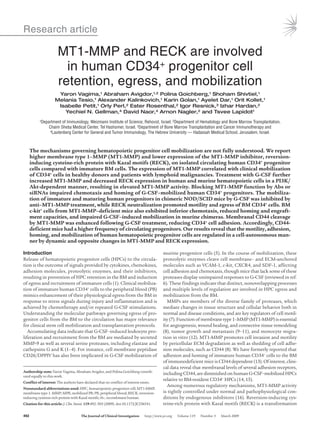
Figure 8
Proposed mechanism of MT1-MMP–mediated HPC mobilization. In
the absence of mobilizing stimuli (e.g., G-CSF), proteolytic activities of
MT1-MMP, MMP-2, and MMP-9 are relatively low due to inhibition by
RECK. Functional membranal CD44 contributes to progenitor cell adhe-
sion to the BM components (retention). G-CSF signaling induces PI3K-
mediated Akt phosphorylation, increasing MT1-MMP and decreasing
RECK expression. The opposed changes in MT1-MMP and RECK lev-
els result in MT1-MMP–mediated CD44 proteolysis as well as MMP-2
and MMP-9 secretion and activation. Collectively, these changes reduce
progenitor cell retention and facilitate their egress and mobilization.
11. research article
502 The Journal of Clinical Investigation http://www.jci.org Volume 119 Number 3 March 2009
antisense, 5′-AGAACCATCGCTCCTTGAAGAC-3′; for RECK, sense, 5′-
AAAAAGAAAAAGCCACGGAACA-3′, antisense, 5′-CACACTAAATTACCC-
GCAAAACAAG-3′; for HPRT, sense, 5′-GCAGTACAGCCCCAAAATGG-3′,
antisense, 5′-GGTCCTTTTCACCAGCAAGCT-3′.
Immunohistochemistry of BM sections. Femurs of chimeric NOD/SCID mice
with 60%–70% engraftment of human CD45+ cells were fixed and immu-
nolabeled as previously described (13) with Abs to human MT1-MMP
(Chemicon), human RECK (R&D Systems), human CD44 (Serotec), or
phospho-Akt (Thr308; Cell Signaling Technology). Staining was per-
formed as previously described (48).
MT1-MMP and MMP-2/9 activity assays. Plasma membrane fractions were
prepared from 5 × 10⁶ BM cells from 3 bones of NOD/SCID chimeric
mice, and MT1-MMP activity was determined by fluorescence intensity for
2.5 hours (37°C) at 395 nm using an assay kit (Chemicon) according to the
manufacturer’s protocol. Active MMP-2 and MMP-9 in BM supernatants
were detected by gelatin zymography as previously described (49).
Trans-Matrigel chemotaxis assay. Enriched human CD34+ (105 cells/well)
or murine BM mononuclear cells (2–3 × 10⁵/well) were used. Where indi-
cated (Figure 4, A and C, and Results), MPB CD34+ cells were pre-incubat-
ed for 30 minutes with 8 μg/ml (400 nM) rhTIMP-2 (R&D Systems) (35),
50 μg/ml anti–MT1-MMP (Chemicon) Abs, or nonimmune rabbit serum
(Sigma-Aldrich) as control IgG and allowed to migrate for 3–4 hours in
serum-free medium to reduce background MMP activity. BM CD34+ cells
were treated with 50 μg/ml anti-RECK (MBL International Corp.) Abs
or mouse anti-human IgG (Serotec) and subjected to migration assay in
serum-containing medium due to their low migration efficiency in the
absence of serum. The number of cells that migrated spontaneously or
toward 125 ng/ml SDF-1 (PeproTech) via transwell inserts of 5-μm pore
size (Corning) precoated with a thick layer (60 μg/insert) of Matrigel (BD)
was determined using FACSCalibur (BD).
siRNA transfection. Pools of 4 siRNA duplexes to target human MT1-MMP
(accession no. NM_004995) or nonspecific siRNA were purchased from
Dharmacon. MPB CD34+ cells were subjected to transfections with 2.5 μg
of siRNA using the amaxa nucleofector kit (amaxa GmbH) according to the
manufacturer’s instructions and further cultured for 24–48 hours in RPMI
supplemented with serum and rhSCF (50 ng/ml), rhIL-6 (20 ng/ml), rhIL-3
(10 ng/ml) (all purchased from PeproTech), and rhG-CSF (100 ng/ml; Fil-
grastim Roche). U937 cells were transfected with siRNA as described above
and treated with 100 ng/ml rhG-CSF for 36 hours. Membranal MT1-MMP
expression was reduced by 30%–50%, as monitored by flow cytometry.
Immunoblot for cleaved CD44. Cell-free supernatants were obtained from
the BM of 4 bones of chimeric NOD/SCID mice, and 50–100 μg total pro-
tein per sample were subjected to 10% SDS-PAGE followed by immunoblot
with 2 μg/ml Hermes-3 anti–pan-CD44 Abs (50).
Adhesion assay. BM mononuclear cells isolated from chimeric NOD/SCID
mice were subjected to the adhesion assay to hyaluronan-coated micro-
plates as previously described (13), labeled with anti-human CD34+ Abs,
and enumerated by flow cytometry. Where indicated in the figures, cells
were treated for 30 minutes with 50 μg/ml anti-CD44 Abs (BU52; The Bind-
ing Site) or for 24 hours with either anti-RECK (MBL International Corp.)
Abs or 200 ng/ml rhG-CSF with or without 50 μg/ml anti–MT1-MMP
(R&D Systems) Abs.
Statistics. Statistical significance (P value of less than 0.05) was deter-
mined using the 2-tailed Student’s t test. The Spearman correlation test
was performed using GraphPad Prism software.
Acknowledgments
We thank Motoharu Seiki for providing us with MT1-MMP KO
mice and Daigo Niiya for technical advice, Stephen J. Weiss for
fruitful discussions, Irit Sagi and Netta Sela for sharing their
expertise on the MT1-MMP activity assay, Sonia Berrih-Aknin for
the correlation analysis, and Loya Abel for her excellent techni-
cal assistance. This study was supported by the Legacy Heritage
Foundation, a European Union FP6 grant, the Helen and Martin
Kimmel Institute for Stem Cell Research, the Minerva Foundation,
and the Gabriella Rich Center for Transplantation Biology.
Received for publication June 19, 2008, and accepted in revised
form December 22, 2008.
Address correspondence to: Tsvee Lapidot, Immunology Depart-
ment, Weizmann Institute of Science, PO Box 26, Hertzl Str.,
Rehovot 76100, Israel. Phone: 972-8-9342481; Fax: 972-8-9344141;
E-mail: tsvee.lapidot@weizmann.ac.il.
1. Lapidot,T.,andPetit,I.2002.Currentunderstanding
of stem cell mobilization: the roles of chemokines,
proteolytic enzymes, adhesion molecules, cytokines,
and stromal cells. Exp. Hematol. 30:973–981.
2. Heissig, B., et al. 2002. Recruitment of stem and
progenitor cells from the bone marrow niche
requires MMP-9 mediated release of kit-ligand.
Cell. 109:625–637.
3. Levesque, J.P., Hendy, J., Takamatsu, Y., Simmons,
P.J.,andBendall,L.J.2003.DisruptionoftheCXCR4/
CXCL12 chemotactic interaction during hemato-
poietic stem cell mobilization induced by GCSF or
cyclophosphamide. J. Clin. Invest. 111:187–196.
4. Kollet, O., et al. 2006. Osteoclasts degrade endoste-
al components and promote mobilization of hema-
topoietic progenitor cells. Nat. Med. 12:657–664.
5. Christopherson, K.W., 2nd, Cooper, S., and Brox-
meyer, H.E. 2003. Cell surface peptidase CD26/
DPPIV mediates G-CSF mobilization of mouse
progenitor cells. Blood. 101:4680–4686.
6. Papayannopoulou, T., and Scadden, D.T. 2008.
Stem-cell ecology and stem cells in motion. Blood.
111:3923–3930.
7. Page-McCaw, A., Ewald, A.J., and Werb, Z. 2007.
Matrix metalloproteinases and the regulation of tis-
sue remodelling. Nat. Rev. Mol. Cell Biol. 8:221–233.
8. Itoh, Y., and Seiki, M. 2006. MT1-MMP: A potent
modifier of pericellular microenvironment. J. Cell.
Physiol. 206:1–8.
9. Hotary, K.B., et al. 2003. Membrane type I matrix
metalloproteinase usurps tumor growth control
imposed by the three-dimensional extracellular
matrix. Cell. 114:33–45.
10. Seiki, M. 2003. Membrane-type 1 matrix metal-
loproteinase: a key enzyme for tumor invasion.
Cancer Lett. 194:1–11.
11. Belkin, A.M., et al. 2001. Matrix-dependent
proteolysis of surface transglutaminase by mem-
brane-type metalloproteinase regulates cancer
cell adhesion and locomotion. J. Biol. Chem.
276:18415–18422.
12. Matias-Roman, S., et al. 2005. Membrane type 1-
matrix metalloproteinase is involved in migration
of human monocytes and is regulated through
their interaction with fibronectin or endothelium.
Blood. 105:3956–3964.
13. Avigdor, A., et al. 2004. CD44 and hyaluronic acid
cooperate with SDF-1 in the trafficking of human
CD34+ stem/progenitor cells to bone marrow.
Blood. 103:2981–2989.
14. Lee, S., et al. 2000. Mobilization kinetics of CD34(+)
cells in association with modulation of CD44 and
CD31 expression during continuous intravenous
administration of G-CSF in normal donors. Stem
Cells. 18:281–286.
15. Sovalat, H., et al. 2003. CD34+ cells and
CD34+CD38- subset from mobilized blood show
different patterns of adhesion molecules com-
pared to those from steady-state blood, bone mar-
row, and cord blood. J. Hematother. Stem Cell Res.
12:473–489.
16. Osenkowski, P., Toth, M., and Fridman, R. 2004.
Processing,shedding,andendocytosisofmembrane
type 1-matrix metalloproteinase (MT1-MMP).
J Cell. Physiol. 200:2–10.
17. Noda, M., and Takahashi, C. 2007. Recklessness
as a hallmark of aggressive cancer. Cancer Sci.
98:1659–1665.
18. Oh, J., et al. 2001. The membrane-anchored MMP
inhibitor RECK is a key regulator of extracellular
matrix integrity and angiogenesis. Cell. 107:789–800.
19. Lapidot, T., et al. 1992. Cytokine stimulation
of multilineage hematopoiesis from immature
human cells engrafted in SCID mice. Science.
255:1137–1141.
20. Larochelle, A., et al. 1996. Identification of primi-
tive human hematopoietic cells capable of repopu-
lating NOD/SCID mouse bone marrow: implica-
tions for gene therapy. Nat. Med. 2:1329–1337.
21. Shaw, R.J., and Cantley, L.C. 2006. Ras, PI(3)K and
mTOR signalling controls tumour cell growth.
Nature. 441:424–430.
22. Lapidot,T.,Dar,A.,andKollet,O.2005.Howdostem
cells find their way home? Blood. 106:1901–1910.
23. Wright, D.E., Bowman, E.P., Wagers, A.J., Butcher,
E.C., and Weissman, I.L. 2002. Hematopoietic stem
cellsareuniquelyselectiveintheirmigratoryresponse
12. research article
The Journal of Clinical Investigation http://www.jci.org Volume 119 Number 3 March 2009 503
to chemokines. J. Exp. Med. 195:1145–1154.
24. Peled, A., et al. 1999. Dependence of human stem
cell engraftment and repopulation of NOD/SCID
mice on CXCR4. Science. 283:845–848.
25. Voermans, C., et al. 2001. In vitro migratory capac-
ity of CD34+ cells is related to hematopoietic recov-
ery after autologous stem cell transplantation.
Blood. 97:799–804.
26. Shankavaram, U.T., et al. 2001. Monocyte mem-
brane type 1-matrix metalloproteinase. Pros-
taglandin-dependent regulation and role in
metalloproteinase-2 activation. J. Biol. Chem.
276:19027–19032.
27. Holmbeck, K., et al. 1999. MT1-MMP-deficient
mice develop dwarfism, osteopenia, arthritis, and
connective tissue disease due to inadequate colla-
gen turnover. Cell. 99:81–92.
28. Takahashi, C., et al. 1998. Regulation of matrix
metalloproteinase-9 and inhibition of tumor inva-
sion by the membrane-anchored glycoprotein
RECK. Proc. Natl. Acad. Sci. U. S. A. 95:13221–13226.
29. Kajita, M., et al. 2001. Membrane-type 1 matrix
metalloproteinase cleaves CD44 and promotes cell
migration. J. Cell Biol. 153:893–904.
30. Nakamura, H., et al. 2004. Constitutive and
induced CD44 shedding by ADAM-like proteases
and membrane-type 1 matrix metalloproteinase.
Cancer Res. 64:876–882.
31. Liu, F., Poursine-Laurent, J., and Link, D.C. 2000.
Expression of the G-CSF receptor on hematopoi-
etic progenitor cells is not required for their mobi-
lization by G-CSF. Blood. 95:3025–3031.
32. Zhang, J., et al. 2006. PTEN maintains haemato-
poietic stem cells and acts in lineage choice and
leukaemia prevention. Nature. 441:518–522.
33. Yilmaz, O.H., et al. 2006. Pten dependence distin-
guishes haematopoietic stem cells from leukaemia-
initiating cells. Nature. 441:475–482.
34. Noda, M., et al. 2003. RECK: a novel suppressor
of malignancy linking oncogenic signaling to
extracellular matrix remodeling. Cancer Metastasis
Rev. 22:167–175.
35. Janowska-Wieczorek, A., et al. 1999. Growth factors
and cytokines upregulate gelatinase expression in
bone marrow CD34(+) cells and their transmigra-
tion through reconstituted basement membrane.
Blood. 93:3379–3390.
36. Spiegel, A., et al. 2007. Catecholaminergic neu-
rotransmitters regulate migration and repopula-
tion of immature human CD34(+) cells through
Wnt signaling. Nat. Immunol. 8:1123–1131.
37. Zhou, Z., et al. 2000. Impaired endochondral ossifi-
cation and angiogenesis in mice deficient in mem-
brane-type matrix metalloproteinase I. Proc. Natl.
Acad. Sci. U. S. A. 97:4052–4057.
38. Winkler,I.G.,Hendy,J.,Coughlin,P.,Horvath,A.,and
Levesque, J.P. 2005. Serine protease inhibitors serpi-
na1 and serpina3 are down-regulated in bone mar-
row during hematopoietic progenitor mobilization.
J. Exp. Med. 201:1077–1088.
39. Nilsson, S.K., et al. 2003. Hyaluronan is synthesized
by primitive hemopoietic cells, participates in their
lodgment at the endosteum following transplan-
tation, and is involved in the regulation of their
proliferation and differentiation in vitro. Blood.
101:856–862.
40. Dimitroff, C.J., Lee, J.Y., Rafii, S., Fuhlbrigge, R.C.,
and Sackstein, R. 2001. CD44 is a major E-selectin
ligand on human hematopoietic progenitor cells.
J. Cell Biol. 153:1277–1286.
41. Vermeulen, M., et al. 1998. Role of adhesion mol-
ecules in the homing and mobilization of murine
hematopoietic stem and progenitor cells. Blood.
92:894–900.
42. Christ, O., Kronenwett, R., Haas, R., and Zoller, M.
2001. Combining G-CSF with a blockade of adhe-
sion strongly improves the reconstitutive capacity
of mobilized hematopoietic progenitor cells. Exp.
Hematol. 29:380–390.
43. Craddock, C.F., et al. 1997. Antibodies to VLA4
integrin mobilize long-term repopulating cells and
augment cytokine-induced mobilization in pri-
mates and mice. Blood. 90:4779–4788.
44. Levesque, J.P., Takamatsu, Y., Nilsson, S.K., Hay-
lock, D.N., and Simmons, P.J. 2001. Vascular
cell adhesion molecule-1 (CD106) is cleaved by
neutrophil proteases in the bone marrow follow-
ing hematopoietic progenitor cell mobilization
by granulocyte colony-stimulating factor. Blood.
98:1289–1297.
45. Nedvetzki, S., et al. 2004. RHAMM, a receptor for
hyaluronan-mediated motility, compensates for
CD44 in inflamed CD44-knockout mice: a differ-
ent interpretation of redundancy. Proc. Natl. Acad.
Sci. U. S. A. 101:18081–18086.
46. Schmits, R., et al. 1997. CD44 regulates hematopoi-
etic progenitor distribution, granuloma formation,
and tumorigenicity. Blood. 90:2217–2233.
47. Oh, J., et al. 2004. Mutations in two matrix metal-
loproteinase genes, MMP-2 and MT1-MMP, are
synthetic lethal in mice. Oncogene. 23:5041–5048.
48. Petit, I., et al. 2002. G-CSF induces stem cell mobi-
lization by decreasing bone marrow SDF-1 and up-
regulating CXCR4. Nat. Immunol. 3:687–694.
49. Kollet, O., et al. 2003. HGF, SDF-1, and MMP-9 are
involved in stress-induced human CD34+ stem cell
recruitment to the liver. J. Clin. Invest. 112:160–169.
50. Nedvetzki, S., et al. 2003. A mutation in a CD44
variant of inflammatory cells enhances the mito-
genic interaction of FGF with its receptor. J. Clin.
Invest. 111:1211–1220.