
Empfohlen
Empfohlen
Weitere ähnliche Inhalte
Was ist angesagt?
Was ist angesagt? (20)
Ähnlich wie To hop 3 thao duoc co tac dung tuong tu corticoid
Ähnlich wie To hop 3 thao duoc co tac dung tuong tu corticoid (20)
Kürzlich hochgeladen
Kürzlich hochgeladen (20)
To hop 3 thao duoc co tac dung tuong tu corticoid
- 1. Efficacy and tolerability of antiasthma herbal medicine intervention in adult patients with moderate-severe allergic asthma Ming-Chun Wen, MD,a Chun-Hua Wei, MD,a Zhao-Qiu Hu, MD, MS,a Kamal Srivastava, MPhil,b Jimmy Ko, MD,b Su-Ting Xi, MD, MS,a Dong-Zhen Mu, MD, MS,d Ji-Bin Du, MD,a Guo-Hua Li, MD,a Sylvan Wallenstein, PhD,c Hugh Sampson, MD,b Meyer Kattan, MD,b and Xiu-Min Li, MDb Weifang, China, and New York, NY Background: Chinese herbal medicine has a long history of human use. A novel herbal formula, antiasthma herbal medicine intervention (ASHMI), has been shown to be an effective therapy in a murine model of allergic asthma. Objective: This study was undertaken to compare the efficacy, safety, and immunomodulatory effects of ASHMI treatment in patients with moderate-severe, persistent asthma with prednisone therapy. Methods: In a double-blind trial, 91 subjects underwent randomization. Forty-five subjects received oral ASHMI capsules and prednisone placebo tablets (ASHMI group) and 46 subjects received oral prednisone tablets and ASHMI placebo capsules (prednisone group) for 4 weeks. Spirometry measurements; symptom scores; side effects; and serum cortisol, cytokine, and IgE levels were evaluated before and after treatment. Results: Posttreatment lung function was significantly improved in both groups as shown by increased FEV1 and peak expiratory flow findings (P < .001). The improvement was slightly but significantly greater in the prednisone group (P < .05). Clinical symptom scores, use of b2-bronchodilators, and serum IgE levels were reduced significantly, and to a similar degree in both groups (P < .001). TH2 cytokine levels were significantly reduced in both treated groups (P < .001) and were lower in the prednisone-treated group (P < .05). Serum IFN-g and cortisol levels were significantly decreased in the prednisone group (P < .001) but significantly increased in the ASHMI group (P < .001). No severe side effects were observed in either group. Conclusion: Antiasthma herbal medicine intervention appears to be a safe and effective alternative medicine for treating asthma. In contrast with prednisone, ASHMI had no adverse effect on adrenal function and had a beneficial effect on TH1 and TH2 balance. (J Allergy Clin Immunol 2005;116:517-24.) Key words: Asthma, clinical trial, Chinese herbal medicine, prednisone, cortisol, TH1/TH2 balance Asthma is characterized by chronic airway inflamma- tion, which adversely affects normal lung function. Corticosteroids, the most potent nonspecific anti-inflam- matory agents, produce substantial improvement in objective lung functions of patients with asthma and are the cornerstone of asthma treatment.1 However, systemic corticosteroids also induce serious systemic adverse effects when given for prolonged periods.2 Corticosteroids also produce overall immune suppression, resulting in increased susceptibility to infections.3 The side effects are significantly reduced with inhaled corticosteroids, but in higher doses, side effects including adrenal suppression and reduction in growth velocity have been reported.4,5 There is a need for development of additional effective treatments with fewer side effects. Recently, there has been a surge in interest in traditional Chinese medicine (TCM) in Western countries, possibly because of the low cost and favorable safety profile. Although a role for TCM in Western medicine has not been established, TCM is in the mainstream of modern medical practice in China for treatment of various diseases, including asthma, either as monotherapy or as complementary therapy to standard Western medications. However, well-controlled clinical trials using TCM for asthma treatment are still rare. In an attempt to develop novel herbal interventions for asthma, we previously tested Chinese herbal formula MSSM-002 (an extract of 14 herbs based on a TCM prescription used to treat childhood asthma in the Pediatric Department of the China-Japan Friendship Hospital in Beijing) on a well-characterized murine model of asthma. We found that MSSM-002 virtually eliminated airway hyperreactivity, markedly reduced the total number of cells and the percentage of eosinophils in bronchoalveolar From a the Weifang Asthma Hospital; b the Department of Pediatrics and c the Department of Community Medicine, Mount Sinai School of Medicine, New York and d the Department of Immunology, Weifang School of Medicine. Supported by National Institutes of Health grant # AT001495-01A1. Disclosure of potential conflict of interest: M.-C. Wen has filed a US patent application (reference #60554775). H. Sampson has received grants/ research support from the National Institutes of Health and has filed a US patent application (reference #60554775). M. Kaltan is on the speakers’ bureau for AstraZeneca. X.-M. Li has received grants/research support from the National Institutes of Health and has filed a US patent application (reference #60554775). Received for publication January 20, 2005; revised May 9, 2005; accepted for publication May 16, 2005. Available online August 8, 2005. Reprint requests: Xiu-Min Li, MD, Pediatric Allergy and Immunology, Mount Sinai School of Medicine, One Gustave L. Levy Place, New York, NY 10029-6574. E-mail: xiu-min.li@mssm.edu; Ming-Chun Wen, MD, Weifang Asthma Hospital, Weifang, N0. 68, Xinhua Road, Weifang, Shandong 261041, China. E-mail: wen637@hotmail.com. 0091-6749/$30.00 Ó 2005 American Academy of Allergy, Asthma and Immunology doi:10.1016/j.jaci.2005.05.029 517 Asthmadiagnosisand treatment
- 2. Abbreviations used ASHMI: Antiasthma herbal medicine intervention PEF: Peak expiratory flow TCM: Traditional Chinese medicine lavage fluid, and inhibited mucus production in lungs of allergen-challenged mice.6 Interestingly, in contrast with corticosteroids, which suppress both TH1 and TH2 responses, MSSM-002 specifically suppressed TH2 responses (IL-4, IL-5, IgE production), but not TH1 responses (IFN-g, IgG2a production).6 We further found that the immunomodulatory effects of MSSM-002 on TH2 cells are caused, at least in part, by downregulation of GATA-3, a TH2 transcription factor, and unlike cortico- steroids, MSSM-002 does not induce apoptosis.7 These findings suggest that MSSM-002 may be of benefit in the treatment of asthma. On the basis of the actions of individual herbs contained in MSSM-002 in our murine asthma model and on TCM formulation concepts,8 we developed a simplified antiasthma herbal medicine intervention (ASHMI).9 ASHMI is an extract of 3 herbs: Ling-Zhi(Ganoderma lucidum), Ku-Shen (RadixSophora flavescentis), and Gan-Cao (Radix Glycyrrhiza uralensis). We found that ASHMI, like MSSM-002, exhibits the same broad spectrum of therapeutic effects on the major pathogenic mechanisms of asthma—airway hyperreactiv- ity, pulmonary inflammation, and airway remodeling—as well as downregulating TH2 responses and direct modu- lation of airway smooth muscle contraction.10,11 In addi- tion, ASHMI significantly suppressed TH2 cytokine production by human PBMCs from patients with asthma. No cytotoxicity was detected at the highest effective dose tested.12 On the basis of these findings, we undertook a study of the clinical effects, safety, and immunomodula- tory effects of ASHMI treatment in patients with asthma compared with standard therapy with prednisone. METHODS Patients A randomized, double-blind, placebo-controlled study was per- formed at Weifang Asthma Hospital from September 2003 to September 2004. Weifang Asthma Hospital is a chronic asthma treatment facility receiving patients nationwide. The 4-week study was conducted in the inpatient unit. The recruiting process involved 3 screening steps: clinical history, clinical testing, and laboratory testing. Patients prescreened and recruited from the outpatient facility were admitted to the hospital for purposes of the study. Ninety-two atopic, nonsmoking patients with asthma (43 men and 49 women, age 18-65 years) meeting the criteria of moderate-severe, persistent asthma13 were recruited into this study. Inclusion criteria included (1) a history of allergic asthma for at least 1 year; (2) a serum IgE level above 100 IU/mL; (3) daily asthma symptoms; (4) exacerbations affecting activity and sleep; (5) nocturnal symptoms more than once a week; (6) FEV1 59% to 72% predicted or peak expiratory flow (PEF) 59% to 72%, PEF or FEV1 variability 30%; (7) daily use of a b2-agonist in the past month; (8) 2 short courses (3-7 days) of oral corticosteroids in the previous 6 months; (9) no use of oral cortico- steroids in the previous 4 weeks; and (10) understanding the research protocol and consent to participate. Exclusion criteria in this study included (1) use of oral corticosteroids within the past 4 weeks; (2) heart, liver, kidney, or other organ diseases; (3) allergy or intolerance to the individual herbs in ASHMI; (4) pregnant and lactating women; and (5) being unable to comply with the research protocol because of severity of asthma (needed additional therapy). The study was approved by the hospital medical ethics committee, and all patients gave written informed consent. Study design There was a 1-week run-in period before initiating treatment. During the run-in period, patients were allowed to use b2-agonist and/or theophylline. Any patient showing exacerbation of symp- toms requiring additional medications was excluded from the study before the study began. The subjects were randomly assigned to receive ASHMI (n = 46) or prednisone (n = 46). Subjects in the ASHMI group received oral ASHMI capsules (4 capsules, three times a day) and placebo tablets similar in appearance to predni- sone. Subjects in the prednisone group received oral prednisone tablets (20 mg once a day in the morning) and ASHMI placebo capsules for 4 weeks. For the duration of the study, leukotriene modifiers, antihistamines, and inhaled and intravenous glucocorti- coids were prohibited. b2-Agonist inhalation was allowed as needed. Subjects requiring additional intervention at any time because of disease severity were withdrawn from the study. Each ASHMI capsule contained 0.3 g dried aqueous extract. The total daily dose of 12 capsules (3.6 g) is equivalent to extracts of a mixture of the raw herbs Ling-Zhi (Ganoderma Lucidum) 20 g, Ku-Shen (Radix Sophorae Flavescentis) 9 g, and Gan-Cao (Radix Glycyrrhiza) 3 g. ASHMI capsules and ASHMI placebo capsules were prepared by Weifang Pharmaceutical Manufacturing Factory, affiliated with Weifang Asthma Hospital. Prednisone placebo tablets were made by Shandong Luoxin Ltd, Weifang. Before and after com- pleting treatment, symptom scores, use of salbutamol (puffs/d), and lung function were evaluated. Serum total IgE, IL-5, IL-13, IFN-g, and cortisol levels also were measured. Symptom scores, adverse events, and b2-agonist use were recorded daily. Grading of adverse events followed the World Health Organization Recommendations for Grading of Acute and Subacute Toxicity.14 Hematology and serum chemistry testing and electrocardiograms were performed before and after the treatment. Clinical and laboratory evaluation Evaluation of symptom scores and use of b2-agonist. Average daily symptom scores were evaluated over a 1-week period before treatment to establish a baseline. The effect of treatment on symptom scores was evaluated by analyzing average daily symptom scores in weeks 1, 2, 3, and 4 of treatment on the basis of 3 categories: day- time symptoms, nocturnal symptoms, and allergic nasal and ocular symptoms. Each category was scored by physicians from 0 to 3, with a maximum possible score of 9.15 The daytime symptom scores (cough, chest tightness, wheezing or dyspnea) were 0, no symptoms; 1, mild symptoms or intermittent occurrence; 2, moderate symptoms with frequent occurrence that may affect normal activity at least 1 time; and 3, persistent symptoms, affecting all activities. Nocturnal symptoms scores were 0, no night awakening; 1, 1 night awakening or early morning awakening caused by dyspnea; 2, 2 night awakenings caused by dyspnea (including early morning awaken- ing); and 3, multiple night awakenings caused by dyspnea. The score of signs and symptoms of allergic rhinitis (nasal pruritus, rhinorrhea, sneezing, and periocular pruritus and tearing) were 0, no symptoms; 1, symptoms 4 d/wk, no effect on comfort level, sleep, and daily J ALLERGY CLIN IMMUNOL SEPTEMBER 2005 518 Wen et al Asthmadiagnosisand treatment
- 3. activity; 2, symptoms 4 d/wk, with effects on comfort level, sleep, and daily activity, or symptoms 4 d/wk without effects on comfort- able level, sleep, and daily activity; and 3, persistent symptoms with effects on comfort level, sleep, and daily activity. Average daily use of b2-agonist was evaluated over a 1-week period before treatment and during the last week of treatment. Evaluation of lung function. FEV1 and PEF measurements were performed by using an HI-701 spirometer (Chest Co. Ltd, Tokyo, Japan). Lung function measurements were recorded the day before treatment was initiated and the day after treatment was discontinued. All measurements were repeated 3 times, and the highest reading for each parameter was used for the study. Patients were not allowed to use theophylline 24 hours before or short-term b2-agonist 6 hours before lung function evaluation. Serum IgE. Venous blood samples were obtained from all patients before and after treatment. Serum IgE was measured by commercial ELISA kit (Shanghai Splendid; Shanghai Refulgence Technological Co. Ltd, Shanghai, China) according to the manufacturer’s instruc- tions. Peripheral eosinophil counts. Twenty microliters of peripheral blood collected from a finger stick were diluted in 380 mL staining buffer (5 mL 2% eosin, 5 mL acetone, and 90 mL double-distilled H2O). The leukocytes were counted by using a hemacytometer. The absolute eosinophil number was calculated.16 Cortisol testing. Serum cortisol levels were determined by radioimmunoassay with a commercial kit (Beijing North Bio- Technology Research Institute, Beijing, China) according to the kit reference manual. To ensure consistency of the results, all blood samples were drawn between 7:30 and 8:30 AM (before treatment and 48 hours after treatment). Serum cytokines. Serum IL-5, IL-13 and IFN-g levels were determined by commercial ELISA kits (IL-5 kit from Diaclone, Besacon, France; IL-13 kit from Yes Biotech Laboratories Ltd, Toronto, Canada; and IFN-g kit from Roche, Basel, Switzerland). ELISA tests were performed according to manuals provided by the manufacturers. Statistical analysis All analyses for baseline and treatment effects were performed by using SigmaStat software (SYSTAT Software, Port Richmond, Calif). P .05 was considered statistically significant, and all tests were 2-tailed. On the basis of the nature of the outcomes, we used nonparametric methods (descriptive statistics of median and range, Wilcoxon signed rank test, and Mann-Whitney-Wilcoxon rank test) for symptom scores and number of times per day b2-agonists were used. We also used these methods for IgE concentrations, as described previ- ously.17,18 For all other variables, we tested the assumptions of equal variances and normality used in the analysis of differences between groups with respect to changes from baseline. We used the indepen- dent sample t test to analyze these changes if these assumptions were satisfied, and the Mann-Whitney-Wilcoxon test otherwise. Analysis of within-group differences from baseline was usually not an issue because the results were very clear, and our policy was to use the paired t test if the analysis of differences between groups (which involved an examination of assumptions) was based on the indepen- dent sample t test. RESULTS Patient characteristics One patient in the ASHMI group acquired an infection in the fourth week of treatment and dropped out of the study. Forty-five patients in the ASHMI group and 46 patients in the prednisone group completed the study. There were no significant differences between the 2 groups in age, sex, asthma duration, or body weight before treatment. The baseline FEV1, PEF measurements, symptom scores, and use of b2-agonist in the 2 groups were not different (Table I). Effect of ASHMI treatment on pulmonary function, symptom scores, and b2-agonist use By week 4 (the last week of treatment), symptom scores (median [range]) were reduced in patients treated with ASHMI (5.0 [4-8] to 2.0 [0-4] P .001; Fig 1, A) and patients treated with prednisone (5.0 [4-7] to 2.0 [0-4]; TABLE I. Patient characteristics and demographics ASHMI (n = 45) Prednisone (n = 46) P value (between- groups) Age, y, mean 6 SD 44.6 6 11.3 45.1 6 12.0 .89 Male/female 21/24 19/27 .76 Duration of disease, y, median (range) 10.0 (1-24) 12 (1-22) .59 Weight, kg, mean 6 SD 64.8 6 7.3 61.7 6 7.1 .13 FEV1%, mean 6 SD 64.9 6 3.6 65.2 6 3.7 .61 PEF%, mean 6 SD 64.6 6 3.5 65.0 6 3.5 .55 Symptom score, median (range) 5 (4-7) 5 (4-8) .70 b2-Agonist, puffs/d, median (range) 4.7 (3.5-5.7) 4.7 (3.4-5.5) .53 FIG 1. Effect of treatment with (A) ASHMI or (B) prednisone on symptom scores. Average daily symptom scores were evaluated over a 1-week period before treatment to establish a baseline (week 0). The effect of treatment on symptom scores was evalu- ated by analyzing average daily symptom scores in weeks 1, 2, 3, and 4 of treatment. ***P .001. J ALLERGY CLIN IMMUNOL VOLUME 116, NUMBER 3 Wen et al 519 Asthmadiagnosisand treatment
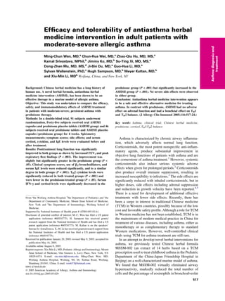
- 4. P .001; Fig 1, B). Improvement in symptom scores was similar between the treatment groups (P = .47; Table II). We also analyzed the changes in symptom score over weeks 1, 2, and 3 of treatment and found that improve- ment in symptom scores occurred earlier in the prednisone treatment group than in ASHMI group. Median symptom scores of ASHMI-treated patients were not significantly reduced until week 3 of treatment (baseline vs weeks 1, 2, and 3 of treatment: 5.4 vs 5.2 [P = .74], 5.1 [P = .54], and 3.6 [P .001], respectively; Fig 1, A), whereas median symptom scores were significantly reduced in patients treated with prednisone by week 1 (baseline vs weeks 1, 2, and 3 of treatment: 5.2 vs 4.2, 3.5, and 2.5, respectively; P .001; Fig 1, B). Effect of ASHMI treatment on pulmonary function and b2-agonist use FEV1 values (means 6 SDs) showed significant im- provement after treatment with ASHMI (64.9 6 3.6 to 84.2 6 5.0; P .001) and prednisone (65.2 6 3.7 to 88.4 6 8.0; P .001; Fig 2, A). PEF values (means 6 SDs) in both treatment groups also showed significant increases (ASHMI, 64.6 6 3.5 to 84.8 6 5.4, P .001; prednisone, 65.0 6 3.5 to 88.1 6 7.0, P .001; Fig 2, B). Increases in FEV1 and PEF were significantly greater in the prednisone group than in the ASHMI group (P = .02 and .04, respectively; Table II). Consistent with decreased symptoms and improvement of pulmonary function, inhaled b2-agonist use in both treatment groups was reduced (ASHMI, 4.7 [3.5-5.7] to 0.9 [0.14-2.3], P .001; prednisone, 4.7 [3.5-5.6] to 0.6 [0.3-1.0], P .001; Fig 2, C). The reduction in b2-agonist use was slightly greater in the prednisone- treated group but was not statistically different (P = .12; Table II). Effect of ASHMI treatment on peripheral eosinophils Before treatment, numbers of peripheral blood eosino- phils in both groups were slightly higher than the normal range (0-0.5 3 109 /L) and were not different between the 2 groups (Fig 3). After treatment, numbers of peripheral eosinophils in both groups were significantly reduced in the ASHMI and prednisone groups (means 6 SD, ASHMI, 0.52 6 0.24 to 0.27 6 0.14 3 109 /L, P .001; prednisone, 0.53 6 0.21 to 0.19 6 0.1 3 109 /L, P .001). The reduction of eosinophils in the treated groups was not statistically different (P .37). Effect of ASHMI treatment on adrenal function Corticosteroid-induced suppression of the hypotha- lamic-pituitary-adrenal axis, marked by depressed cortisol levels, has been implicated as an adverse side effect of systemic steroid use.19,20 In this study, pretreatment cortisol levels were slightly below normal (6-23 mg/dL) in both groups (Fig 4). After treatment, subjects in the prednisone treatment group showed a significant reduc- tion in serum cortisol levels after treatment (5.1 6 3.0 to 3.7 6 2.3 mg/dL; P .001). In contrast, patients in the ASHMI treatment group showed increased levels of serum cortisol (5.4 6 2.8 to 7.7 6 2.3 mg/dL; P .001; Fig 4), which were within the normal range. The difference between groups was statistically significant (P .001). Effect of ASHMI treatment on serum IgE and serum cytokine levels Marked reductions in IgE levels (median [range]) were observed in the ASHMI (950 [552-1349] to 476 [73-913] U/mL; P .001; Fig 5, A) and prednisone treatment groups (948 [368-1356] to 310 [60-619] U/mL; P .001; Fig 5, A) Reduction in IgE between the 2 treatment groups was not significantly different (P = .10). Significantly reduced levels of serum IL-5 were found in both the ASHMI (95.02 6 43.8 to 55.2 6 23.5 pg/mL; P .001; Fig 5, B) and prednisone (103.9 6 49.6 to 41 6 19.1 pg/mL; P .001; Fig 5, B) treatment groups. Similarly, serum IL-13 levels were also reduced in the ASHMI (133.8 6 25.9 to 103.0 6 23.0 pg/mL; P .001; Fig 5, C) and prednisone (130.9 6 24.9 to 85.8 6 19.5 pg/ mL; P .001; Fig 5, C) treatment groups. For both IL-5 and IL-13, reduction in the prednisone group was sig- nificantly greater than in the ASHMI group (P = .04 and .02, respectively). Prednisone treatment also resulted in reduction of serum levels of the TH1 cytokine IFN-g (403.7 6 144.1 to 275.7 6 135.4 pg/mL; P .001; Fig 5, D). In contrast, serum IFN-g levels in the ASHMI treatment group were significantly elevated after treat- ment (402.8 6 142.6 to 585.6 6 150.8 pg/mL; Fig 5, D). The difference between the groups was significant (P .001). Safety of ASHMI treatment Treatment with ASHMI and prednisone was well tolerated. Neither group showed abnormal findings in hematology, serum chemistry tests, or electrocardiograms. No serious adverse effects were observed in either group. TABLE II. Changes in lung function parameters, symptom scores, and b-agonist use posttreatment Change after treatment P values (between-groups)ASHMI (n = 45) Prednisone (n = 46) FEV1%, mean 6 SD [ 19.4 6 5.5 % [ 23.2 6 8.9 % .02 PEF%, mean 6 SD [ 20.1 6 5.6 % [ 23.0 6 7.5 % .04 Symptom scores, median (range) Y 3.0 (Y 5-2) Y 3.0 (Y 6-0) .47 b2-Agonist use, median (range) Y 3.8 (Y 4.8-1.9) puffs/d Y 4.0 (Y 4.7-2.8) puffs/d .12 J ALLERGY CLIN IMMUNOL SEPTEMBER 2005 520 Wen et al Asthmadiagnosisand treatment
- 5. Both groups showed an increase in weight (2.8 6 1.3 kg for prednisone group and 0.8 6 1.4 kg for ASHMI group), and the difference between the groups was statistically significant (P .001). Of patients receiving ASHMI, 5.08% (3 of 45) had gastric discomfort, whereas 15.51% (9 of 46) patients in the prednisone group reported gastric discomfort. DISCUSSION In this study, we found that ASHMI significantly reduced symptom scores, increased lung function as determined by increased FEV1 and PEF, reduced use of b2-agonist, and reduced peripheral blood eosinophil numbers and serum IgE levels. Although the improvement in FEV1 levels and PEF in the ASHMI-treated group was slightly but significantly less than in the prednisone- treated group, reduction in use of b2-agonist, eosinophil counts, and serum IgE levels was comparable with the prednisone-treated group. These therapeutic benefits are most likely because of ASHMI suppression of inflamma- tion-induced airway hyperreactivity, because we previ- ously demonstrated in a murine model of allergic asthma that ASHMI completely blocked airway hyperreactivity and markedly reduced eosinophilic inflammation in the lung.10 Direct effects on airway smooth muscle reactivity also may have been involved, because ex vivo studies using murine tracheal rings showed that ASHMI inhibited airway smooth muscle contractility and enhanced muscle relaxation.11 Antiasthma herbal medicine intervention treatment did not cause any severe adverse effects, although a few patients had mild gastric discomfort. ASHMI did not significantly affect body weight, whereas patients receiv- ing prednisone showed a significant weight gain after 4 weeks of treatment. We found that pretreatment serum cortisol levels were below normal in both groups. Although there are still conflicting opinions regarding the role of endogenous cortisol in asthma, previous studies found that cortisol levels were lower in patients with asthma than in normal controls.21-23 The lower basal levels of endogenous cortisol in these patients upon entry may have been associated with their asthmatic status and previous use of corticosteroids. As expected, serum cortisol levels were reduced by prednisone treatment. In contrast, ASHMI treatment significantly increased serum cortisol levels, which, after 4 weeks of treatment, were within the normal range. This result might be attributed to glycyrrhizin (a component in Gan-Cao), which affects the conversion of cortisol to cortisone by inhibition of 11-b- hydroxysteroid dehydrogenase enzyme activity.24 These findings show that, although ASHMI and prednisone were almost equally effective in treating asthma, ASHMI had no negative effect on adrenal function. The majority of patients with allergic asthma also have concomitant FIG 2. Effect of 4 weeks of treatment with ASHMI or prednisone on (A) FEV1, (B) PEF, or (C) b-agonist use. ***P .001. FIG 3. Effect of 4 weeks of treatment with ASHMI or prednisone on eosinophil numbers in peripheral blood. ***P .001. FIG 4. Effect of 4 weeks of treatment with ASHMI or prednisone on serum cortisol levels (7:30-8:30 AM) determined by radioimmuno- assay. Data are means 6 SDs. ***P .001. J ALLERGY CLIN IMMUNOL VOLUME 116, NUMBER 3 Wen et al 521 Asthmadiagnosisand treatment
- 6. rhinitis,25,26 both of which are associated with elevated serum IgE levels and TH2-type inflammatory pathways.27 This was also the case in the ASHMI study population. In this study, we did not attempt to separate the effects of medications on allergic rhinitis from effects on asthma. The severity of allergic rhinitis was a factor in the total score as per the reference for symptom scoring.15 Further investigation is required to address the effect of ASHMI on allergic rhinitis. Taken together, the findings of this study show that ASHMI is effective and well-tolerated in nonsteroid-dependent patients with moderate-severe, persistent asthma. Increased serum total IgE levels in our study population before therapy were in accordance with previous publica- tions.28-30 These increased serum total IgE levels were significantly reduced in both ASHMI-treated and predni- sone-treated groups. A previous study showed that Gan- Cao, one of the components of ASHMI, decreased IgE levels.31 The effect of corticosteroids on IgE is still a matter of debate. Zieg et al32 reported that subjects treated with 40 mg prednisone per day for 7 days actually had a rise in their IgE levels. However, the course of treatment in that study was shorter compared with our study (4 weeks). Interestingly, Settipane et al33 found a transient increase in IgE levels in atopic patients with asthma after 15 days of oral prednisone, followed by a significant decrease after 3 weeks of treatment. Thus, it is possible that decreases in IgE may not be apparent until several weeks after commencement of oral steroid therapy. The relationship between cytokine imbalance and the expression of both atopy and asthma is of considerable interest and importance. A TH1-TH2 imbalance has been hypothesized in allergic asthma, with a shift in immune responses away from a TH1 (IFN-g) pattern toward a TH2 (IL-4, IL-5, and IL-13) profile. In a cohort study, patients with severe asthma exhibited significantly reduced IFN-g production in response to allergen compared with control subjects and subjects with resolved asthma.34 In addition, all patients with asthma, irrespective of disease activity, showed increased generation of IL-5 in comparison with control subjects.34 Numerous studies have shown that IL-4, IL-5, and IL-13 secretion after repeated antigen encounter is the major driving force behind persistent allergic asthma.35,36 It is conceivable that correcting the imbalance between TH1 and TH2 cytokines could con- tribute to disease resolution.34 In this study, we measured ASHMI treatment effects on serum cytokine levels. The asthma literature reveals extreme variability in serum cytokine levels, in particular IL-5 and IFN-g. In our study, serum cytokine levels before treatment were comparable with the findings of Alexander et al37 and Tang et al.30 ASHMI treatment significantly reduced serum IL-13 and IL-5 levels. Also, in contrast with prednisone, which significantly reduced IFN-g, ASHMI increased IFN-g, thus demonstrating an immunomodulatory effect. These findings suggest that ASHMI, and perhaps other antiasthma herbal formulas, may offer some clinical advantages over corticosteroids. It appears that the immu- notherapeutic effects of Chinese herbal formulas observed in the animal studies7 were reproduced in this clinical study. Although the molecular mechanisms of herbal formula regulation of TH1-TH2 responses remain to be determined, we previously found that the herbal formula MSSM-002’s immunomodulatory actions on TH2 cells was caused, at least in part, by downregulation of GATA- 3, a TH2 transcription factor. Unlike most other herbal products used by patients with asthma throughout the world, ASHMI does not contain Ma Huang, a classic asthma remedy and a source of ephedrine. Ephedrine is associated with adverse effects on the cardiovascular and central nervous system.38 All 3 herbs in ASHMI have a long history of human use in China and are considered to be safe when used according to TCM practice either alone or in formulas.39 Clinical, pharmacological, and toxicity studies supporting the clinical use and safety of these 3 herbs in various human FIG 5. Effect of 4 weeks of treatment with ASHMI or prednisone on (A) serum IgE titers, (B) IL-13, (C) IL-5, and (D) IFN-g as determined by ELISA. Data are medians (A) or means 6 SDs (B-D). ***P .001. J ALLERGY CLIN IMMUNOL SEPTEMBER 2005 522 Wen et al Asthmadiagnosisand treatment
- 7. diseases, including asthma and other immune disorders, have been published.40-45 For example, Ling-Zhi has been used for allergic conditions such as asthma39,45 and allergic rhinitis.41 Ku-Shen has been used for allergy associated conditions such as eczema, pruritus, and asthma39,40,43,46 and nonallergic conditions such as hep- atitis B,42 acute dysentery, and jaundice.39 Gan-Cao has been used for asthmalike ailments such as shortness of breath and productive cough and other conditions such as adrenal insufficiency,44 peptic ulcers, and epigastric spasm.39,44 The mechanisms underlying the remarkable effects of ASHMI on chronic asthma are largely unknown but are likely a result of synergistic or additive effects of the complex nature of its constituents. In conclusion, ASHMI appears to be a safe and effective alternative or complementary medicine for treating moderate-persistent asthma. The onset of action of ASHMI was somewhat slower than that of prednisone but suggests that longer-term therapy with ASHMI may prove to be more effective than indicated by this 4-week trial. In contrast with prednisone, ASHMI had no negative effects on adrenal gland function in this study and had a beneficial immunoregulatory effect on TH1 and TH2 balance. We thank Dr Chunfeng Qu, Dr Jing-Wu Chen, Ms Hao Qin, and Dr Guo-Hua Li for their help in preparing this manuscript. US Provisional Patent Application (reference number 60554775) regard- ing ASHMI has been filed. REFERENCES 1. Leonard P, Sur S. Asthma: future directions. Med Clin North Am 2002; 86:1131-56. 2. National Heart LaBI. Guidelines for the diagnosis and management of asthma. National Heart, Lung, and Blood Institute. National Asthma Education Program. Expert Panel Report. J Allergy Clin Immunol 1991; 88:425-534. 3. Gea-Banacloche JC, Opal SM, Jorgensen J, Carcillo JA, Sepkowitz KA, Cordonnier C. Sepsis associated with immunosuppressive medica- tions: an evidence-based review. Crit Care Med 2004;32(suppl 11): S578-90. 4. Eid N, Morton R, Olds B, Clark P, Sheikh S, Looney S. Decreased morning serum cortisol levels in children with asthma treated with inhaled fluticasone propionate. Pediatrics 2002;109:217-21. 5. Visser MJ, van der Veer E, Postma DS, Arends LR, de Vries TW, Brand PL, et al. Side-effects of fluticasone in asthmatic children: no effects after dose reduction. Eur Respir J 2004;24(3):420-5. 6. Li XM, Huang CK, Zhang TF, Teper AA, Srivastava K, Schofield BH, et al. The chinese herbal medicine formula MSSM-002 suppresses allergic airway hyperreactivity and modulates TH1/TH2 responses in a murine model of allergic asthma. J Allergy Clin Immunol 2000;106:660-8. 7. Srivastava K, Teper AA, Zhang TF, Li S, Walsh MJ, Huang CK, et al. Immunomodulatory effect of the anti-asthma Chinese herbal formula, MSSM-002 on Th2 cells. J Allergy Clin Immunol 2004;113:268-76. 8. Bensky D, Barolet R. Introduction. In: Bensky D, Barolet R, editors. Chinese herbal medicine: formulas strategies. Seattle (WA): Eastland Press; 1993. p. 466-7. 9. Li XM, Zhang TF, Sampson H, Zou ZM, Beyer K, Wen MC, et al. The potential use of Chinese herbal medicines in treating allergic asthma. Ann Allergy Asthma Immunol 2004;93(suppl 1):S35-44. 10. Busse PJ, Wen MC, Huang CK, Srivastava K, Zhang TF, Schofield B, et al. Therapeutic effects of the Chinese herbal formula, MSSM-03d, on persistent airway hyperreactivity and airway remodeling [abstract]. J Allergy Clin Immunol 2004;113:S220-1. 11. Srivastava K, Zou ZM, Sampson HA, Dansky H, Li XM. Direct modulation of airway reactivity by the Chinese anti-asthma herbal formula ASHMI [abstract]. J Allergy Clin Immunol 2005;115:S7. 12. Ko J, Busse PJ, Shek L, Noone SA, Sampson HA, Li XM. Effect of Chinese herbal formulas on T cell responses in patients with peanut allergy or asthma [abstract]. J Allergy Clin Immunol 2005;115:S34. 13. Global Initiative for Asthma. Global strategy for asthma management and prevention. National Institutes of Health. Bethesda (Md): National Heart, Lung, and Blood Institute; 2002. 14. Franklin HR, Simonetti GP, Dubbelman AC, Bokkel Huinink WW, Taal BG, Wigbout G, et al. Toxicity grading systems: a comparison between the WHO scoring system and the Common Toxicity Criteria when used for nausea and vomiting. Ann Oncol 1994;5:113-7. 15. The Chinese respiratory Medical Science Institute. Asthma Educational Management Project Scheme. The Chinese Respiratory Medical Science Institute, The Chinese Pediatrics of Medical Science Institute, Asthma Study Group, 8-9. 2003. 16. Zhang S. Eosinophil counting: clinical test. Chengdu (China): Sichuan Ke ji publishers; 1994. p. 13-8. 17. Backer V, Ulrik CS, Wendelboe D, Bach-Mortensen N, Hansen KK, Laursen EM, et al. Distribution of serum IgE in children and adolescents aged 7 to 16 years in Copenhagen, in relation to factors of importance. Allergy 1992;47:484-9. 18. Barbee RA, Halonen M, Lebowitz M, Burrows B. Distribution of IgE in a community population sample: correlations with age, sex, and allergen skin test reactivity. J Allergy Clin Immunol 1981;68: 106-11. 19. Sorkness CA, LaForce C, Storms W, Lincourt WR, Edwards L, Rogenes PR. Effects of the inhaled corticosteroids fluticasone propionate, triam- cinolone acetonide, and flunisolide and oral prednisone on the hypotha- lamic-pituitary-adrenal axis in adult patients with asthma. Clin Ther 1999;21:353-67. 20. Zora JA, Zimmerman D, Carey TL, O’Connell EJ, Yunginger JW. Hypothalamic-pituitary-adrenal axis suppression after short-term, high- dose glucocorticoid therapy in children with asthma. J Allergy Clin Immunol 1986;77:9-13. 21. Fei GH, Liu RY, Zhang ZH, Zhou JN. Alterations in circadian rhythms of melatonin and cortisol in patients with bronchial asthma. Acta Pharmacol Sin 2004;25:651-6. 22. Kauffmann F, Guiochon-Mantel A, Neukirch F. Is low endogenous cortisol a risk factor for asthma? Am J Respir Crit Care Med 1999;160: 1428. 23. Landstra AM, Postma DS, Boezen HM, van Aalderen WM. Role of serum cortisol levels in children with asthma. Am J Respir Crit Care Med 2002;165:708-12. 24. Fong HHS, Farnsworth NR, Mahady GB. Radix Glycyrrhizae. In: WHO monographs on selected medicinal plants. Vol 1. Geneva: World Health Organization; 1999. p. 183-94. 25. Togias A. Rhinitis and asthma: evidence for respiratory system integra- tion. J Allergy Clin Immunol 2003;111:1171-83. 26. Leynaert B, Neukirch F, Demoly P, Bousquet J. Epidemiologic evidence for asthma and rhinitis comorbidity. J Allergy Clin Immunol 2000; 106(suppl 5):S201-5. 27. Bousquet J, Chanez P, Lacoste JY, Barneon G, Ghavanian N, Enander I, et al. Eosinophilic inflammation in asthma. N Engl J Med 1990;323: 1033-9. 28. Yang M, Wu T, Cheng L, Wang F, Wei Q, Tanguay RM. Plasma antibodies against heat shock protein 70 correlate with the incidence and severity of asthma in a Chinese population. Respir Res 2005;6:18. 29. Wang Q, Lin J, Sun H, Li J, Zhao W. The study of T helper cell subsets and relationship to relative cytokines and ventilatory function in asthmatic patients. Chin J Tuberc Respir Dis 2000;23:147-51. 30. Tang A, Li J, Meng X, An F, Zhao K. Relationship between IL-4, Sicam-1, IFN-gama and IgE in allergic bronchial asthma. Zhong Guo Shiyong Neika Za Zhi (Chinese) 1999;39:665-6. 31. Jin SL. Study of effects and mechanisms of anti-anaphylaxis of LLA SFA GUF. J Harbin Med Sch 1995;16:81-4. 32. Zieg G, Lack G, Harbeck RJ, Gelfand EW, Leung DY. In vivo effects of glucocorticoids on IgE production. J Allergy Clin Immunol 1994;94: 222-30. 33. Settipane GA, Pudupakkam RK, McGowan JH. Corticosteroid effect on immunoglobulins. J Allergy Clin Immunol 1978;62:162-6. J ALLERGY CLIN IMMUNOL VOLUME 116, NUMBER 3 Wen et al 523 Asthmadiagnosisand treatment
- 8. 34. Busse WW, Rosenwasser LJ. Mechanisms of asthma. J Allergy Clin Immunol 2003;111(suppl 3):S799-804. 35. Salek-Ardakani S, Song J, Halteman BS, Jember AG, Akiba H, Yagita H, et al. OX40 (CD134) controls memory T helper 2 cells that drive lung inflammation. J Exp Med 2003;198:315-24. 36. Larche M, Robinson DS, Kay AB. The role of T lymphocytes in the pathogenesis of asthma. J Allergy Clin Immunol 2003;111: 450-63. 37. Alexander AG, Barkans J, Moqbel R, Barnes NC, Kay AB, Corrigan CJ. Serum interleukin 5 concentrations in atopic and non-atopic patients with glucocorticoid-dependent chronic severe asthma. Thorax 1994;49: 1231-3. 38. Haller CA, Benowitz NL. Adverse cardiovascular and central nervous system events associated with dietary supplements containing ephedra alkaloids. N Engl J Med 2000;343:1833-8. 39. The State Pharmacopoeia Commission of the People’s Republic of China. Pharmacopoeia of the People’s Republic of China. Beijing, China: Chemical Industry Press; 2000. 40. Hope BE, Massey DG, Fournier-Massey G. Hawaiian materia medica for asthma. Hawaii Med J 1993;52:160-6. 41. Hu KM, He YM. Correlation between complex prescription with lucid ganoderma in treating rhinoallergosis and constitute. Shanghai J Tradit Chin Med 2000;8:39-41. 42. Liu J, Zhu M, Shi R, Yang M. Radix Sophorae flavescentis for chronic hepatitis B: a systematic review of randomized trials. Am J Chin Med 2003;31:337-54. 43. Massey DG, Chien YK, Fournier-Massey G. Mamane: scientific therapy for asthma? Hawaii Med J 1994;53:350-1, 63. 44. Peirce A. The American Pharmaceutical Association practical guide to natural medicines. 1st ed. New York: William Morrow; 1999. 45. Yu QH, Wang PX, Liu L, Liu FJ, Liu LH, Liu MP, et al. Immuno- herbology of traditional Chinese medicine: immunology, pharmacology and clinic: Ling Zhi. In: Luo HS, Luo DH, editors. Beijing: Beijing Medical University and Chinese Academy of Medical Sciences Peking Union Medical College Union Publisher; 1999. p. 54-60. 46. Yu QH, Wang PX, Liu L, Liu FJ, Liu LH, Liu MP, et al. Immuno- herbology of traditional Chinese medicine: immunology, pharmacology and clinic: Ku-Shen. In: Luo HS, Luo DH, editors. Beijing: Beijing Medical University and Chinese Academy of Medical Sciences Peking Union Medical College Union Publisher; 1999. p. 208-13. Correction With regard to the December 2004 article entitled ‘‘Atopic characteristics of children with recurrent wheezing at high risk for the development of childhood asthma’’ (2004;114:1282-7): The first sentence of the second paragraph of the Results section should have appeared as follows: ‘‘Of the 244 participants, 100% met the mAPI criteria on the basis of a history of either a positive parental history or personal history of atopic dermatitis.’’ J ALLERGY CLIN IMMUNOL SEPTEMBER 2005 524 Wen et al Asthmadiagnosisand treatment