Extracellular matrix
•
9 gefällt mir•3,302 views

EXTRACELLULAR MATRIX AND CELL INTERACTIONS

Empfohlen
Weitere ähnliche Inhalte
Was ist angesagt?
Was ist angesagt? (20)
Ähnlich wie Extracellular matrix
Ähnlich wie Extracellular matrix (20)
Mehr von DR MUKESH SAH
Mehr von DR MUKESH SAH (20)
Kürzlich hochgeladen
Kürzlich hochgeladen (20)
Extracellular matrix
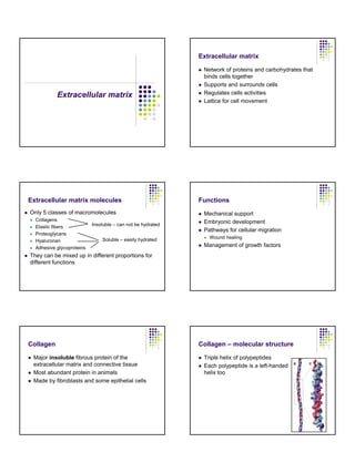
- 1. Extracellular matrix Extracellular matrix z Network of proteins and carbohydrates that binds cells together z Supports and surrounds cells z Regulates cells activities z Lattice for cell movement Extracellular matrix molecules z Only 5 classes of macromolecules z Collagens z Elastic fibers z Proteoglycans z Hyaluronan z Adhesive glycoproteins Insoluble – can not be hydrated Soluble – easily hydrated z They can be mixed up in different proportions for different functions Functions z Mechanical support z Embryonic development z Pathways for cellular migration z Wound healing z Management of growth factors Collagen z Major insoluble fibrous protein of the extracellular matrix and connective tissue z Most abundant protein in animals z Made by fibroblasts and some epithelial cells Collagen – molecular structure z Triple helix of polypeptides z Each polypeptide is a left-handed helix too
- 2. Types of collagen z Fibrillar z Forms structures such as tendon or cartilage z Sheet forming z Connecting z Supports and organizes fibrous collagen z Transmembrane Fibrillar collagen I z Basic unit – triple helix 300 nm long z 2 α1 and 1 α2 molecules z Collagen fibrils form by lateral interactions of triple helices z Stabilized by covalent bonds z Displacement by 67 nm (gives the striated look) The basic structural unit of collagen Assembly of collagen fibers z Synthesized in secretory pathway as procollagen z Glycosylation in ER and Golgi z Helix formation in ER z Disulfide bonds that are cleaved later z Assembly outside ! Sheet forming collagen z Polymerizes into sheets z Forms basal membranes – collagen IV z Collagen VIII – Descemet’s membrane of cornea Collagen IV z A helix interrupted about 24 times by segments that can not form a helix z Globular domains at both C- and N-termini
- 3. Collagen IV z Nonhelical domains introduce flexibility z C-terminus globular domains associate with each other z Helical domains associate laterally to form branching strands Connecting collagens z Link fibrillar and sheet forming collagens to into networks and connect them to other structures z Collagen VI – short helices interspersed with globular domains z Align collagen I into parallel structures Connecting collagens z Collagen IX has two rigid helices connected by the flexible kink z The globular N-terminus binds to proteoglycans in the extracellular matrix z Links collagen II to glycosoaminoglycans (provides cushion for compression as in cartilage) Elastic fibers z Found throughout the body z Most prominent in skin z Composite of fibrillin fibrils and elastin z Synthesized only by fetal and juvenile fibroblasts z Whatever is made by puberty has to last until the end z Loss is responsible for wrinkles Fibrillin z Tread like protein z Forms 10 nm microfibrils z Found in elastic fibers and basal lamina z Linear molecule with independently folded domains Elastin z A polymer of tropoelastins z Tropoelastins form a family of closely related proteins z Products of a single gene and alternative splicing
- 4. Elastin z Continuous random network of elastin polypeptides z Helical domains separate random chains rich in hydrophobic residues Soluble components of extracellular matrix z Proteoglycans z Form highly hydrated gel responsible for volume of extracellular matrix z Hyaluronan z Hydrated polysaccharide z Makes matrix resilient to compression z Multiadhesive matrix proteins z Long flexible molecules that bind other matrix components and cells Proteoglycans z Diverse group of proteins with multiple polysaccharide chains z Found in connective tissues and extracellular matrix z Also attached to the surface of many cells z Responsible for volume of extracellular matrix z Highly hydrated Proteoglycans z Consist of multiple glycosaminoglycans (GAGs) posttranslationally added to a core protein Proteoglycans z Very diverse z Different type of core protein (aggrecan, syndecan) z Different composition of GAGs (chondroitin sulfate, heparin, heparan sulfate) z Different lengths GAG synthesis z GAGs are posttranslational modifications of a core protein z Core protein is synthesized in secretory pathway z Polysaccharides are added in ER by glycosyl transferases z Elongated and modified in Golgi
- 5. Functions of proteogylcans z Structural – elastic space fillers z Limit diffusion of macromolecules z Impede passage of microorganisms z Act as lubricants in the joints z Regulate cell motility and adhesion Other (nonstructural) functions of proteoglycans z Sequestration of growth factors z Present hormones to cell-surface receptors Proteoglycans z Assemble into aggregates with hyaluronan as a core of the aggregate Hyaluronan a.k.a. hyaluronic acid, hyaluronate z Major component of proteoglycans z Extremely long, negatively charged polysaccharide z Resists compression z Swollen gel creates turgor pressure z Forms viscous, hydrated gels z Large number of anionic residues on the surface bind water Hyaluronan z Hyaluronan keeps cells apart from one another z Facilitates cell migration z Surrounds migrating and proliferating cells z Inhibits cell-cell adhesion Adhesive glycoproteins z Long flexible molecules with domains for binding z Collagen z Other matrix proteins z Polysaccharides z Cell surface molecules z Signaling molecules
- 6. Adhesive glycoproteins z Attach cells to the extracellular matrix z Regulate cell attachment z Migration z Cell shape z Organize components of the matrix z Most bind to integrins – cellular adhesion receptors Laminins z Adhesive glycoproteins present in basal lamina z Basal lamina guides cells during development z Cells migrate along laminin containing surfaces z Cross-shaped proteins z As long as basal lamina is thick z 3 subunits z High affinity binding sites for z Heparan sulfate z Collagen IV z Cellular adhesion receptors Basal lamina z A thin planar assembly of extracellular matrix proteins z Basis is formed by collagen IV and laminin Interaction of the basal lamina with adjacent cells z Collagen IV and laminin interact with cell surface integrins and bind adjacent cells to basal lamina z Basal lamina guides cells during development z Cells migrate along laminin containing surfaces Basal lamina is structured differently in different tissues z Polarized cells z Filter that regulates passage of nutrients z Smooth muscle z Maintenance of integrity Basal lamina is structured differently in different tissues z Kidney glomerulus z Separates two cell layers z Double thickness lamina – both layers produce the basal lamina z Filters blood to form urine
- 7. Fibronectins z Attach cells to matrices that contain fibrous collagen z Essential for migration and cellular differentiation Tenascin z Expressed in embryonic tissues, wounds and tumors z Plays a role in development z Modular protein with 6 arms z Binds to cells via integrins