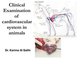
8 clinical examination of cardiovascular system in animals
•
3 gefällt mir•1,223 views

This document summarizes the clinical examination of the cardiovascular system in animals. It describes examining the heart through inspection, palpation, percussion, and auscultation. Auscultation is the primary method and involves listening for normal and abnormal heart sounds. Abnormal sounds include murmurs which can be systolic, diastolic, or continuous. The heart rate and rhythm are also examined. Additional examination involves the jugular vein, capillaries, and peripheral circulation.

Empfohlen
Weitere ähnliche Inhalte
Was ist angesagt?
Was ist angesagt? (20)
Ähnlich wie 8 clinical examination of cardiovascular system in animals
Ähnlich wie 8 clinical examination of cardiovascular system in animals (20)
Mehr von karima Akool AlSalihi
Mehr von karima Akool AlSalihi (20)
Kürzlich hochgeladen
Kürzlich hochgeladen (20)
8 clinical examination of cardiovascular system in animals
- 2. The cardiovascular system ,consist of two main structural units 1. The Heart 2. Blood vessels both of which are jointly involved in Maintaining the circulation of the blood and thereby ensuring normal exchange of oxygen, carbon dioxide dioxide, electrolytes, fluids, nutrients, and waste products between the blood and the tissue fluids and cells.
- 4. Examination of the Heart 1. The position of the heart within the thorax The heart occupies the ventral half of the thorax, in the lower two thirds of the thoracic cavity between the third to the sixth pairs of ribs, suspended at its base by the large blood vessels which ,traverse the mediastinum and its apex lying in the midline towards the sternum, slightly towards the left
- 6. Anatomy of the heart:
- 7. Methods of physical examination of the heart: The heart is examined by A. Inspection B. Palpation C. Percussion D. Auscultation All of these techniques can provide information, but auscultation is the fundamental component of the examination of the heart.
- 8. Inspection: Visible evidence of the contact of the heart with the chest may be obtained by observing the so-called “apex beat” of the heart, causing movement of the chest wall over a limited area of the fifth or sixth intercostal space
- 9. Palpation: Palpation may done to detect palpable vibration, or thrill, exist with the cardiac cycle. Palpation is performed by placing the fingers and the palm of the hand over the anatomical area of the heart
- 10. Percussion: Percussion is of limited value in the examination of the heart because most of the heart underlies the muscles of the forelimbs and the thoracic wall overlying the heart cannot be resonated
- 11. Auscultation Auscultation of the heart will performed to determine the 1-Normal and abnormal sounds 2-The heart rate, 3-The heart rhythm 4-The intensity of heart sounds.
- 12. Rules of auscultation: 1-Optimum auscultation sites are the fourth and fifth left intercostal space. 2-Manual extension of the forelimbs can facilitate auscultation of the heart. 3-chest piece of the stethoscope must be pushed firmly against the skin under the left elbow and the triceps muscle, with light pressure. 4- Examination should be conducted in a quiet environment
- 13. Heart sounds 1- Normal Heart sounds: There are four sounds of the heart. The first and second sounds are clearly audible in all normal animals. A third and fourth sounds may be heard in normal large animals, particularly in horses. First sound like “Lubb”: It is associated with closure of atrioventricular valves (mitral and tricuspid).
- 14. Second sound like “Dubb”: It is associated with closure of aortic and pulmonary valves. Third sound associated with rapid filling of the ventricle in early diastole. Forth sound associated with atrial contraction.
- 15. 2- Abnormal Heart sounds: Abnormal heart sounds may originate from the heart due to 1-Endocardial lesions and related to the cardiac cycle, or 2-From overpower normal heart sound and not related to the cardiac cycle.
- 16. Cardiac murmurs: Cardiac murmurs are audible vibrations transmitted to the surface of the thoracic wall, caused by turbulent blood flow inside the heart due to endocardial lesions such as insufficiency of valvular closure and abnormal orifices. Cardiac murmurs can be described according to the cardiac cycle as Systolic, Diastolic or Continuous.
- 17. Systolic murmur: Occurs during systole due to insufficiency in the atrioventricular valves
- 18. Diastolic murmur: Occurs during diastole due to insufficiency in mitral or pulmonic valves
- 19. Continuous murmur: begins in systole and continues into diastole. It is occurs in the presence of abnormal orifices in the heart, such as ductus arteriosus, a defect where there is continuous shunting of blood from aorta to the main pulmonary artery. Pericardial frictional rub: This is audible vibration heard over the thorax, produced by friction occurring with movement of the heart in an inflamed pericardial sac due to pericarditis The sound occur with each heart cycle
- 20. Tinkling sound: This sound heard in the cardiac area, indicate the presence of gas on the surface of fluid enclosed in the pericardial sac. The sound will be as fluid- splashing “washing machine”. Muffled sound: This sound occur due to an increase in the volume of pericardial fluid (pericardial effusion)
- 21. Heart rate The heart rate is determined by counting the number of beats per 1 minute . Animal Range Horse, adult 28 – 46 Foal 60 – 100 Cattle, adult 60 – 80 Calf (up to 1 year) 80 – 100 Sheep, Goat 70 – 90 Dog, small breeds 90 – 120 Dog, large breeds 65 – 90 Cat 110 – 130 Camel 25 - 50
- 22. Abnormal heart rate: Tachycardia: it is a marked increase in the heart rate, Bradycardia: it is a marked slowing of heartbeat Heart rhythm The normal rhythm of the heartbeat is in three-time and described as ( LUBB-DUPP-pause ).
- 23. Heart intensity The intensity or “loudness” of the heart sounds depends on 1-The strength of the heartbeat 2-The thickness of the thoracic wall, 3-The presence of any factor which interferes with or enhances the transmission of the sounds from the heart to the stethoscope
- 24. Examination of jugular vein Jugular vein present in subcutaneous in the jugular furrow on both side of neck Examination of the jugular vein provides a valuable indication of the functioning of the right side of the heart. Examination include …. A-Jugular pulsation: Several events in the cardiac cycle cause pulsation in the jugular vein. It may be physiological (Half of neck) or pathological (Full neck)
- 25. Normal or negative jugular pulsation “Physiological jugular pulsation”: It occurs during the early part of systolic phase of the cardiac cycle when the blood being temporarily unable to enter the contracting right atrium ,its dammed back in the jugular vein
- 27. True, or positive jugular pulsation “Pathological jugular pulsation”: It is consists of typical pressure wave, run forward from the thoracic inlet to the angle of mandible. It occurs during cardiac systole in the case of the tricuspid incompetence because the blood in the right ventricle is regurgitated back through incompletely closed valvular orifice into the right atrium and jugular vein.
- 28. False jugular pulsation: It is associated with transmission of the pulse from the underlying carotid artery through the jugular vein. It is noted in sever emaciated animals and can be easily differentiated from true jugular pulsation by occluding the jugular vein with the thumb, if the pulsations continue then they are associated with a transmitted carotid pulse
- 29. B- Jugular engorgement or Venous stasis Negative Jugular engorgement If the vein is compressed digitally in the middle of the neck it becomes distended above the pressure point because of the accumulation of blood,whereas the section below the pressure point will become empty
- 30. positive Jugular engorgement When the jugular vein is become visibly distended from upper and lower part after compressed digitally (Venous stasis ) Its occur in right-sided congestive heart failure, pericarditis, endocarditis, and myocarditis,
- 31. Examination of capillaries Sclera is the best place to examine the peripheral capillaries. The capillaries on the sclera may be normal “contain enough quantity of venous blood”, or abnormal as follows….
- 32. Engorged: when the sclera appeared redden and the capillaries filled and corrugated with blood. Empty: when the sclera appeared pale or whitish and the capillaries empty from blood. Capillary refilling time: Capillary refilling time is examined through oral mucous membrane(Gingiva) (Gum) . It is a useful indicator of perfusion of peripheral tissues and the state of the cardiovascular system Normal refilling time of capillaries is 1-2 second