The alimentary canal of Scoliodon comprises:
the mouth,
buccal cavity,
pharynx,
oesophagus,
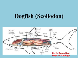
stomach,
intestine and
rectum opening in the cloaca through anus.
2. Alimentary Canal:
The alimentary canal of Scoliodon
comprises:
the mouth,
buccal cavity,
pharynx,
oesophagus,
stomach,
intestine and
rectum opening in the cloaca through
anus.
3.
4. a. Mouth:
It is a ventral crescentic opening guarded
by upper and lower lips which are folds of
integument.
b. Buccal Cavity:
The mouth leads into a spacious dorso-
ventrally compressed buccal cavity. It is
bordered with jaws. The buccal cavity is
lined with a thick mucous membrane.
5. Denticles are all alike in shape,
homodont, and are borne in several
parallel rows on the inner margin of the
upper and lower jaws.
c. Pharynx:
The buccal cavity merges posteriorly
with the large pharynx lined by
endoderm.
The cavity of pharynx is lined with
mucous membrane containing numerous
dermal denticles.
6. d. Oesophagus:
The pharynx narrows posteriorly to
form the short and wide oesophagus.
The oesophagus has thick muscular
walls with an internal lining of
mucous membrane raised into
longitudinal folds.
7. e. Stomach:
The oesophagus widens posteriorly to
form the large muscular stomach.
The stomach is bent on itself and forms
a J-shaped organ, the long, wider
distensible proximal limb which is called
the cardiac stomach, while the short
narrow distal limb is called the pyloric
stomach.
8.
9. f. Intestine:
The bursa entiana continues into the
intestine. The intestine is a wide tube
running straight backwards into
abdominal cavity and its middle region is
like cardiac stomach in diameter.
Its narrow anterior part receives the bile
and pancreatic ducts.
10.
11. g. Rectum:
The short and narrow rectum is the last
part of the alimentary canal. The
tubular rectal (caecal) gland opens
dorsally into the rectum.
Its function is not known. The rectum
opens into the cloaca through anus. Into
the cloaca also open the urinogenital
ducts.
12. Glands of the Alimentary Canal:
a. Liver, Rectal Gland, and Spleen:
The liver is an elongated yellowish gland,
consisting of two lobes which extend back
along the greater part of the abdominal
cavity.
The liver produces bile, stores glycogen and
fat. It also destroys worn-out red blood
corpuscles since Kupffer cells are present.
The gall bladder, which stores the bile
secreted by the liver.
13. b. Pancreas:
The pancreas is a compact bilobed gland
situated in the angle between the cardiac
and pyloric stomach.
The pancreatic duct opens into the
intestine just opposite to the opening of
the bile-duct
14. c. Rectal Gland:
The rectal or caecal gland is a short thick
hollow diverticulum arising from the
dorsal wall of the rectum.
It is richly vascularised and formed of
lymphoid tissue.
15. d. Spleen:
It is a large lymphoid organ attached
with the cardiac and pyloric stomach.
It produced lymphocytes and, thus,
has no physiological relation with the
alimentary canal.
16. Food and Physiology of Digestion:
Scoliodon is carnivorous and feeder. The
digestion starts only in the cardiac
stomach. The cardiac stomach secretes
the gastric juice which contains pepsin
and hydrochloric acid that converts
proteins into proteoses and peptones.
The gastric juice is not able to digest
chitin.
17. The pancreas secretes trypsin, amylopsin,
and lipase. As the semi-digested food
enters the intestine it is acted upon “by
the bile and the pancreatic juice.
The bile makes the food alkaline and,
thus, helps the action of pancreatic juice.
The trypsin acts on the remaining
proteins, the amylopsin converts starch
into sugar and lipase turns fats into fatty
acids.
18. The digested food is absorbed into
the blood over the extensive
surfaces of the intestine and scroll-
valve.
20. The respiration is aquatic. It breathes
by means of gills borne in a series of
gill-pouches on either lateral side of the
pharynx.
Water enters the mouth and after
passing through the buccal cavity,
pharynx, gill- pouches bearing gill-
lamallae, goes out through the external
gill-slits after bathing the gills.
21.
22. Respiratory Organs:
There are five pairs of gill- pouches
bearing gills, arranged in a series
behind the hyoid arch in the lateral
walls of the pharynx.
Each gill-pouch has two sets of gill-
lamellae. The branchial lamellae have
a rich blood supply.
23. Mechanism of Respiration:
1. Inspiration:
The floor of the buccopharyngeal
cavity is depressed by the contraction
of hypobranchial (hypoglossal) muscles
due to which the visceral arches expand
the wall of the pharynx.
24. 2. Expiration:
During expiration the mouth becomes
closed by the action of adductor muscle.
The respiratory movements are caused
by pharyngeal muscles which are
innervated by V, VII, IX and X cranial
nerves and the hypoglossal spinal nerve.
25. Physiology of Respiration:
the highest concentration of oxygen and
the lowest of carbon dioxide, thus, an
efficient exchange of oxygen and carbon
dioxide takes place between the blood
and sea-water.
26. The oxygen of the water passes by endosmosis
through the thin capillary walls into the
blood, and at the same time the carbon
dioxide of the blood passes into the water by a
process of exosmosis.
The oxygen is conveyed by the blood to all the
parts of the body, while carbon dioxide
brought to the gills in the venous blood is
eliminated by the water of the outgoing
respiratory current.
28. Heart:
Heart is a bent muscular tube and
consists of the receiving parts,
comprising of a sinus venosus and a
dorsally placed auricle and the
forwarding parts, consisting of a
ventricle and a conus arteriosus.
The heart is situated on the ventral
side of the body between two series of
gill pouches.
29.
30. The auricle is a large, triangular and
thin walled chamber situated dorsal to
the ventricle but in front of the sinus
venosus.
The auricle communicates with the
ventricle through a slit-like auriculo-
ventricular aperture guarded by two
lipped valves.
31. The receiving chambers (sinus venosus and
auricle) receive the venous blood from all
parts of the body.
The function of the heart is to receive the
deoxygenated blood from all parts of the body
and to pump it for aeration to the gills.
Such a type of heart is designated as the
venous or branchial heart, because only the
deoxygenated blood circulates through it.
33. The nervous system of Scoliodon
includes:
(i) The central nervous system,
(ii) The peripheral nervous system, and
(iii) The autonomous nervous system.
(i) Central nervous system:
The central nervous system consists of
brain and the spinal cord.
(a) Brain:
34. Brain is highly organised and shows
many advancements over that of the
agnathans.
The brain is divided into three
primary parts:
(a) The forebrain or prosencephalon,
(b) The midbrain or mesencephalon
(c) The hindbrain or rhomben-
cephalon.
35.
36. 1. The forebrain consists of a
massive undivided cerebral
hemisphere. The cerebral
hemisphere is relatively larger than
that of other fishes.
From the anterior end of cerebral
hemisphere arise two stout olfactory
peduncles; each terminates into a
large bilobed olfactory lobe
37. The olfactory lobes lie close to the olfactory
capsules. Each olfactory nerve is composed of
many bundles of nerve fibres.
The surface of the cerebrum is smooth and the
walls are thick. A small opening on the mid-
ventral surface of the cerebrum.
The posterior part of forebrain (diencephalon)
is very short. The roof of the diencephalon is
thin, non-nervous and contains the anterior
choroid plexus.
38. 2. The mid-brain is large and consists of
two round optic lobes. The optic lobes are
situated behind the diencephalon.
3. The hindbrain consists of a highly
developed cerebellum and a medulla
oblongata.
The cerebellum is divided into three lobes
by two well-marked transverse furrows.
39. The medulla oblongata is triangular
and the anterior end gives a pair of
hollow corpora restiformia with trace
of convolutions in adults. The
medulla controls respiration.
The hind- brain controls swimming
movements. The cerebellum is
divided into three lobes by two well-
marked transverse furrows.
40. (b) Spinal cord:
The spinal cord in Scoliodon shows
definite advancement towards the
plan of higher vertebrates. The grey
matter is arranged into the dorsal
and the ventral horns. The dorsal
horns are united to form a single
broad region; as a result the grey
matter assumes a shape of an
inverted ‘T’.
41. (ii) Peripheral nervous system:
The peripheral nervous system
includes the cranial nerves and spinal
nerves.
(a) Cranial nerves:
There are ten pairs of cranial nerves
in all the fishes.
The first pair of cranial nerves is the
olfactory nerves which originate from
the olfactory lobes and innervate the
olfactory sacs.
42. The second pair of cranial nerves is the
optic nerves which, after the origin from
the optic thalami, form the optic chiasma
and supply the eyes.
The third cranial nerve is called
oculomotor nerve which originates from
the ventral surface of the mesencephalon
and supplies the anterior, superior and
inferior recti and the inferior oblique
muscles of each eye ball.
43. The fourth cranial nerve is called
trochlear or pathetic nerve which
arises from the dorsolateral surface
of the midbrain and supplies the
superior oblique eye muscle.
44. The fifth cranial nerve is the
trigeminal which has three branches:
(1) Ophthalmicus superficial which
supplies to the skin of the snout.
(2) The maxillaris which is divided
into maxillaris superior supplying
nerves to the skin of the upper jaw
and maxillaris inferior innervating
the posterior part of the upper jaw.
45.
46.
47. (3) The mandibularis innervating the
muscles of the lower jaw.
Another nerve called ophthalmicus
pro-fundus becomes secondarily
associated with the trigeminal to
supply nerves to the eye ball and the
dorsal surface of the snout
48. The sixth cranial nerve is the
abducens which supplies the
posterior rectus muscle of the eye
ball.
The seventh cranial nerve is known
as facial which divides into two
branches:
(1) The ophthalmicus superficialis
branch like that of the fifth cranial
nerve.
49. (2) A bundle of mixed nerves which
subdivides into three routes:
The eighth cranial nerve is called auditory
which gives the vestibular and saccular
branches to the internal ear.
The ninth cranial nerve is the glosso-
pharyngeal which, in the region of the first
gill cleft, divides into a small pretrematic
nerve and a large post-trematic nerve.
50. These nerves supply branches to the
pharynx, pharyngeal muscles and the
mucous membrane surrounding the
first gill slit.
The tenth cranial nerve is the vagus
which arises by multiple roots and
gives off many branches.
51. (b) Spinal nerves:
The spinal nerves arise from the
spinal cord. Each has one dorsal and
one ventral root. The dorsal root
bears a ganglionic swelling.
After emerging out through the
vertebral column, the dorsal and the
ventral roots unite to form a common
mixed nerve.