Measuring visual acuity and contrast sensitivity by optomotor reflex in rodents

There is a growing need for behavioral readouts to monitor disease progression and to assess the success of a potential therapy. In vision research, observing the optomotor reflex (OMR) is an important and widely established method for assessing visual acuity and contrast sensitivity in rodents. These tests can be performed with freely moving animals without any need for anaesthesia or restraints. In addition, since OMR is a reflex-based behavior, observing it does not require any training of the animal. In this webinar, sponsored by Striatech and supported in part by Stoelting, researchers will present objective and bias-free results obtained using a newly developed automated optomotor system. For more information, please visit: https://insidescientific.com/webinar/measuring-visual-acuity-contrast-sensitivity-optomotor-reflex-striatech

Empfohlen
Weitere ähnliche Inhalte
Was ist angesagt?
Was ist angesagt? (20)
Ähnlich wie Measuring visual acuity and contrast sensitivity by optomotor reflex in rodents
Ähnlich wie Measuring visual acuity and contrast sensitivity by optomotor reflex in rodents (20)
Mehr von InsideScientific
Mehr von InsideScientific (20)
Kürzlich hochgeladen
Kürzlich hochgeladen (20)
Measuring visual acuity and contrast sensitivity by optomotor reflex in rodents
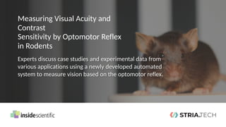
- 1. Measuring Visual Acuity and Contrast Sensitivity by Optomotor Reflex in Rodents Experts discuss case studies and experimental data from various applications using a newly developed automated system to measure vision based on the optomotor reflex.
- 2. Measuring Visual Acuity and Contrast Sensitivity by Optomotor Reflex in Rodents Kaushikaram Subramanian, PhD Volker Enzmann, PhD Research Director Department of Ophthalmology Inselspital, University of Bern Post Doctoral Researcher Max Planck Institute of Molecular Cell Biology & Genetics Thomas Münch, PhD Director of Research & Development Striatech
- 3. InsideScientific is an online educational environment designed for life science researchers. Our goal is to aid in the sharing and distribution of scientific information regarding innovative technologies, protocols, research tools and laboratory services
- 4. To access webinar content, Q&A reports, FAQ documents, and information on lab workshops, subscribe to our mail list
- 5. The Striatech Founders Experts in applied ophthalmology & vision research 5 Dr. Thomas Münch | Director for Research and Development Neuroscientist t.muench@stria.tech Dr. Boris Benkner | Managing Director Neuroscientist b.benkner@stria.tech Marion Mutter | M. Sc., MBA Chief Financial Officer Neuroscientist International Management m.mutter@stria.tech
- 6. Determining vision in laboratory rodents by optomotor reflex measurements 6
- 7. 7 • Reflex to stabilize the image of the environment in the eye • OMR initiates a head movement that automatically follows an environmental movement The Optomotor Reflex (OMR) Only possible if the animal can (still) see!
- 8. 8 Measuring visual acuity and contrast sensitivity based on OMR ✔ ✔ ✘ Visual acuityScientist Animal Step-by-step procedure until visual threshold is found
- 9. 9 Measuring visual acuity and contrast sensitivity based on OMR ✔ ✔ ✘ Visual acuity Contrast sensitivityScientist Animal Step-by-step procedure until visual threshold is found
- 10. 10 ADVANTAGES • No training required • No fixation or surgery required • Reflex exists directly after eye opening • Longitudinal experiments possible • Very robust • For testing visual acuity and contrast sensitivity OMR Pros & Cons
- 11. 11 ADVANTAGES • No training required • No fixation or surgery required • Reflex exists directly after eye opening • Longitudinal experiments possible • Very robust • For testing visual acuity and contrast sensitivity CAUTION • Reflex → No cortical vision is tested • Depending on disease model, no effect on optomotor reflex by moderate retinal damage OMR Pros & Cons
- 12. DOWNSIDES OF MANUAL MEASUREMENTS • Experimenter bias • Non-consistent data • Time consuming 12 Manual measurements vs. automated analysis Problems can be solved with a reliable automated analysis.
- 14. How the OptoDrum works 14 1 Animal is placed in arena on elevated platform, surrounded by computer monitors. 2 Stripe pattern slowly rotates around animal, triggering optomotor reflex. 3 Camera observes animal behavior. 4 Behavior is automatically detected and analyzed by OptoDrum software. 5 Stimulus pattern is continuously and automatically adjusted during experiment to find animal‘s visual threshold (visual acuity or contrast sensitivity). 1 2 3 4 5 OptoDrum
- 15. What do the results look like? Example: Retinal degeneration (rd10) 15 rd10 mice Model for human Retinitis pigmentosa disease Benkner et al. Behav Neurosci. (2013)
- 16. What do the results look like? Example: Retinal degeneration (rd10) 16 Part I: Measuring contrast sensitivity Contrast threshold (CTHR): weakest contrast that still triggers the optomotor reflex Contrast sensitivity: 1 / CTHR Contrast sensitivity ✔ ✔ ✘ Benkner et al. Behav Neurosci. (2013)
- 17. What do the results look like? Example: Retinal degeneration (rd10) 17 Part I: Measuring contrast sensitivity 0.05 cyc/° Contrast sensitivity ✔ ✔ ✘ Benkner et al. Behav Neurosci. (2013)
- 18. What do the results look like? Example: Retinal degeneration (rd10) 18 Part I: Measuring contrast sensitivity 0.05 cyc/° 0.15 cyc/° 0.3 cyc/° Contrast sensitivity ✔ ✔ ✘ Benkner et al. Behav Neurosci. (2013)
- 19. What do the results look like? Example: Retinal degeneration (rd10) 19 Part I: Measuring contrast sensitivity Contrast sensitivity ✔ ✔ ✘ Benkner et al. Behav Neurosci. (2013)
- 20. What do the results look like? Example: Retinal degeneration (rd10) 20 Part II: Measuring visual acuity Visual acuity threshold: highest resolution (finest stripes) that still triggers the optomotor reflex (usually measured at maximal contrast) Contrast sensitivity ✔ ✔ ✘ Benkner et al. Behav Neurosci. (2013)
- 21. What do the results look like? Example: Retinal degeneration (rd10) 21 Part II: Measuring visual acuity Visual acuity ✔ ✔ ✘ Benkner et al. Behav Neurosci. (2013)
- 22. What do the results look like? Example: Retinal degeneration (rd10) 22 Part II: Measuring visual acuity Visual acuity ✔ ✔ ✘ Benkner et al. Behav Neurosci. (2013)
- 23. What do the results look like? Example: Retinal degeneration (rd10) 23 Part II: Measuring visual acuity Visual acuity ✔ ✔ ✘ Benkner et al. Behav Neurosci. (2013)
- 24. What do the results look like? Example: Retinal degeneration (rd10) 24 Contrast sensitivity function Visual acuity ✔ ✔ ✘ Benkner et al. Behav Neurosci. (2013)
- 25. Applications of optomotor reflex measurements 25 •Retinal degeneration o rd10 animals •Basic biological questions about vision o Visual benefits of mouse rod nuclear architecture •Pharmacological and toxicological studies o Mimicking age-related macular degeneration with NaIO3 •Multiple Sclerosis o Optic nerve damages Dr. Thomas Münch Prof. Dr. Volker Enzmann Dr. Kaushikaram Subramanian
- 26. Relevance of the Rod Nuclear Architecture for Animal’s Vision & Behavior Copyright 2019 K. Subramanian and InsideScientific. All Rights Reserved. Kaushikaram Subramanian, PhD Post Doctoral Researcher Max Planck Institute of Molecular Cell Biology & Genetics
- 27. 27 Relevance of the rod nuclear architecture for animal’s vision & behavior Consequences for vision Tissue optical properties Cellular architecture
- 28. Tissue optical properties 28 Consequences for vision Cellular architecture Relevance of the rod nuclear architecture for animal’s vision & behavior
- 29. Solovei et al. Cell (2009, 2013) Nuclear Architecture Conventional architecture pig mice euchromatinheterochromatin 29
- 30. Solovei et al. Cell (2009, 2013) diurnal pig, cow, macaque, human… 50 more species. Conventional architecture pig Rod cell euchromatinheterochromatin 30 Nuclear Architecture
- 31. Solovei et al. Cell (2009, 2013) diurnal pig, cow, macaque, human… 50 more species. Conventional architecture pig nocturnal Inverted architecture Mouse, rat, cat, rabbit, deer… 50 more species. mice Rod cell euchromatinheterochromatin 31 Nuclear Architecture
- 32. Solovei et al. Cell (2009, 2013) 0 14 28 adult Development (days after birth) Lamin A/C & Lamin B receptor (LBR) 32 Cellular Architecture
- 33. Solovei et al. Cell (2009, 2013) euchromatin heterochromatin 0 14 28 adult Development (days after birth) LBR over expression Developmental arrest chromocenters Lamin A/C & Lamin B receptor (LBR) 33 Cellular Architecture
- 34. 34 Relevance of the rod nuclear architecture for animal’s vision & behavior Consequences for vision Tissue optical properties Cellular architecture
- 35. 35 Tissue optical properties – The vertebrate eye & retina Mouse Retina
- 36. 36 Image transmission through the retina 760 x 640 um Sinusoidal pattern transmitted through retina transmitted image
- 37. 37 Subramanian et al., eLife 2019 Retinal Contrast Transmission
- 38. 38 Subramanian et al., eLife 2019 Retinal Contrast Transmission
- 39. 39 Subramanian et al., eLife 2019 Nuclear inversion causes improved retinal transparency Retinal Contrast Transmission
- 40. Tissue optical properties 40 Relevance of the rod nuclear architecture for animal’s vision & behavior Consequences for vision Cellular architecture
- 41. 41 Consequences for vision – Optomotor measurement
- 42. 42 Subramanian et al., eLife 2019 Contrast Sensitivity Assessment
- 43. Photopic 43 Photopic Contrast Sensitivity WT TG-LBR
- 44. 44 Subramanian et al., eLife 2019 Photopic and Scotopic Contrast Sensitivity WT TG-LBR Photopic Scotopic
- 45. 45 Subramanian et al., eLife 2019 Retinal Transmission - realistic images Early detection of stimulus by WT in dim light environments WT TG-LBR
- 46. 46 Acknowledgements Kreysing Lab Collaborators Irina Solovei, LMU Martin Weigert, Myers Lab, CSBD Oliver Bosch, Ader Lab, CRTD Striatech GmbH MPI - Facilities Thank you!
- 47. Quantification of Visual Acuity by OMR in the Degenerated Mouse Retina – Pros and Cons Copyright 2020 V. Enzmann and InsideScientific. All Rights Reserved. Volker Enzmann, PhD Research Director Department of Ophthalmology Inselspital, University of Bern
- 48. Animal models of retinal degeneration 48 GENETIC MODELS • rds mouse (peripherin mutation) • rd mouse (phosphodiesterase 6B) • RCS rat (Mertk mutation) PHARMACOLOGICAL MODELS • Methylnitrosourea (MNU) in different species • Sodium iodate (NaIO3) in different species • Induced Experimental autoimmune encephalomyelitis (EAE) KO MODELS • RPE65-/- mouse • Rho-/- mouse LASER-INDUCED MODELS • Argon laser-induced CNV in monkey • Diode laser-induced damage in different species Measurement of function is always important to validate the animal model and quantify changes after treatment.
- 49. 49 Quantification of visual acuity by OMR in the degenerated mouse retina Induced Experimental autoimmune encephalomyelitis Diode laser-induced damage Sodium iodate (NaIO3)
- 50. 50 Quantification of visual acuity by OMR in the degenerated mouse retina Induced Experimental autoimmune encephalomyelitis Diode laser-induced damage in different species Sodium iodate (NaIO3)
- 51. 51 • Selective toxin for retinal pigment epithelium (RPE, necrosis) followed by photoreceptor death (apoptosis) • No model for age-related macular degeneration (AMD), but mimics symptoms of AMD Sodium iodate (NaIO3) – Basics • Concentration-dependent / Time-dependent o Morphological changes: diminished RPE autofluorescence, degenerated RPE layer, diminished retina thickness (outer nuclear layer, ONL) o Functional deficits: decrease in electroretinogram (ERG), optomotor reflex
- 52. 52 Sodium iodate (NaIO3) – Function Measurements Optomotor Reflex Optical Coherence Tomography camera
- 53. 53 Sodium iodate (NaIO3 ) – Function Correlation between morphological and functional changes UninjuredD3D10 B L Untreated D3 Untreated D10 Untreated BL 35m g-Kg N aIO 3 D 3 35m g-Kg N aIO 3 D10 35m g-Kg N aIO 3 BL 50m g-Kg N aIO 3 D3 50m g-Kg N aIO 3 D10 50m g-Kg N aIO 30.0 0.2 0.4 0.6 Time (days) Visualacuity(C/°) * *p<0.05 and **p<0.01 vs the corresponding BL group ** ** Optomotor Reflex Optical Coherence Tomography
- 54. Sodium iodate (NaIO3) 54 Quantification of visual acuity by OMR in the degenerated mouse retina Induced Experimental autoimmune encephalomyelitis Diode laser-induced damage
- 55. 55 Laser-induced Retinal Damage GFAP/PCNA d3H&E d3 •Model of photoreceptor degeneration •Focal damage in outer nuclear layer (ONL) •Müller cell activation •Scar formation •No regeneration in the mouse •Diode laser 532 nm •Output power: 120 mW •Aerial diameter: 100 µm •Pulse duration: 60 ms Mouse experimental set-up:
- 56. 56 Laser model & OMR No correlation between morphological and functional changes •532 nm diode laser, up to 30 spots (100 µm) around the optic nerve head •Optomotor measurements at baseline & 7, 14, 21, and 28 days after laser damage •No significant difference n.s. Optomotor Reflex Optical Coherence Tomography
- 57. 57 Conclusions •Functional measurements depend on damage extent •Detection of functional deficits in models w/ focal damage are difficult •Methods needs to be evaluated •Alternatives: o Cued water maze o Electroretinogram (ERG) o Focal ERG
- 58. Diode laser-induced damage Sodium iodate (NaIO3) 58 Quantification of visual acuity by OMR in the degenerated mouse retina Induced experimental autoimmune encephalomyelitis
- 59. Optic neuropathy – No animal model Causes and Symptoms •Optic neuropathy (also called optic neuritis) is inflammation of the optic nerve •Temporary vision loss and pain are the two main symptoms •Most people who suffer an episode of this condition fully recover their vision. •Risk factors: adults aged 20-45 / women twice as likely as men Visual field defects Disc swelling because of inflammation
- 60. Is Experimental autoimmune encephalomyelitis (EAE) a model for optic neuropathy? Experimental autoimmune encephalomyelitis (EAE) model •Most commonly used experimental model for multiple sclerosis (MS) •Immunization w/ Myelin Oligodendrocyte Glycoprotein (MOG 35-55) emulsified in CFA (Complete Freund’s Adjuvant) •Augmented with 200 mg anti-MOG Ab (8-18C5)
- 61. Is Experimental autoimmune encephalomyelitis (EAE) a model for optic neuropathy? Induction of EAE •Day 0 o i.p. injection of pertussis toxin (PTX) o s.c. injection of MOG peptide •Day 2 o i.p. injection of PTX •Day 10 o i.v. injection of anti-MOG Ab Disease Course
- 62. 62 Murine EAE disease course w/ and w/o monoclonal MOG-IgG (8-18C5) Visual acuity correlates with disease severity1 Functional measurement in EAE Augmentation of EAE with the injection of monoclonal MOG-IgG at day 10 1One measurement per animal was included in the analysis (last available measurement in controls, first measurement after onset of clinical signs in diseased mice). | Spearman’s rho r=0.33, p=0.01 Control Non-augmented EAE Augmented EAE
- 63. 63 Therapeutic intervention ameliorates EAE score Visual function is preserved in treated mice1 Quantification of treatment effects Treatment w/ neonatal Fc receptor (FcRn) as a potential therapeutic option for optic neuropathy 1Significant decrease of visual acuity at follow-up in untreated animals vs. their baseline measurement (p=0.024, Wilcoxon’s signed rank test, line) and vs. treated animals at follow-up (p=0.005, Mann-Whitney test, dashed line). ns= not significant.
- 64. 64 Conclusions Pros •Freely moving animals without anesthesia •Fast and reliable measurement •Automated detection Cons •High variability of the values •Optomotor response vs. Optokinetic reflex •No cell type-specific measurement
- 65. 65 Acknowledgements Experimental Ophthalmology, Dept. of Ophthalmology: Ana Maria Quintela Pousa, PhD Federica Maria Conedera, MSc Stephanie Lötscher Neuroimmunological Laboratory, Dept. of Neurology Anke Salmen, MD Jana Remlinger, MSc Adrian Madrasz Funding partners: Sutter-Stöttner-Stiftung Thank you for the attention!
- 66. Kaushikaram Subramanian, PhD Volker Enzmann, PhD Research Director Department of Ophthalmology Inselspital, University of Bern Post Doctoral Researcher Max Planck Institute of Molecular Cell Biology & Genetics Thomas Münch, PhD Director of Research & Development Striatech For additional information on the products and applications presented during this webinar, please contact the speakers below. Thank you!