SlideShare ist ein Scribd-Unternehmen logo
1 von 117
SUBARRACHINOID HAEMORRHAGE
1
By
Dr. Vinayak Hiremath
PG-INTERNAL MEDICINE
MRMC,GULBARGA
vinayak551989@gmail.com
 SAH = extravasation of blood into the
subarachnoid space
CLASSIFICATION OF STROKE
3
Stroke
Primary Hemorrhagic
(20% of Strokes)
Primary Ischemic
(80% of Strokes)
Thrombotic
50%
Embolic
30%
Intracerebral
Hemorrhage 15%
Subarachnoid
Hemorrhage 5%
INTRODUCTION
 Subarachnoid hemorrhage (SAH) is a pathologic
condition that exists when blood enters the
subarachnoid space
 The most common cause of SAH is trauma
 The most common cause of spontaneous SAH is an
aneurysmal bleed (65-80%)
Anatomy of Subarachnoid space
 In the central nervous system, the subarachnoid
cavity (subarachnoid space) is the interval between
the arachnoid membrane and pia mater.
 It is occupied by spongy tissue consisting of trabeculae
(delicate connective tissue filaments that extend from
the arachnoid mater and blend into the pia mater) and
intercommunicating channels in which the
cerebrospinal fluid is contained.
 This cavity is small on the surface of the hemispheres
of the brain. On the summit of each gyrus the pia
mater and the arachnoid are in close contact, but in
the sulci between the gyri, triangular spaces are left, in
which the subarachnoid trabecular tissue is found.
 At certain parts of the base of the brain, the arachnoid
is separated from the pia mater by wide intervals,
which communicate freely with each other and are
named subarachnoid cisternae; in these the
subarachnoid tissue is less abundant.
 The subarachnoid space is the location of the interface
between the vascular tissue and the cerebrospinal
fluid and is active in the blood brain barrier.
When blood vessels of the brain are weak, abnormal, or under unusual pressure, a
hemorrhagic stroke can occur. In hemorrhagic strokes, bleeding may occur within the
brain, as an intracerebral hemorrhage. Or bleeding may occur between the inner and
middle layer of tissue covering the brain (in the subarachnoid space), as a subarachnoid
hemorrhage.
EPIDEMIOLOGY
 Incidence about 9-10/100,000/yr
 Higher in Japan (3 times) and Finland
 Mean age of onset 51 years (6th decade)
 Facts and figures
 Men predominate until age 40, then more
women (55%); some studies – 3:2 ratio
 Seasonal (winter/spring), diurnal (late morning)
and day (Sunday)
 30 % rupture during sleep
 About 50 % of patients with an aneurysm have
warning prior to SAH (6-20 days)
Case Fatality
 The Overall case fatality varied from 32-67%
 Population-based study in England with essentially
complete case ascertainment
 24 hour mortality: 21%
 7 days: 37%
 30 days: 44%
Pobereskin JNNP 2001;70:340-3
OVERVIEW
 Highest mortality occurs immediately after the
hemorrhage and then decreases rapidly
 Rebleeding is estimated to occur in 50% of
ruptured aneurysms within 6 months of the
first hemorrhage, and afterwards at 3% per year
 50-60% of patients die after rebleeding and 25%
are left disabled
Probably the best natural history data come from S
Pakarinen: (Incidence, aetiology, and prognosis of primary subarachnoid
hemorrhage: a study based on 589 cases diagnosed in a defined urban population
during a defined period. Neurol Scand (Suppl) 1967; 29:1-128)
 Unselected series, involving the entire city of Helsinki,
from 1954 to 1961; minimal surgical withdrawals; all
sudden deaths were autopsied
 Mortality at 1 day was 32%
 Mortality at 1 week was 43%
 Mortality at 1 month was 56%
 Mortality at 6 months was 60%
ETIOLOGY
CATEGORY CAUSES
TRAUMA CLOSED, PENETRATING, ELECTRIC,ETC…
VASCULAR ANEURYSMS, ATHEROSCLEROSIS, AVM, VASCULITIDES
IDIOPATHIC BENIGN PERIMESENCEPHAIC SUBARACHNOID
HEMORRHAGE
BLOOD DYSCRASIAS LEUKEMIAS, HEMOPHILIAS, THROMBOCYTOPENIAS
INFECTIONS DENGUE, LEPTOSPIROSIS, BACTERIAL MENINGITIS
TOXINS AMPHETAMINES, COCAINE, NICOTINE,
ANTICOAGULANTS
NEOPLASMS GLIOMAS, MENINGIOMAS, HEMANGIOBLASTOMA, ETC
Risk Factors
 Hypertension
 Cigarette smoking
 Oral contraceptives
 Alcohol consumption (debatable)
 Diurnal variations in blood pressure
 Pregnancy and parturition
 Slight increased risk during lumbar puncture
and/or cerebral angiography in patient with
cerebral aneurysm
 Slight increased risk with advancing age
 Following cocaine abuse
Higher chance
if:
Female
3rd trimester of
pregnancy
Middle-aged
Abuse of
stimulant drugs
–Connective
tissue disorder
–Family history
PATHOPHYSIOLOGY
Modifiable Risk
Factors
>HPN
>Smoking
>excessive intake of
foods high in fats and
cholesterol
Non-modifiable Risk
Factors>
Advanced Age
>Gender
>Heredity
Triggering Factors
>Sudden extreme
emotion
Arterio venous
malformation
Cerebral aneurysm
rupture
Bleeding into the brain tissue
and subarachnoid space
Blood Clots in the
Subarachnoid Space
Brain Compression
Blood supply
interruption
Tissue Necrosis
Neuronal Death
Increase Intracranial
Pressure
T
total Paralysis
Regional Paralysis
Epileptic Seizure :
increase intraocular
pressure= blindness
Death
Coma
PATHOPHYSIOLOGY
CEREBRAL BLOOD
FLOW AND
METABOLISM
RAISED ICT
IMPAIRED
VASODILATORY
AUTOREGULATION
VASOSPASM WITH
GLOBAL ISCHEMIA
BLOOD (‘TOXIC’)
BLOCKAGE OF THE
ARACHNOID
GRANULATIONS
SYMPATHETIC
SURGE
MI
PULMONARY
EDEMA
 CEREBRAL BLOOD FLOW/VOLUME/METABOLISM
CBF is globally decreased after SAH
Mean CBF decreases with time after SAH;
it reaches a nadir in 10 to 14 days, after which it slowly
increases toward normal, In patients with poor grades, CBF
and cerebral metabolism may remain depressed for weeks ;
Cerebral blood volume is markedly increased in patients
with severe neurological deficits
 INTRACRANIAL PRESSURE RESPONSES
Mean ICP was 10 mm Hg in patients with clinical grades
1 and 2, 18 mm Hg in patients with clinical grades 2 and
3, and 29 mm Hg in those with clinical grades 3 to 5.
Vasospasm, which was more common in patients with a
poor clinical grade and larger SAH, was associated with a
significant rise in ICP from a mean of 16 mm Hg in those
without vasospasm to 29 mm Hg in those with
vasospasm
An aneurysm is an abnormal widening or
ballooning of a portion of an artery due to
weakness in the wall of the blood vessel
Internal carotid artery
Posterior
communicating artery
aneurysm
27
Mechanism of formation of aneurysms
 One theory is – that they result from a developmental defect
in the media & elastica
 Other theory says that aneurysmal process is initiated by the
focal destruction of internal elastic membrane caused by
hemodynamic forces acting at bifurcations & branching of
arteries
As a result of local weakness in vessel wall intima bulges out
covered only by adventetia
 The sac gradually enlarges and subsequently rupture
29
30
 For patients of SAH who arrive alive at hospital, the
mortality rate over the next month is about 45%. Of those
who survive, more than 50% are left with major
neurologic deficits as a result of
Initial hemorrhage,
Cerebral vasospasm with infarction, or
Hydrocephalus.
 If the patient survives but the aneurysm is not
obliterated, the rate of rebleeding is about
 20% in the first 2 weeks,
 30% in the first month, and about
 3% per year afterwards
 The annual risk of rupture for aneurysms <10 mm in
size is 0.1%, and for aneurysms >10 mm in size is 0.5–
1%;
 the surgical morbidity rate far exceeds these
percentages
31
32
 GIANT ANEURYSMS, those >2.5 cm in diameter
account for 5% of cases.
The three most common locations are
-The terminal internal carotid artery,
-Middle cerebral artery (MCA) bifurcation, and
-Top of the basilar artery.
 Their risk of rupture is ~6% in the first year after
identification and may remain high indefinitely.
 They often cause symptoms by compressing the adjacent
brain or cranial nerves.
 Saccular aneurysms occur at the bifurcations of the large to
medium-sized intracranial arteries.
Rupture is into the
 subarachnoid space,
 in the basal cisterns and often into
 the parenchyma of the adjacent brain.
 Approximately 85% of aneurysms occur in the anterior circulation,
mostly on the circle of Willis.
 About 20% of patients have multiple aneurysms,
- - many at mirror sites bilaterally.
33
34
Aneurysms are seldom seen in childern on
autopsy and so SAH in childern is rare
 Beyond childhood they gradually increase in size & the
frequency of rupture increases between 35-65 yrs of age
 There is increased incidence of
 polycystic kidneys,
 fibromuscular dysplasia of extracranial arteries,
 moyamoya, AV malformations &
 coarctation of aorta
 Numerous studies have documented familial occurrence of
saccular aneurysms, lending support to the idea that
genetic factors may play a role in their development 35
 As an aneurysm develops, it typically forms a neck with a
dome.
 At the site of rupture (most often the dome) the wall thins,
and the tear that allows bleeding is often 0.5 mm long.
 Aneurysm size and site are important in
predicting risk of rupture.
 Those >7 mm in diameter and
 Those at the top of the basilar artery and
 At the origin of the posterior
communicating artery are at greater risk of
rupture.
36
37
38
CLINICAL
MANIFESTATIONS-
The main symptom is a severe headache that starts suddenly
(often called thunderclap headache). It is often worse near the
back of the head. Many persons often describe it as the "worst
headache ever" and unlike any other type of headache pain.
The headache may start after a popping or snapping feeling in
the head.
Other symptoms:
and alertness
Eye discomfort in bright
Mood and personality changes, including and
irritability
(especially
Nausea and vomiting
often with neck stiffness, and
vomiting is common
40
Pain at other sites
 Occipital and posterior cervical pain may signal a
posterior inferior cerebellar artery (PICA) or anterior
inferior cerebellar artery aneurysm.
 Pain in or behind the eye and in the low temple can
occur with an expanding MCA aneurysm
41
Loss of consciousness
 At the moment of aneurysmal rupture with major
SAH, the ICP suddenly rises.
 This may account for the sudden transient loss of
consciousness that occurs in nearly half of patients
which may be preceded by headache.
 In 10% of cases, aneurysmal bleeding is severe
enough to cause loss of consciousness for several
days.
42
 meningismus
 neck stiffness
 photophobia
 back pain
 Numbness in part of the body
 Stiff neck
 focal neurologic deficits:
 oculomotor palsy
 hemiparesis
 ocular hemorrhage
What causes symptoms & signs?
Blood leaking from the
aneurysm
Local pressure effects of the
aneurysm
Associated ICH
Emboli
CLINICAL MANIFESTATIONS- cont.
FOCAL NEUROLOGICAL DEFICITS
 Although sudden headache in the absence of focal
neurologic symptoms is the hallmark of aneurysmal rupture,
FOCAL NEUROLOGIC DEFICITS MAY OCCUR.
 Anterior communicating artery or MCA bifurcation
aneurysms may rupture into the adjacent brain or subdural
space and form a hematoma large enough to produce mass
effect.
The common deficits that result include hemiparesis,
aphasia, and abulia (progressive drowsiness or slow
mentation)
48
Anatomic – clinical corellation of
focal neurological Deficits
 Third nerve palsy (ptosis, diplopia,dilatation of pupil &
divergent squint) indicates an aneurysm at the junction of
post. Comunicating &post. Cerebral arteries
 A sixth nerve palsy may indicate an aneurysm in the
cavernous sinus .but uni or bilateral palsy could be because
of raised ICP
 Unilateral blindness indicates aneurysm lying
anteromedially in the circle of willis usually at the origin of
opthalmic artery
49
Anatomic – clinical corellation of
focal neurological Deficits- cont
 Transient paresis of one or both lower limbs indicates ant
comunicating A. aneurysm that has interfered with
circulation in ant. Cerebral arteries.
 Hemiperesis or aphasia suggests aneurysm at the first
bifurcation of middle cerebral artery
50
51
The side of aneurysmal rupture may be
indicated by
monocular pain
unilateral preponderace of headache
or
 unilateral preretinal(subhyaloid)
hemorrhage (Terson Syndrome)
D/D
 Other conditions which may present as severe
headache need to be differentiated, like:-
Cerebral venous thrombosis,
Diffuse vasospasm (call-flemming syndrome),
 Pituatry apoplexy,
Hypertensive encephalopathy,
Intracranial or extracranial arterial dissection
 CSF examination assumes great importance in
differentiating these conditions from SAH 52
D/D- SAH to be differentiated from
THUNDERCLAP HEADACHE is a variant of
migraine that simulates a SAH.
Headache of explosive onset may also be
caused by ingestion of sympathomimetic
drugs or ingestion of tyramine containg foods
in a pt. who is taking MAO inhibitors
Sudden severe headache may also be a
symptom of phaeochromocytoma
53
D/D
 Before concluding that a patient with sudden, severe
headache has thunderclap migraine or any other type
of severe headache,
a definitive workup for aneurysm is always required in
the form of CT scan, Lumbar puncture, and if the
diagnosis still remains doubtful, then cerebral
angiography
54
DELAYED NEUROLOGIC DEFICITS
There are four major causes of delayed
neurologic deficits:
1. RERUPTURE/REBLEED,
2.HYDROCEPHALUS
3.VASOSPASM
4.HYPONATREMIA.
55
2.HYDROCEPHALUS
 Acute hydrocephalus can cause stupor and
coma and can be mitigated by placement of
an external ventricular drain.
More often, subacute hydrocephalus may
develop over a few days or weeks and causes
progressive drowsiness or slowed mentation
(abulia) with incontinence.. It may clear
spontaneously or require temporary
ventricular drainage.
56
Hydrocephalus-cont.
Chronic hydrocephalus may develop
weeks to months after SAH and present
as normal pressure hydrocephalus
(NPH) manifested by gait difficulty,
incontinence, or impaired mentation.
Subtle signs may be a lack of initiative
in conversation or a failure to recover
independence 57
3.VASOSPASM
 Causes symptomatic ischemia and infarction in ~30% of
patients and is the major cause of delayed morbidity and
death.
 Signs of ischemia appear 4–14 days after the hemorrhage,
most often at 7 days.
 The severity and distribution of vasospasm determine
whether infarction will occur.
 Vasospasm is believed to result from direct effects of
clotted blood and its breakdown products on the arteries
within the subarachnoid space.
58
VASOSPASM- cont.
 Spasm of major arteries produces focal symptoms
referable to the appropriate vascular territory.
 All of these focal symptoms may present abruptly,
fluctuate, or develop over a few days.
 In most cases, focal spasm is preceded by a decline in
mental status.
 Patient may develop fluctuating hemiperisis or aphasia
 Severe cerebral edema in patients with infarction from
vasospasm may increase the ICP enough to reduce
cerebral perfusion pressure.
59
4.HYPONATREMIA
 Hyponatremia. Hyponatremia may be profound and can develop
quickly in the first 2 weeks following SAH. There is both
natriuresis and volume depletion with SAH, so that patients
become both hyponatremic and hypovolemic.
 Both atrial natriuretic peptide (ANP)and brain
natriuretic peptide (BNP) have a role in
producing this "cerebral salt-wasting syndrome.“
 There may some role of antidiuretic hormone also, causing water
retention
 Typically it clears over the course of 1–2 weeks and, in the setting
of SAH, should not be treated with free-water restriction as this
may increase the risk of stroke. 60
SYSTEMIC CHANGES ASSOCIATED WITH SAH ECG changes suggestive of subendicardial or myocardial ischemia
 Elevation of troponis/CPK (MB) levels
 In some patients cardiac dysfunction may be severe enough to
cause fall in EF % and heart failure
 There is evidence that structural myocardial lesions produced by
circulating catecholamines and excessive discharge of sympathetic
neurons may occur after SAH, causing these ECG changes and a
reversible cardiomyopathy sufficient to cause shock or congestive
heart failure.
 Hyponatremia
 D. Insipidus
 Albuminuria,
 Glycosuria
 leukocytosis
61
INVESTIGATIONS
62
NON CONTRAST CT SCAN.
 CT Scan is Initial investigative procedure
 The extent and location of subarachnoid blood on
noncontrast CT scan help
 locate the underlying aneurysm,
 identify the cause of any neurologic deficit, and
 predict delayed vasospasm.
A high incidence of symptomatic vasospasm in the MCA
and ACA has been found when early CT scans show
subarachnoid clots >5 x 3 mm in the basal cisterns or layers
of blood >1 mm thick in the cerebral fissures.
 CT scans less reliably predict vasospasm in the vertebral,
basilar, or posterior cerebral arteries. 63
More than 95% of cases have enough blood to be
visualized on a high-quality NONCONTRAST CT SCAN
obtained within 72 h.
64
CT scan reveals subarachnoid hemorrhage in the right sylvian fissure; no evidence of
hydrocephalus is apparent.
(A)Noncontrast CT demonstrates subarachnoid hemorrhage (arrows). (B)3-D
reconstruction image from a CT angiogram demonstrates an aneurysm (arrow) from the
anterior communicating artery as the cause of the bleed.
IMAGING STUDIES-cont.
 Exact site of offending aneurysm can be inferred from the
location of main clot in CT scan
 Collection of blood in the anterior inter-hemispheric fissure
indicates rupture of an ant. Communicating artery
 Blood in sylvian fissure – middle cerebral artery
 Blood in ant. Perimesencephalic cistern- post.
Communicating artery or distal basilar artery aneurysm
 MRI can also detect blood in the proton density sequence
67
Lab investigations-cont
 If the scan fails to establish the diagnosis of SAH and no
mass lesion or obstructive hydrocephalus is found,
 Then a lumbar puncture should be performed to establish
the presence of subarachnoid blood.
 Usually CSF becomes grossly bloody within 30 minutes of
bleed with RBC counts upto 1 million/c mm or more
 Lysis of the red blood cells and subsequent conversion of
hemoglobin to bilirubin stains the spinal fluid yellow within
6–12 h.
 This xanthochromic spinal fluid peaks in intensity at 48 h
and lasts for 1–4 weeks, depending on the amount of
subarachnoid blood 68
Angiography
 Once the diagnosis of hemorrhage from a ruptured
saccular aneurysm is suspected, four-vessel
conventional x-ray angiography (both carotids and both
vertebrals) is generally performed
 to localize and define the anatomic details of the
offending aneurysm and
 to determine if other unruptured aneurysms exist).
 CT angiography is an alternative method for locating
the aneurysm and may be sufficient to plan definitive
therapy. 69
Angiography-cont.
 in 5-10% pts with aneurysmal bleed , there may not be
any evidence of aneurysmal rupture on angiography.
It may be because
in some cases obliteration of lesion occurs
In some lesions were somewhat more benign
 Pts without evidence of an offending aneurysm or an
arteriovenous malformationhave a better prognosis
70
Angiography-cont.
 Perimesencephalic hemorrhage also caries a better
prognosis
In this cisterns surrounding midbrain & upper pons
gets filled up with blood
Mild headache, no vasospasm
 Usually no aneurysm is found at the expected site i.e.
at the top of basillary artery
71
Lab Inv. – cont.
 Close monitoring (daily or twice daily) of electrolytes
is important because hyponatremia can occur
precipitously during the first 2 weeks following SAH
 An asymptomatic troponin elevation is common.
72
ECG Changes
 The electrocardiogram (ECG) frequently shows ST-
segment and T-wave changes similar to those
associated with cardiac ischemia.
 Prolonged QRS complex, increased QT interval, and
prominent "peaked" or deeply inverted symmetric T
waves are usually secondary to the intracranial
hemorrhage ( cerebral T waves).
 Serious ventricular dysrhythmias are unusual
73
74
ECHO Echocardiography reveals a pattern of regional wall
motion abnormalities that follow the distribution of
sympathetic nerves rather than the major coronary
arteries
 The sympathetic nerves themselves appear to be injured
by direct toxicity from the excessive catecholamine
release.
75
TREATMENT
The medical management of SAH focuses on
 Protecting the airway,
 Managing blood pressure
 Preventing rebleeding prior to treatment,
 Managing vasospasm,
 Treating hydrocephalus,
 Treating hyponatremia, and
 Preventing pulmonary embolus.
76
 Because rebleeding is common, all patients who are not candidates for
early aneurysm repair are put on bed rest in a quiet room and are given
stool softeners to prevent straining.
 If Headache or neck pain is severe, mild sedation and analgesia are
prescribed.
 Extreme sedation is avoided because it can obscure changes in
neurologic status.
 Adequate hydration is necessary to avoid a decrease in blood volume
predisposing to brain ischemia.
So adequate amount of fluid is administered so as to maintain above
normal circulating blood volume & central venous pressure
77
Managing raised ICP
 Intracranial hypertension following aneurysmal rupture
occurs secondary to
subarachnoid blood,
parenchymal hematoma,
acute hydrocephalus, or
 loss of vascular autoregulation.
Perilesional edema if infarct occurs
78
Managing raised ICP- cont.
 Patients who are stuporous should undergo emergent
ventriculostomy to measure ICP and to treat high ICP
in order to prevent cerebral ischemia.
 Medical therapies designed to combat raised ICP e.g.,
mild hyperventilation, mannitol, and sedation can also
be used as needed. (Mannitol dose-25-100gm
q4h)
 High ICP refractory to treatment is a poor prognostic
sign 79
MANAGING BP
 Prior to definitive treatment of the ruptured aneurysm,
care is required to maintain adequate cerebral perfusion
pressure
while avoiding excessive elevation/fall of arterial pressure.
 If the patient is alert, it is reasonable to lower the blood
pressure to <150 systolic using nicardipine, labetolol, or
esmolol.
 If the patient has a depressed level of consciousness, then
ICP(Intracranial pressure) should be measured and the
cerebral perfusion pressure targeted to 60–70 mmHg.
80
Managing Seizure Activity
 Seizures are uncommon at the onset of aneurysmal
rupture.
 The quivering, jerking, and extensor posturing that often
accompany loss of consciousness with SAH are probably
related to
 the sharp rise in ICP or, perhaps,
 acute generalized vasospasm rather than seizure.
 Use of anti-seizure drugs is controvercial.
However, phenytoin is often given as prophylactic therapy
since a seizure may promote rebleeding.
81
MANAGING VASOSPASM Vasospasm remains the leading cause of morbidity
and mortality following aneurysmal SAH.
 Treatment with the calcium channel antagonist
nimodipine (60 mg PO every 4 h) improves outcome,
perhaps by preventing ischemic injury
 Nimodipine can cause significant hypotension in
some patients, which may worsen cerebral ischemia in
patients with vasospasm
82
Managing Hydrocephalus Acute hydrocephalus can cause stupor or coma.
 It may clear spontaneously or require temporary
external ventricular drainage.
 When chronic hydrocephalus develops, permanent
ventricular shunting is the treatment of choice.
83
Role of Steroids
 Glucocorticoids may help reduce the head and neck
ache caused by the irritative effect of the subarachnoid
blood.
 There is no good evidence that they reduce cerebral
edema, are neuroprotective, or reduce vascular injury,
and
 Their routine use therefore is not recommended
84
Adjunctive Therapies and Measures
 Keep the patient's core body temperature at 37.2°C
 Consider antiemetics for nausea or vomiting.
 Elevate the head of the bed 30° to facilitate intracranial
venous drainage. Emergent ventricular drainage by the
neurosurgeon may be necessary.
 Maintain the patient's serum glucose level at 80-120
mg/dL; use sliding or continuous infusion of insulin if
necessary.
 Fluids and hydration
 Do not over hydrate patients because of the risks of
hydrocephalus.
 Patients with subarachnoid hemorrhage (SAH) may also
have hyponatremia from cerebral salt wasting.
INTERVENTIONAL MANAGEMENT
 Early aneurysm repair prevents
rerupture/rebleeding and
 allows the safe application of techniques
to improve blood flow (e.g., induced
hypertension and hypervolemia)
An aneurysm can be "clipped" by a
neurosurgeon or "coiled" by an
endovascular surgeon.
86
87
Hunt - Hess scale
is helpful in assessing the patient
before deciding for intervntional
management
HUNT-HESS SCALE
 GRADE1:
 Mild headache, normal mental status, no cranial nerve or
motor findings –(GCS* score 15, no motor deficits)
 GRADE 2:
Severe headache, normal mental status, may have cranial nerve
deficit –(GCS score 13–14, no motor deficits)
 GRADE 3:
Somnolent, confused, may have cranial nerve or mild motor
DEFICIT- (GCS SCORE 13–14, WITH MOTOR DEFICITS)
 GRADE 4 :
Stupor, moderate to severe motor deficit, may have intermittent
reflex posturing- (GCS score 7–12, with or without motor
deficits)
 GRADE 5:
Coma, reflex posturing or flaccid (GCS score 3–6, with or
without motor deficits)
88
Management based on HH scaleGrades 1 & 2
 Current approach is to go for intervention within 24
hrs
This will prevent rebleed and will allow measures to be
taken to improve cerebral circulation
 Grade 3 patients- if their condition allows , they too
would be benefitted by the procedure
 Grade4- the outcome is generally poor but some times by
putting a ventricular drain and improving the grade, pt89
90
SURGICAL REPAIR
•Involves placing a metal clip across the aneurysm
neck, thereby immediately eliminating the risk of
rebleeding.
•This approach requires craniotomy and brain
retraction, which is associated with neurologic
morbidity.
91
Endovascular techniques
involve placing platinum
coils, or other embolic
material, within the
aneurysm via a catheter that
is passed from the femoral
. Endovascular techniques-CONT
 The aneurysm is packed tightly to enhance thrombosis
and over time is walled-off from the circulation
 The benefit of endovascular therapy is durable.
 However, some aneurysms have a morphology that is
not amenable to endovascular treatment.
 Thus, surgery remains an important treatment option
 But for aneurysms which are not approachable by
surgery, endovascular technique would become the
only choice
92
INTERNATIONAL SUBARACHNOID
ANEURYSM TRIAL (ISAT)
 The only prospective randomized trial of surgery
versus endovascular treatment for ruptured aneurysm,
the International Subarachnoid Aneurysm Trial
(ISAT), was terminated early when 24% of patients
treated with endovascular therapy were dead or
dependent at 1 year compared to 31% treated with
surgery, a significant 23% relative reduction.
 After 5 years, risk of death was lower in the coiling
group,
 Although the proportion of survivors who were
independent was the same in both groups
 Risk of rebleeding was low, but more common in the
coiling group
93
GRADE CT APPEARANCE
1 NONE
2 < 1mm thickness
3 > 1mm thickness
4 Diffuse with
parenchymal/ventricular extension
FISHERS
GRADE CT APPEARANCE
0 NONE
1 MINIMAL
2 MINIMAL WITH IVH
3 THICK
4 THICK WITH IVH
CLAASSENS MODIFICATION
GRADE GCS NEURODEFICIT
1 15 ABSENT
2 13-14 ABSENT
3 13-14 PRESENT
4 7-12 ABSENT/PRESENT
5 <7 ABSENT/PRESENT
WORLD FEDERATION OF NEUROSURGEONS (WFNS)
GRADE CLINICAL PRESENTATION
1 CONSCIOUS
2 DROWSY
3 DROWSY WITH DEFICIT
4 MAJOR NEURODEFICIT
5 MORIBUND
BOTTERELL ET AL
Discharge Plan
 Activity
 patient will need to have someone with for the next several
days to watch for worsening of symptoms (see below) and to
allow to rest.
 Start with light activity around the house for the first 3 days
at home.
 Gradually increase activity starting with short walks 1-2 times
per day.
 Avoid contact sports, skating, bike riding, or other such
activities for 6 weeks.
 Encourage pt to do passive range of motion
 Nutrition :
 Instruct the relative to feed pt on time with proper food low in
Na
 Low in cholesterol low in fat and give citrus fruits ,moderate
in fluid intake and increase fiber diet to improve health..
Medications
 Take medications as prescribed and
gradually decrease pain medications as pain
improves.
Instruct pt and their relative to follow
medication regimen
Educate and instruct the patient and her
family to monitor BP and PR before giving
medication
Follow up with primary care physician for
all medical issues.
Genetics of Intracranial
Aneurysms
Ref. Neurosurgery clinics of North America, July 1998, pp 485-493
Observations to suggest genetic involvement:
Association of various heritable disorders with ICA
The familial aggregation of ICA in the absence of known systemic
disorder
Heritable Disorders associated with ICA:
Heritable CT disorder account for at least 5% of the cases of ICA
True frequency may be higher due to variability in phenotypic
expression and negative family history due to new mutation
The most important ones are:
Ehlers-Danlos type IV
Marfan’s syndrome
NF1
ADPKD
Ehlers-Danlos Syndrome Type IV
 Defeciency of collagen type 3
 Mutation in the gene encoding for pro 1(III) chain of type III collagen
 Joint hypermobility, hyperelastic/fragile skin, abnormal scaring
 Type IV is least common and the most lethal
 Prevalence 1/50,000-500,000
 Vascular catastrophes account for the great majority of deaths
 Clues to the diagnosis
 Facial appearance: thin nasal bridge, thin lips, lobeless ears,
prematurely aged appearance
 Bruising tendency, MVP, spontaneous pneumothorax, abnormal
scarring, varicose veins
 ICA:
 Prevalence?, 14 in 202 patient
 Saccular or fusiform, located in the cavernous sinus
 Thin walled, difficult to repair
 Marfan’s syndrome
 Mutation in gene encoding fibrillin-1 (glycoprotein, major
component of microfbrils in ECM and elastic tissues)
 Prevalence 1/10,000-20,000
 Abnormalities of the skeleton, cardiovascular system, eyes,
spinal meninges
 Variability in the phenotypic expression
 ICA:
 Saccular or fusiform, dissecting aneurysms
 Propensity to the proximal internal carotid artery
 Fragility of the connective tissue is not a major problem
 Ectasia and tortuosity of the vessels
Neurofibromatosis type 1
 Mutation in NF1 gene encoding neurofibromin which
may have regulatory role in the development of
connective tissue
 Prevalence 1/3000-5000
 Café-au-lait spot, lish nodule, neurofibroma
 Vascular complication; stenosis rupture, aneurysm,
fistula
 ICA:
 Saccular or fusiform
 Vascular fragility, distorted anatomy
 Association with intracranial arterial occlusive disease
ADPKD
 Mutation in two genes PKD1 and PKD2
 PKD1 linked disease is more severe
 Polycystin encoded by PKD1 is an integral membrane protein, play
a role in the integrity of ECM of CT
 Most common monogenetic disorder
 Prevalence 1/400-1000
 Cysts in kidney, liver, spleen, pancreas, ovaries
 Cardiovascular abnormalities: MVP, aortic and cervicocephalic
dissection, coronary and AAA
 ICA:
 Saccular, fusiform, dissecting
 Vascular fragility
 Ruputre occurs in earlier age, increased risk of developing new ICA
 ICA in 25% of patient with ADPKD at autopsy, 10% on MRA
 Family screening with MRA yields ICA in 20-25%
 Cause of death in one-fifth
 ADPKD account for 2-7% of all ICA
Familial ICA
 First described by Chamber et al. In 1954
 With the exception of ADPKD, Ehlers-Danlos IV,
Pompe disease, Idiopathic nonarteriosclerotic
cerebral calcification there is no association with
heritable CT disease
 7-20% of first or second-degree relatives of patient
with SAH have ICA
 First-degree relatives of patient with SAH have twice
to fivefold increase risk of having ICA
 First-degree relatives have threefold increased risk
compared to second-degree relatives
 Inheritance pattern is unknown, most likely
multifactorial, or AD
ICA:
 Rupture at younger age (5 year earlier), 71% has
ruptured by age of 50 Vs 42% in nonfamilial ICA
 Anterior communicating artery aneurysms are
underrepresented, MCA is overrepresented
 Rupture at smaller size
 Increase female preponderance
 Larger multiplicity
 Siblings are more likely to harbor aneurysms at the
same side at mirror sites
 Severity of hemorrhage is similar to nonfamilial ones
Screening for familial ICA
Why?
 Poor prognosis once ruptured
 Low surgical risk for the nonruptured (5% morbidity, 2% mortality)
 Caveat: aneurysms may develop over short period of time
 Indication for surgery for asymptomatic ICA is still unclear (critical size)
Who?
 Families with two or more affected members
 Restricted to first-degree relatives (yield 9-29%)
 Monozygotic twin
When?
 Screening between the ages of 35-65
 Youngest patient with familial ICA is 6 years old
 De novo aneurysms occur at a rate of 2% per year
 Repeat screening at 6-month to 5-year intervals
Screening for familial ICA …/cont’d
How?
 MRA is the most widely used
 Critical size for detection 3-5 mm
 Helical CT angiography
 Coventional angiography
ICA gene
 Current approaches
 Screen the human genome for ICA gene by testing a
large number of distinct highly pleomorphic genetic
markers
 Analyze variations in the sharing of marker alleles
among affected sibling pairs
 Candidate gene sequence analysis e.g., PKD1 or COL3A1
Heritable Disorders Associated with
Intracranial Aneurysms
Disorder Inheritance
Pattern
Locus Gene Gene Product
Achondroplasia AD 4p16.3 FGFR3 Fibroblast growth
factor receptor 3
Alkaptonuria AR 3q2 AKU ?
ADPKD AD 16p13.3 PKD1 Polycystin
Cohen syndrome AR 8q22 CHS1 ?
Ehlers-Danlos
Syndrome type I
AD 9q COL5A1 Ollagen type V
Ehlers-Danlos
Syndrome type IV
AD 2q31 COL3A1 Collagen type III
Fabry disease AR XL-R Xq22.1 -galactosidase A
Kahn syndrome AR ? ? ?
Marfan’s syndrome AD 15q21.1 FBN1 Fibrillin-1
NF1 AD 17q11.2 NF1 Neurofibromin
Noonan syndrome AD 12q22 NS1 ?
Heritable Disorders Associated with
Intracranial Aneurysms …/cont’d
Disorder Inheritance
Pattern
Locus Gene Gene Product
Osler-Rendu-
Weber disease
AD 9q34.1 HHT1 Endoglin
Osteogenesis
imperfecta type 1
AD 17q22.1 COL1A1 Collagen type 1
Pompe disease AR 17q23 GAA -Glucosidase
Pseudoxanthoma
elasticum
AD & AR ? ? ?
Rambaud
syndrome
AR ? ? ?
Tuberous sclerosis AD 9q34/16p13.3 TSC1/TSC2 ?/Tuberin
Wermer syndrome AD 11q13 MEN1 ?
3M syndrome AR ? ? ?
1-Antitrypsin
deficiency
AcoD 14q32.1 P1 1-Antitrypsin
 How well a patient with subarachnoid
hemorrhage does depends on a number of
different factors, including:
 Location and amount of bleeding
 Complications
 Older age and more severe symptoms can lead
to a poorer outcome.
 People can recover completely after treatment.
But some people die even with treatment.
Prognosis:
Possible
Complications
TO SUM-UP
 SAH is a neurolgical emergency
 It is less common but an important cause of stroke(5%)
 saccular/berry aneurysmal rupture is the commonest cause of
SAH
 Most common presenttion is an excruciating headache which
needs to be differentiated from other causes of severe headache
including thunderclap migrane
 CT scan is very helpful in making a dianosis and differentiating it
from other headaches of severe intensity
 If CT is negative but there s strong suspecion of SAH then one
should go for CSF examination
 If othe CT scan & CSF are inconclusive then cerebral
angiography is the last resort
 Complications include rerupture, hydrocephalus, vasospasm
and hyponatremia
112
113
 Management icludes – bed rest, maintaing a clear airway,
managing BP, giving adequate amount of fluids,
monitoring hyponatremia preventing vasospasm and
instituting measures to lower ICP
 one third of patients are prone to develop rebleed which
can be effectively taken care of by interventional modalities
i.e. surgical (clipping)/ endovascular technique (coiling)
 Randomzed trial (ISAT) has shown coiling to superior to
clipping
References
 MS Greenberg. Handbook of Neurosurgery. Seventh
Edition. New York: Thieme Medical Publishers; 2010.
 AH Kaye. Essential Neurosurgery. Third Edition. Oxford:
Blackwell Publishing Ltd; 2005.
 MG Yasargil. Microneurosurgery. New York: Thieme
Medical Publishers Inc; 1994.
 Zivin J. Hemorrhagic cerebrovascular disease. In: Goldman
L, Schafer AI, eds. Goldman's Cecil Medicine. 24th ed.
Philadelphia, PA: Elsevier Saunders; 2011:chap 415.
 http://www.strokecenter.org/professionals/brain-
anatomy/blood-vessels-of-the-brain/
References:
 Naggara ON, White PM, Guilbert F, et al. Endovascular
treatment of intracranial unruptured aneurysms:
systematic review and meta-analysis of the literature on
safety and efficacy. Radiology. 2010;256:887-897.
 Reinhardt MR. Subarachnoid hemorrhoid. J Emerg Nurs.
2010;36:327-329.
 Tateshima S, Duckwiler G. Vascular diseases of the
nervous system: intracranial aneurysms and
subarachnoid hemorrhage. In: Daroff RB, Fenichel GM,
Jankovic J, Mazziotta JC. Bradley’s Neurology in Clinical
Practice. 6th ed. Philadelphia, PA: Elsevier Saunders;
2012:chap 51C.
. Avoiding Pitfalls in the Diagnosis of Subarachnoid Hemorrhage. JA
Edlow and LR Caplan. N Engl J Med 342:29-36, 2000
. Unruptured Intracranial Aneurysms - Risk of Rupture and Risks of
Surgical Intervention. ISUIA Investigators. N Engl J Med; 339: 1725-
33, 1998
. Intracranial Aneurysms and Subarachnoid Hemorrhage: An
Overview. B Weir and RL Macdonald. Chapter 214 (pp 2191-2213) in
Neurosurgery, 2nd edition, volume II, 1996, editors RH Wilkins
and SS Rengachary, published by McGraw-Hill
. Relation of Cerebral Vasospasm to Subarachnoid Hemorrhage
Visualized by Computerized Tomographic Scanning. CM Fisher JP
Kistler and JM Davis. Neurosurgery, 6:1-9, 1980
. Factors Influencing the Outcome of Aneurysm Rupture in Poor
Grade Patients: A Prospective Series. L Disney, B Weir, M Grace et
al Neurosurgery 23: 1-9 1988
Thank you!

Weitere ähnliche Inhalte

Was ist angesagt?

CEREBROVASCULAR ACCIDENT (CVA)
CEREBROVASCULAR ACCIDENT (CVA)CEREBROVASCULAR ACCIDENT (CVA)
CEREBROVASCULAR ACCIDENT (CVA)Willie Mtwana
 
Triple-H Therapy for Cerebral Vasospasm following Aneurysmal Subarachnoid Hem...
Triple-H Therapy for Cerebral Vasospasm following Aneurysmal Subarachnoid Hem...Triple-H Therapy for Cerebral Vasospasm following Aneurysmal Subarachnoid Hem...
Triple-H Therapy for Cerebral Vasospasm following Aneurysmal Subarachnoid Hem...Dhaval Shukla
 
SAH (Subarachnoid Haemorrhage)
SAH (Subarachnoid Haemorrhage)SAH (Subarachnoid Haemorrhage)
SAH (Subarachnoid Haemorrhage)Jigar Padalia
 
Management of seizures
Management of seizuresManagement of seizures
Management of seizuresPraveen Nagula
 
CEREBRAL VENOUS THROMBOSIS
CEREBRAL VENOUS THROMBOSISCEREBRAL VENOUS THROMBOSIS
CEREBRAL VENOUS THROMBOSISNija Panchal
 
2022 Stroke Lecture (Updated Guidelines).pdf
2022 Stroke Lecture (Updated Guidelines).pdf2022 Stroke Lecture (Updated Guidelines).pdf
2022 Stroke Lecture (Updated Guidelines).pdfMeccar Moniem Elino
 
Cerebral venous thrombosis- Treatment
Cerebral venous thrombosis- TreatmentCerebral venous thrombosis- Treatment
Cerebral venous thrombosis- TreatmentRoopchand Ps
 
subarachnoid hemorrhage
subarachnoid hemorrhagesubarachnoid hemorrhage
subarachnoid hemorrhagenadoy1122
 
Cerebrovascular accident(cva) stroke
Cerebrovascular accident(cva) strokeCerebrovascular accident(cva) stroke
Cerebrovascular accident(cva) strokeMUHAMMED MUHSIN KK
 
Management of head injury
Management of head injuryManagement of head injury
Management of head injuryGeorge Owusu
 
Cerebrovascular disease (CVA / Stroke)
Cerebrovascular disease (CVA / Stroke)Cerebrovascular disease (CVA / Stroke)
Cerebrovascular disease (CVA / Stroke)Richard Brown
 
Herniation Syndromes
Herniation SyndromesHerniation Syndromes
Herniation SyndromesCSN Vittal
 

Was ist angesagt? (20)

CEREBROVASCULAR ACCIDENT (CVA)
CEREBROVASCULAR ACCIDENT (CVA)CEREBROVASCULAR ACCIDENT (CVA)
CEREBROVASCULAR ACCIDENT (CVA)
 
INCREASED INTRACRANIAL PRESSURE
INCREASED INTRACRANIAL PRESSUREINCREASED INTRACRANIAL PRESSURE
INCREASED INTRACRANIAL PRESSURE
 
Triple-H Therapy for Cerebral Vasospasm following Aneurysmal Subarachnoid Hem...
Triple-H Therapy for Cerebral Vasospasm following Aneurysmal Subarachnoid Hem...Triple-H Therapy for Cerebral Vasospasm following Aneurysmal Subarachnoid Hem...
Triple-H Therapy for Cerebral Vasospasm following Aneurysmal Subarachnoid Hem...
 
Status epilepticus
Status epilepticusStatus epilepticus
Status epilepticus
 
SAH (Subarachnoid Haemorrhage)
SAH (Subarachnoid Haemorrhage)SAH (Subarachnoid Haemorrhage)
SAH (Subarachnoid Haemorrhage)
 
Management of seizures
Management of seizuresManagement of seizures
Management of seizures
 
CEREBRAL VENOUS THROMBOSIS
CEREBRAL VENOUS THROMBOSISCEREBRAL VENOUS THROMBOSIS
CEREBRAL VENOUS THROMBOSIS
 
Head injury
Head injuryHead injury
Head injury
 
2022 Stroke Lecture (Updated Guidelines).pdf
2022 Stroke Lecture (Updated Guidelines).pdf2022 Stroke Lecture (Updated Guidelines).pdf
2022 Stroke Lecture (Updated Guidelines).pdf
 
Cerebral venous thrombosis- Treatment
Cerebral venous thrombosis- TreatmentCerebral venous thrombosis- Treatment
Cerebral venous thrombosis- Treatment
 
subarachnoid hemorrhage
subarachnoid hemorrhagesubarachnoid hemorrhage
subarachnoid hemorrhage
 
Cerebrovascular accident(cva) stroke
Cerebrovascular accident(cva) strokeCerebrovascular accident(cva) stroke
Cerebrovascular accident(cva) stroke
 
Subarachnoid haemorrhage
Subarachnoid haemorrhageSubarachnoid haemorrhage
Subarachnoid haemorrhage
 
Hypertensive Crisis
Hypertensive CrisisHypertensive Crisis
Hypertensive Crisis
 
Stroke
StrokeStroke
Stroke
 
Management of head injury
Management of head injuryManagement of head injury
Management of head injury
 
Cerebrovascular disease (CVA / Stroke)
Cerebrovascular disease (CVA / Stroke)Cerebrovascular disease (CVA / Stroke)
Cerebrovascular disease (CVA / Stroke)
 
Herniation Syndromes
Herniation SyndromesHerniation Syndromes
Herniation Syndromes
 
Subdural hematoma
Subdural hematomaSubdural hematoma
Subdural hematoma
 
Stroke mimics
Stroke mimicsStroke mimics
Stroke mimics
 

Andere mochten auch

Sah info for patient's families
Sah info for patient's familiesSah info for patient's families
Sah info for patient's familiesmypster sekhon
 
Head injuries
Head injuriesHead injuries
Head injuriesIvan Kato
 
Basilar Skull Fracture
Basilar Skull FractureBasilar Skull Fracture
Basilar Skull FractureTodd Peterson
 
Neurodegenerative disorders MRI approach
Neurodegenerative disorders MRI approachNeurodegenerative disorders MRI approach
Neurodegenerative disorders MRI approachArif S
 
Pathophysiology of left basal ganglia hemorrhage
Pathophysiology of left basal ganglia hemorrhagePathophysiology of left basal ganglia hemorrhage
Pathophysiology of left basal ganglia hemorrhageAbigail Abalos
 
Subarachnoid hemorrhage and Vasospasm
Subarachnoid hemorrhage and VasospasmSubarachnoid hemorrhage and Vasospasm
Subarachnoid hemorrhage and VasospasmAndrew Ferguson
 
Subarachnoid Hemorrhage
Subarachnoid Hemorrhage Subarachnoid Hemorrhage
Subarachnoid Hemorrhage Ionut Caravan
 
Cerebrovascular Disease
Cerebrovascular DiseaseCerebrovascular Disease
Cerebrovascular Diseasemycomic
 
การพยาบาลผู้ป่วยที่มีปัญหาเกี่ยวกับกระดูก (Orthopedics nursing problem)
การพยาบาลผู้ป่วยที่มีปัญหาเกี่ยวกับกระดูก (Orthopedics nursing problem)การพยาบาลผู้ป่วยที่มีปัญหาเกี่ยวกับกระดูก (Orthopedics nursing problem)
การพยาบาลผู้ป่วยที่มีปัญหาเกี่ยวกับกระดูก (Orthopedics nursing problem)Aphisit Aunbusdumberdor
 
Schematic Pathophysiology Cva
Schematic Pathophysiology CvaSchematic Pathophysiology Cva
Schematic Pathophysiology CvaBorjee Heramiz
 
Schematic Pathophysiology Cva 1233470514641540 2
Schematic Pathophysiology Cva 1233470514641540 2Schematic Pathophysiology Cva 1233470514641540 2
Schematic Pathophysiology Cva 1233470514641540 2Edward Patrick Lasquite
 
Stoke pathophysiology-1228539935337551-8
Stoke pathophysiology-1228539935337551-8Stoke pathophysiology-1228539935337551-8
Stoke pathophysiology-1228539935337551-8jilly17
 
Post mortem examination(autopsy)
Post mortem examination(autopsy)Post mortem examination(autopsy)
Post mortem examination(autopsy)Avinash Bhondwe
 
Anatomy of skull fractures
Anatomy of skull fracturesAnatomy of skull fractures
Anatomy of skull fracturesYassera Awan
 

Andere mochten auch (18)

Sah info for patient's families
Sah info for patient's familiesSah info for patient's families
Sah info for patient's families
 
Subarachnoid hemorrhage
Subarachnoid hemorrhageSubarachnoid hemorrhage
Subarachnoid hemorrhage
 
Head injuries
Head injuriesHead injuries
Head injuries
 
Head injuries
Head injuriesHead injuries
Head injuries
 
Basilar Skull Fracture
Basilar Skull FractureBasilar Skull Fracture
Basilar Skull Fracture
 
Head injuries
Head injuriesHead injuries
Head injuries
 
Neurodegenerative disorders MRI approach
Neurodegenerative disorders MRI approachNeurodegenerative disorders MRI approach
Neurodegenerative disorders MRI approach
 
Pathophysiology of left basal ganglia hemorrhage
Pathophysiology of left basal ganglia hemorrhagePathophysiology of left basal ganglia hemorrhage
Pathophysiology of left basal ganglia hemorrhage
 
Subarachnoid hemorrhage and Vasospasm
Subarachnoid hemorrhage and VasospasmSubarachnoid hemorrhage and Vasospasm
Subarachnoid hemorrhage and Vasospasm
 
Subarachnoid Hemorrhage
Subarachnoid Hemorrhage Subarachnoid Hemorrhage
Subarachnoid Hemorrhage
 
Cerebrovascular Disease
Cerebrovascular DiseaseCerebrovascular Disease
Cerebrovascular Disease
 
การพยาบาลผู้ป่วยที่มีปัญหาเกี่ยวกับกระดูก (Orthopedics nursing problem)
การพยาบาลผู้ป่วยที่มีปัญหาเกี่ยวกับกระดูก (Orthopedics nursing problem)การพยาบาลผู้ป่วยที่มีปัญหาเกี่ยวกับกระดูก (Orthopedics nursing problem)
การพยาบาลผู้ป่วยที่มีปัญหาเกี่ยวกับกระดูก (Orthopedics nursing problem)
 
Schematic Pathophysiology Cva
Schematic Pathophysiology CvaSchematic Pathophysiology Cva
Schematic Pathophysiology Cva
 
Schematic Pathophysiology Cva 1233470514641540 2
Schematic Pathophysiology Cva 1233470514641540 2Schematic Pathophysiology Cva 1233470514641540 2
Schematic Pathophysiology Cva 1233470514641540 2
 
Stoke pathophysiology-1228539935337551-8
Stoke pathophysiology-1228539935337551-8Stoke pathophysiology-1228539935337551-8
Stoke pathophysiology-1228539935337551-8
 
Intracerebral hemorrhage
Intracerebral hemorrhageIntracerebral hemorrhage
Intracerebral hemorrhage
 
Post mortem examination(autopsy)
Post mortem examination(autopsy)Post mortem examination(autopsy)
Post mortem examination(autopsy)
 
Anatomy of skull fractures
Anatomy of skull fracturesAnatomy of skull fractures
Anatomy of skull fractures
 

Ähnlich wie Subarachnoid hemorrhage

STROKE LECTURE By Arlyn M. Valencia, M.D. Associate Professo University Of Ne...
STROKE LECTURE By Arlyn M. Valencia, M.D. Associate Professo University Of Ne...STROKE LECTURE By Arlyn M. Valencia, M.D. Associate Professo University Of Ne...
STROKE LECTURE By Arlyn M. Valencia, M.D. Associate Professo University Of Ne...Arlyn Valencia, M.D.
 
intracranial hemorrhage- by KEMISA HASSEN ZAINABU IIHS_jinjaUGANDA (2).pptx
intracranial hemorrhage- by KEMISA HASSEN ZAINABU IIHS_jinjaUGANDA (2).pptxintracranial hemorrhage- by KEMISA HASSEN ZAINABU IIHS_jinjaUGANDA (2).pptx
intracranial hemorrhage- by KEMISA HASSEN ZAINABU IIHS_jinjaUGANDA (2).pptxHASSENZAINABUKEMISA
 
Cerebrovascular diseases.pptx
Cerebrovascular diseases.pptxCerebrovascular diseases.pptx
Cerebrovascular diseases.pptxtesa10
 
Intracranial anurysm
Intracranial anurysmIntracranial anurysm
Intracranial anurysmEDWINjose43
 
Sub-arachnoid hemorrhage
Sub-arachnoid hemorrhageSub-arachnoid hemorrhage
Sub-arachnoid hemorrhageMaulik Panchal
 
I LOVE NEUROSURGERY INITIATIVE: Subarachnoid Hemorrhage
I LOVE NEUROSURGERY INITIATIVE: Subarachnoid HemorrhageI LOVE NEUROSURGERY INITIATIVE: Subarachnoid Hemorrhage
I LOVE NEUROSURGERY INITIATIVE: Subarachnoid Hemorrhagewalid maani
 
SUB-Arachnoid Haemorrhage.pdf
SUB-Arachnoid Haemorrhage.pdfSUB-Arachnoid Haemorrhage.pdf
SUB-Arachnoid Haemorrhage.pdfShapi. MD
 
Managment of acute ischemic stroke
Managment of acute ischemic strokeManagment of acute ischemic stroke
Managment of acute ischemic strokeHussein Ali Ramadhan
 
Pediatric stroke radiology
Pediatric stroke radiologyPediatric stroke radiology
Pediatric stroke radiologyDr. Mohit Goel
 
Carotid endarterectomy
Carotid endarterectomyCarotid endarterectomy
Carotid endarterectomyLiew Boon Seng
 
Carotid artery stenosis
Carotid artery stenosisCarotid artery stenosis
Carotid artery stenosisbarjacob
 
CARDIOEMBOLIC S-1.pptx
CARDIOEMBOLIC S-1.pptxCARDIOEMBOLIC S-1.pptx
CARDIOEMBOLIC S-1.pptxKemi Adaramola
 

Ähnlich wie Subarachnoid hemorrhage (20)

SAH by dr,swapna
SAH  by dr,swapnaSAH  by dr,swapna
SAH by dr,swapna
 
STROKE LECTURE By Arlyn M. Valencia, M.D. Associate Professo University Of Ne...
STROKE LECTURE By Arlyn M. Valencia, M.D. Associate Professo University Of Ne...STROKE LECTURE By Arlyn M. Valencia, M.D. Associate Professo University Of Ne...
STROKE LECTURE By Arlyn M. Valencia, M.D. Associate Professo University Of Ne...
 
Neuro Critical Care
Neuro Critical CareNeuro Critical Care
Neuro Critical Care
 
Stroke_024211.pptx
Stroke_024211.pptxStroke_024211.pptx
Stroke_024211.pptx
 
intracranial hemorrhage- by KEMISA HASSEN ZAINABU IIHS_jinjaUGANDA (2).pptx
intracranial hemorrhage- by KEMISA HASSEN ZAINABU IIHS_jinjaUGANDA (2).pptxintracranial hemorrhage- by KEMISA HASSEN ZAINABU IIHS_jinjaUGANDA (2).pptx
intracranial hemorrhage- by KEMISA HASSEN ZAINABU IIHS_jinjaUGANDA (2).pptx
 
Cerebrovascular diseases.pptx
Cerebrovascular diseases.pptxCerebrovascular diseases.pptx
Cerebrovascular diseases.pptx
 
Intracranial anurysm
Intracranial anurysmIntracranial anurysm
Intracranial anurysm
 
Sub-arachnoid hemorrhage
Sub-arachnoid hemorrhageSub-arachnoid hemorrhage
Sub-arachnoid hemorrhage
 
I LOVE NEUROSURGERY INITIATIVE: Subarachnoid Hemorrhage
I LOVE NEUROSURGERY INITIATIVE: Subarachnoid HemorrhageI LOVE NEUROSURGERY INITIATIVE: Subarachnoid Hemorrhage
I LOVE NEUROSURGERY INITIATIVE: Subarachnoid Hemorrhage
 
SUB-Arachnoid Haemorrhage.pdf
SUB-Arachnoid Haemorrhage.pdfSUB-Arachnoid Haemorrhage.pdf
SUB-Arachnoid Haemorrhage.pdf
 
Cerebral aneurysm
Cerebral aneurysmCerebral aneurysm
Cerebral aneurysm
 
Ihd
IhdIhd
Ihd
 
Medicine 5th year, 4th lecture (Dr. Mohammed Tahir)
Medicine 5th year, 4th lecture (Dr. Mohammed Tahir)Medicine 5th year, 4th lecture (Dr. Mohammed Tahir)
Medicine 5th year, 4th lecture (Dr. Mohammed Tahir)
 
Managment of acute ischemic stroke
Managment of acute ischemic strokeManagment of acute ischemic stroke
Managment of acute ischemic stroke
 
stroke
 stroke stroke
stroke
 
Pediatric stroke radiology
Pediatric stroke radiologyPediatric stroke radiology
Pediatric stroke radiology
 
IHD
IHDIHD
IHD
 
Carotid endarterectomy
Carotid endarterectomyCarotid endarterectomy
Carotid endarterectomy
 
Carotid artery stenosis
Carotid artery stenosisCarotid artery stenosis
Carotid artery stenosis
 
CARDIOEMBOLIC S-1.pptx
CARDIOEMBOLIC S-1.pptxCARDIOEMBOLIC S-1.pptx
CARDIOEMBOLIC S-1.pptx
 

Kürzlich hochgeladen

Sonagachi Call Girls Services 9907093804 @24x7 High Class Babes Here Call Now
Sonagachi Call Girls Services 9907093804 @24x7 High Class Babes Here Call NowSonagachi Call Girls Services 9907093804 @24x7 High Class Babes Here Call Now
Sonagachi Call Girls Services 9907093804 @24x7 High Class Babes Here Call NowRiya Pathan
 
Call Girls Frazer Town Just Call 7001305949 Top Class Call Girl Service Avail...
Call Girls Frazer Town Just Call 7001305949 Top Class Call Girl Service Avail...Call Girls Frazer Town Just Call 7001305949 Top Class Call Girl Service Avail...
Call Girls Frazer Town Just Call 7001305949 Top Class Call Girl Service Avail...narwatsonia7
 
See the 2,456 pharmacies on the National E-Pharmacy Platform
See the 2,456 pharmacies on the National E-Pharmacy PlatformSee the 2,456 pharmacies on the National E-Pharmacy Platform
See the 2,456 pharmacies on the National E-Pharmacy PlatformKweku Zurek
 
Bangalore Call Girls Marathahalli 📞 9907093804 High Profile Service 100% Safe
Bangalore Call Girls Marathahalli 📞 9907093804 High Profile Service 100% SafeBangalore Call Girls Marathahalli 📞 9907093804 High Profile Service 100% Safe
Bangalore Call Girls Marathahalli 📞 9907093804 High Profile Service 100% Safenarwatsonia7
 
Call Girls Kanakapura Road Just Call 7001305949 Top Class Call Girl Service A...
Call Girls Kanakapura Road Just Call 7001305949 Top Class Call Girl Service A...Call Girls Kanakapura Road Just Call 7001305949 Top Class Call Girl Service A...
Call Girls Kanakapura Road Just Call 7001305949 Top Class Call Girl Service A...narwatsonia7
 
Hemostasis Physiology and Clinical correlations by Dr Faiza.pdf
Hemostasis Physiology and Clinical correlations by Dr Faiza.pdfHemostasis Physiology and Clinical correlations by Dr Faiza.pdf
Hemostasis Physiology and Clinical correlations by Dr Faiza.pdfMedicoseAcademics
 
Call Girl Service Bidadi - For 7001305949 Cheap & Best with original Photos
Call Girl Service Bidadi - For 7001305949 Cheap & Best with original PhotosCall Girl Service Bidadi - For 7001305949 Cheap & Best with original Photos
Call Girl Service Bidadi - For 7001305949 Cheap & Best with original Photosnarwatsonia7
 
Book Call Girls in Yelahanka - For 7001305949 Cheap & Best with original Photos
Book Call Girls in Yelahanka - For 7001305949 Cheap & Best with original PhotosBook Call Girls in Yelahanka - For 7001305949 Cheap & Best with original Photos
Book Call Girls in Yelahanka - For 7001305949 Cheap & Best with original Photosnarwatsonia7
 
Call Girls Jayanagar Just Call 7001305949 Top Class Call Girl Service Available
Call Girls Jayanagar Just Call 7001305949 Top Class Call Girl Service AvailableCall Girls Jayanagar Just Call 7001305949 Top Class Call Girl Service Available
Call Girls Jayanagar Just Call 7001305949 Top Class Call Girl Service Availablenarwatsonia7
 
VIP Call Girls Mumbai Arpita 9910780858 Independent Escort Service Mumbai
VIP Call Girls Mumbai Arpita 9910780858 Independent Escort Service MumbaiVIP Call Girls Mumbai Arpita 9910780858 Independent Escort Service Mumbai
VIP Call Girls Mumbai Arpita 9910780858 Independent Escort Service Mumbaisonalikaur4
 
Call Girls Whitefield Just Call 7001305949 Top Class Call Girl Service Available
Call Girls Whitefield Just Call 7001305949 Top Class Call Girl Service AvailableCall Girls Whitefield Just Call 7001305949 Top Class Call Girl Service Available
Call Girls Whitefield Just Call 7001305949 Top Class Call Girl Service Availablenarwatsonia7
 
Kolkata Call Girls Services 9907093804 @24x7 High Class Babes Here Call Now
Kolkata Call Girls Services 9907093804 @24x7 High Class Babes Here Call NowKolkata Call Girls Services 9907093804 @24x7 High Class Babes Here Call Now
Kolkata Call Girls Services 9907093804 @24x7 High Class Babes Here Call NowNehru place Escorts
 
Russian Call Girls in Pune Riya 9907093804 Short 1500 Night 6000 Best call gi...
Russian Call Girls in Pune Riya 9907093804 Short 1500 Night 6000 Best call gi...Russian Call Girls in Pune Riya 9907093804 Short 1500 Night 6000 Best call gi...
Russian Call Girls in Pune Riya 9907093804 Short 1500 Night 6000 Best call gi...Miss joya
 
Call Girls ITPL Just Call 7001305949 Top Class Call Girl Service Available
Call Girls ITPL Just Call 7001305949 Top Class Call Girl Service AvailableCall Girls ITPL Just Call 7001305949 Top Class Call Girl Service Available
Call Girls ITPL Just Call 7001305949 Top Class Call Girl Service Availablenarwatsonia7
 
Ahmedabad Call Girls CG Road 🔝9907093804 Short 1500 💋 Night 6000
Ahmedabad Call Girls CG Road 🔝9907093804  Short 1500  💋 Night 6000Ahmedabad Call Girls CG Road 🔝9907093804  Short 1500  💋 Night 6000
Ahmedabad Call Girls CG Road 🔝9907093804 Short 1500 💋 Night 6000aliya bhat
 
VIP Call Girls Lucknow Nandini 7001305949 Independent Escort Service Lucknow
VIP Call Girls Lucknow Nandini 7001305949 Independent Escort Service LucknowVIP Call Girls Lucknow Nandini 7001305949 Independent Escort Service Lucknow
VIP Call Girls Lucknow Nandini 7001305949 Independent Escort Service Lucknownarwatsonia7
 
High Profile Call Girls Jaipur Vani 8445551418 Independent Escort Service Jaipur
High Profile Call Girls Jaipur Vani 8445551418 Independent Escort Service JaipurHigh Profile Call Girls Jaipur Vani 8445551418 Independent Escort Service Jaipur
High Profile Call Girls Jaipur Vani 8445551418 Independent Escort Service Jaipurparulsinha
 
call girls in Connaught Place DELHI 🔝 >༒9540349809 🔝 genuine Escort Service ...
call girls in Connaught Place  DELHI 🔝 >༒9540349809 🔝 genuine Escort Service ...call girls in Connaught Place  DELHI 🔝 >༒9540349809 🔝 genuine Escort Service ...
call girls in Connaught Place DELHI 🔝 >༒9540349809 🔝 genuine Escort Service ...saminamagar
 

Kürzlich hochgeladen (20)

Sonagachi Call Girls Services 9907093804 @24x7 High Class Babes Here Call Now
Sonagachi Call Girls Services 9907093804 @24x7 High Class Babes Here Call NowSonagachi Call Girls Services 9907093804 @24x7 High Class Babes Here Call Now
Sonagachi Call Girls Services 9907093804 @24x7 High Class Babes Here Call Now
 
Escort Service Call Girls In Sarita Vihar,, 99530°56974 Delhi NCR
Escort Service Call Girls In Sarita Vihar,, 99530°56974 Delhi NCREscort Service Call Girls In Sarita Vihar,, 99530°56974 Delhi NCR
Escort Service Call Girls In Sarita Vihar,, 99530°56974 Delhi NCR
 
Call Girls Frazer Town Just Call 7001305949 Top Class Call Girl Service Avail...
Call Girls Frazer Town Just Call 7001305949 Top Class Call Girl Service Avail...Call Girls Frazer Town Just Call 7001305949 Top Class Call Girl Service Avail...
Call Girls Frazer Town Just Call 7001305949 Top Class Call Girl Service Avail...
 
See the 2,456 pharmacies on the National E-Pharmacy Platform
See the 2,456 pharmacies on the National E-Pharmacy PlatformSee the 2,456 pharmacies on the National E-Pharmacy Platform
See the 2,456 pharmacies on the National E-Pharmacy Platform
 
Bangalore Call Girls Marathahalli 📞 9907093804 High Profile Service 100% Safe
Bangalore Call Girls Marathahalli 📞 9907093804 High Profile Service 100% SafeBangalore Call Girls Marathahalli 📞 9907093804 High Profile Service 100% Safe
Bangalore Call Girls Marathahalli 📞 9907093804 High Profile Service 100% Safe
 
Call Girls Kanakapura Road Just Call 7001305949 Top Class Call Girl Service A...
Call Girls Kanakapura Road Just Call 7001305949 Top Class Call Girl Service A...Call Girls Kanakapura Road Just Call 7001305949 Top Class Call Girl Service A...
Call Girls Kanakapura Road Just Call 7001305949 Top Class Call Girl Service A...
 
Hemostasis Physiology and Clinical correlations by Dr Faiza.pdf
Hemostasis Physiology and Clinical correlations by Dr Faiza.pdfHemostasis Physiology and Clinical correlations by Dr Faiza.pdf
Hemostasis Physiology and Clinical correlations by Dr Faiza.pdf
 
Call Girl Service Bidadi - For 7001305949 Cheap & Best with original Photos
Call Girl Service Bidadi - For 7001305949 Cheap & Best with original PhotosCall Girl Service Bidadi - For 7001305949 Cheap & Best with original Photos
Call Girl Service Bidadi - For 7001305949 Cheap & Best with original Photos
 
sauth delhi call girls in Bhajanpura 🔝 9953056974 🔝 escort Service
sauth delhi call girls in Bhajanpura 🔝 9953056974 🔝 escort Servicesauth delhi call girls in Bhajanpura 🔝 9953056974 🔝 escort Service
sauth delhi call girls in Bhajanpura 🔝 9953056974 🔝 escort Service
 
Book Call Girls in Yelahanka - For 7001305949 Cheap & Best with original Photos
Book Call Girls in Yelahanka - For 7001305949 Cheap & Best with original PhotosBook Call Girls in Yelahanka - For 7001305949 Cheap & Best with original Photos
Book Call Girls in Yelahanka - For 7001305949 Cheap & Best with original Photos
 
Call Girls Jayanagar Just Call 7001305949 Top Class Call Girl Service Available
Call Girls Jayanagar Just Call 7001305949 Top Class Call Girl Service AvailableCall Girls Jayanagar Just Call 7001305949 Top Class Call Girl Service Available
Call Girls Jayanagar Just Call 7001305949 Top Class Call Girl Service Available
 
VIP Call Girls Mumbai Arpita 9910780858 Independent Escort Service Mumbai
VIP Call Girls Mumbai Arpita 9910780858 Independent Escort Service MumbaiVIP Call Girls Mumbai Arpita 9910780858 Independent Escort Service Mumbai
VIP Call Girls Mumbai Arpita 9910780858 Independent Escort Service Mumbai
 
Call Girls Whitefield Just Call 7001305949 Top Class Call Girl Service Available
Call Girls Whitefield Just Call 7001305949 Top Class Call Girl Service AvailableCall Girls Whitefield Just Call 7001305949 Top Class Call Girl Service Available
Call Girls Whitefield Just Call 7001305949 Top Class Call Girl Service Available
 
Kolkata Call Girls Services 9907093804 @24x7 High Class Babes Here Call Now
Kolkata Call Girls Services 9907093804 @24x7 High Class Babes Here Call NowKolkata Call Girls Services 9907093804 @24x7 High Class Babes Here Call Now
Kolkata Call Girls Services 9907093804 @24x7 High Class Babes Here Call Now
 
Russian Call Girls in Pune Riya 9907093804 Short 1500 Night 6000 Best call gi...
Russian Call Girls in Pune Riya 9907093804 Short 1500 Night 6000 Best call gi...Russian Call Girls in Pune Riya 9907093804 Short 1500 Night 6000 Best call gi...
Russian Call Girls in Pune Riya 9907093804 Short 1500 Night 6000 Best call gi...
 
Call Girls ITPL Just Call 7001305949 Top Class Call Girl Service Available
Call Girls ITPL Just Call 7001305949 Top Class Call Girl Service AvailableCall Girls ITPL Just Call 7001305949 Top Class Call Girl Service Available
Call Girls ITPL Just Call 7001305949 Top Class Call Girl Service Available
 
Ahmedabad Call Girls CG Road 🔝9907093804 Short 1500 💋 Night 6000
Ahmedabad Call Girls CG Road 🔝9907093804  Short 1500  💋 Night 6000Ahmedabad Call Girls CG Road 🔝9907093804  Short 1500  💋 Night 6000
Ahmedabad Call Girls CG Road 🔝9907093804 Short 1500 💋 Night 6000
 
VIP Call Girls Lucknow Nandini 7001305949 Independent Escort Service Lucknow
VIP Call Girls Lucknow Nandini 7001305949 Independent Escort Service LucknowVIP Call Girls Lucknow Nandini 7001305949 Independent Escort Service Lucknow
VIP Call Girls Lucknow Nandini 7001305949 Independent Escort Service Lucknow
 
High Profile Call Girls Jaipur Vani 8445551418 Independent Escort Service Jaipur
High Profile Call Girls Jaipur Vani 8445551418 Independent Escort Service JaipurHigh Profile Call Girls Jaipur Vani 8445551418 Independent Escort Service Jaipur
High Profile Call Girls Jaipur Vani 8445551418 Independent Escort Service Jaipur
 
call girls in Connaught Place DELHI 🔝 >༒9540349809 🔝 genuine Escort Service ...
call girls in Connaught Place  DELHI 🔝 >༒9540349809 🔝 genuine Escort Service ...call girls in Connaught Place  DELHI 🔝 >༒9540349809 🔝 genuine Escort Service ...
call girls in Connaught Place DELHI 🔝 >༒9540349809 🔝 genuine Escort Service ...
 

Subarachnoid hemorrhage

  • 1. SUBARRACHINOID HAEMORRHAGE 1 By Dr. Vinayak Hiremath PG-INTERNAL MEDICINE MRMC,GULBARGA vinayak551989@gmail.com
  • 2.  SAH = extravasation of blood into the subarachnoid space
  • 3. CLASSIFICATION OF STROKE 3 Stroke Primary Hemorrhagic (20% of Strokes) Primary Ischemic (80% of Strokes) Thrombotic 50% Embolic 30% Intracerebral Hemorrhage 15% Subarachnoid Hemorrhage 5%
  • 4. INTRODUCTION  Subarachnoid hemorrhage (SAH) is a pathologic condition that exists when blood enters the subarachnoid space  The most common cause of SAH is trauma  The most common cause of spontaneous SAH is an aneurysmal bleed (65-80%)
  • 5. Anatomy of Subarachnoid space  In the central nervous system, the subarachnoid cavity (subarachnoid space) is the interval between the arachnoid membrane and pia mater.
  • 6.  It is occupied by spongy tissue consisting of trabeculae (delicate connective tissue filaments that extend from the arachnoid mater and blend into the pia mater) and intercommunicating channels in which the cerebrospinal fluid is contained.
  • 7.  This cavity is small on the surface of the hemispheres of the brain. On the summit of each gyrus the pia mater and the arachnoid are in close contact, but in the sulci between the gyri, triangular spaces are left, in which the subarachnoid trabecular tissue is found.
  • 8.  At certain parts of the base of the brain, the arachnoid is separated from the pia mater by wide intervals, which communicate freely with each other and are named subarachnoid cisternae; in these the subarachnoid tissue is less abundant.
  • 9.  The subarachnoid space is the location of the interface between the vascular tissue and the cerebrospinal fluid and is active in the blood brain barrier.
  • 10.
  • 11.
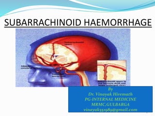
  • 12. When blood vessels of the brain are weak, abnormal, or under unusual pressure, a hemorrhagic stroke can occur. In hemorrhagic strokes, bleeding may occur within the brain, as an intracerebral hemorrhage. Or bleeding may occur between the inner and middle layer of tissue covering the brain (in the subarachnoid space), as a subarachnoid hemorrhage.
  • 13. EPIDEMIOLOGY  Incidence about 9-10/100,000/yr  Higher in Japan (3 times) and Finland  Mean age of onset 51 years (6th decade)  Facts and figures  Men predominate until age 40, then more women (55%); some studies – 3:2 ratio  Seasonal (winter/spring), diurnal (late morning) and day (Sunday)  30 % rupture during sleep  About 50 % of patients with an aneurysm have warning prior to SAH (6-20 days)
  • 14. Case Fatality  The Overall case fatality varied from 32-67%  Population-based study in England with essentially complete case ascertainment  24 hour mortality: 21%  7 days: 37%  30 days: 44% Pobereskin JNNP 2001;70:340-3
  • 15. OVERVIEW  Highest mortality occurs immediately after the hemorrhage and then decreases rapidly  Rebleeding is estimated to occur in 50% of ruptured aneurysms within 6 months of the first hemorrhage, and afterwards at 3% per year  50-60% of patients die after rebleeding and 25% are left disabled
  • 16. Probably the best natural history data come from S Pakarinen: (Incidence, aetiology, and prognosis of primary subarachnoid hemorrhage: a study based on 589 cases diagnosed in a defined urban population during a defined period. Neurol Scand (Suppl) 1967; 29:1-128)  Unselected series, involving the entire city of Helsinki, from 1954 to 1961; minimal surgical withdrawals; all sudden deaths were autopsied  Mortality at 1 day was 32%  Mortality at 1 week was 43%  Mortality at 1 month was 56%  Mortality at 6 months was 60%
  • 17. ETIOLOGY CATEGORY CAUSES TRAUMA CLOSED, PENETRATING, ELECTRIC,ETC… VASCULAR ANEURYSMS, ATHEROSCLEROSIS, AVM, VASCULITIDES IDIOPATHIC BENIGN PERIMESENCEPHAIC SUBARACHNOID HEMORRHAGE BLOOD DYSCRASIAS LEUKEMIAS, HEMOPHILIAS, THROMBOCYTOPENIAS INFECTIONS DENGUE, LEPTOSPIROSIS, BACTERIAL MENINGITIS TOXINS AMPHETAMINES, COCAINE, NICOTINE, ANTICOAGULANTS NEOPLASMS GLIOMAS, MENINGIOMAS, HEMANGIOBLASTOMA, ETC
  • 18. Risk Factors  Hypertension  Cigarette smoking  Oral contraceptives  Alcohol consumption (debatable)  Diurnal variations in blood pressure  Pregnancy and parturition  Slight increased risk during lumbar puncture and/or cerebral angiography in patient with cerebral aneurysm  Slight increased risk with advancing age  Following cocaine abuse
  • 19. Higher chance if: Female 3rd trimester of pregnancy Middle-aged Abuse of stimulant drugs –Connective tissue disorder –Family history
  • 20. PATHOPHYSIOLOGY Modifiable Risk Factors >HPN >Smoking >excessive intake of foods high in fats and cholesterol Non-modifiable Risk Factors> Advanced Age >Gender >Heredity Triggering Factors >Sudden extreme emotion Arterio venous malformation Cerebral aneurysm rupture Bleeding into the brain tissue and subarachnoid space
  • 21. Blood Clots in the Subarachnoid Space Brain Compression Blood supply interruption Tissue Necrosis Neuronal Death Increase Intracranial Pressure T total Paralysis Regional Paralysis Epileptic Seizure : increase intraocular pressure= blindness Death Coma
  • 22. PATHOPHYSIOLOGY CEREBRAL BLOOD FLOW AND METABOLISM RAISED ICT IMPAIRED VASODILATORY AUTOREGULATION VASOSPASM WITH GLOBAL ISCHEMIA BLOOD (‘TOXIC’) BLOCKAGE OF THE ARACHNOID GRANULATIONS SYMPATHETIC SURGE MI PULMONARY EDEMA
  • 23.  CEREBRAL BLOOD FLOW/VOLUME/METABOLISM CBF is globally decreased after SAH Mean CBF decreases with time after SAH; it reaches a nadir in 10 to 14 days, after which it slowly increases toward normal, In patients with poor grades, CBF and cerebral metabolism may remain depressed for weeks ; Cerebral blood volume is markedly increased in patients with severe neurological deficits
  • 24.  INTRACRANIAL PRESSURE RESPONSES Mean ICP was 10 mm Hg in patients with clinical grades 1 and 2, 18 mm Hg in patients with clinical grades 2 and 3, and 29 mm Hg in those with clinical grades 3 to 5. Vasospasm, which was more common in patients with a poor clinical grade and larger SAH, was associated with a significant rise in ICP from a mean of 16 mm Hg in those without vasospasm to 29 mm Hg in those with vasospasm
  • 25. An aneurysm is an abnormal widening or ballooning of a portion of an artery due to weakness in the wall of the blood vessel
  • 27. 27
  • 28.
  • 29. Mechanism of formation of aneurysms  One theory is – that they result from a developmental defect in the media & elastica  Other theory says that aneurysmal process is initiated by the focal destruction of internal elastic membrane caused by hemodynamic forces acting at bifurcations & branching of arteries As a result of local weakness in vessel wall intima bulges out covered only by adventetia  The sac gradually enlarges and subsequently rupture 29
  • 30. 30  For patients of SAH who arrive alive at hospital, the mortality rate over the next month is about 45%. Of those who survive, more than 50% are left with major neurologic deficits as a result of Initial hemorrhage, Cerebral vasospasm with infarction, or Hydrocephalus.
  • 31.  If the patient survives but the aneurysm is not obliterated, the rate of rebleeding is about  20% in the first 2 weeks,  30% in the first month, and about  3% per year afterwards  The annual risk of rupture for aneurysms <10 mm in size is 0.1%, and for aneurysms >10 mm in size is 0.5– 1%;  the surgical morbidity rate far exceeds these percentages 31
  • 32. 32  GIANT ANEURYSMS, those >2.5 cm in diameter account for 5% of cases. The three most common locations are -The terminal internal carotid artery, -Middle cerebral artery (MCA) bifurcation, and -Top of the basilar artery.  Their risk of rupture is ~6% in the first year after identification and may remain high indefinitely.  They often cause symptoms by compressing the adjacent brain or cranial nerves.
  • 33.  Saccular aneurysms occur at the bifurcations of the large to medium-sized intracranial arteries. Rupture is into the  subarachnoid space,  in the basal cisterns and often into  the parenchyma of the adjacent brain.  Approximately 85% of aneurysms occur in the anterior circulation, mostly on the circle of Willis.  About 20% of patients have multiple aneurysms, - - many at mirror sites bilaterally. 33
  • 34. 34
  • 35. Aneurysms are seldom seen in childern on autopsy and so SAH in childern is rare  Beyond childhood they gradually increase in size & the frequency of rupture increases between 35-65 yrs of age  There is increased incidence of  polycystic kidneys,  fibromuscular dysplasia of extracranial arteries,  moyamoya, AV malformations &  coarctation of aorta  Numerous studies have documented familial occurrence of saccular aneurysms, lending support to the idea that genetic factors may play a role in their development 35
  • 36.  As an aneurysm develops, it typically forms a neck with a dome.  At the site of rupture (most often the dome) the wall thins, and the tear that allows bleeding is often 0.5 mm long.  Aneurysm size and site are important in predicting risk of rupture.  Those >7 mm in diameter and  Those at the top of the basilar artery and  At the origin of the posterior communicating artery are at greater risk of rupture. 36
  • 37. 37
  • 39. The main symptom is a severe headache that starts suddenly (often called thunderclap headache). It is often worse near the back of the head. Many persons often describe it as the "worst headache ever" and unlike any other type of headache pain. The headache may start after a popping or snapping feeling in the head. Other symptoms: and alertness Eye discomfort in bright Mood and personality changes, including and irritability (especially Nausea and vomiting
  • 40. often with neck stiffness, and vomiting is common 40
  • 41. Pain at other sites  Occipital and posterior cervical pain may signal a posterior inferior cerebellar artery (PICA) or anterior inferior cerebellar artery aneurysm.  Pain in or behind the eye and in the low temple can occur with an expanding MCA aneurysm 41
  • 42. Loss of consciousness  At the moment of aneurysmal rupture with major SAH, the ICP suddenly rises.  This may account for the sudden transient loss of consciousness that occurs in nearly half of patients which may be preceded by headache.  In 10% of cases, aneurysmal bleeding is severe enough to cause loss of consciousness for several days. 42
  • 43.  meningismus  neck stiffness  photophobia  back pain
  • 44.  Numbness in part of the body  Stiff neck
  • 45.
  • 46.  focal neurologic deficits:  oculomotor palsy  hemiparesis  ocular hemorrhage
  • 47. What causes symptoms & signs? Blood leaking from the aneurysm Local pressure effects of the aneurysm Associated ICH Emboli
  • 48. CLINICAL MANIFESTATIONS- cont. FOCAL NEUROLOGICAL DEFICITS  Although sudden headache in the absence of focal neurologic symptoms is the hallmark of aneurysmal rupture, FOCAL NEUROLOGIC DEFICITS MAY OCCUR.  Anterior communicating artery or MCA bifurcation aneurysms may rupture into the adjacent brain or subdural space and form a hematoma large enough to produce mass effect. The common deficits that result include hemiparesis, aphasia, and abulia (progressive drowsiness or slow mentation) 48
  • 49. Anatomic – clinical corellation of focal neurological Deficits  Third nerve palsy (ptosis, diplopia,dilatation of pupil & divergent squint) indicates an aneurysm at the junction of post. Comunicating &post. Cerebral arteries  A sixth nerve palsy may indicate an aneurysm in the cavernous sinus .but uni or bilateral palsy could be because of raised ICP  Unilateral blindness indicates aneurysm lying anteromedially in the circle of willis usually at the origin of opthalmic artery 49
  • 50. Anatomic – clinical corellation of focal neurological Deficits- cont  Transient paresis of one or both lower limbs indicates ant comunicating A. aneurysm that has interfered with circulation in ant. Cerebral arteries.  Hemiperesis or aphasia suggests aneurysm at the first bifurcation of middle cerebral artery 50
  • 51. 51 The side of aneurysmal rupture may be indicated by monocular pain unilateral preponderace of headache or  unilateral preretinal(subhyaloid) hemorrhage (Terson Syndrome)
  • 52. D/D  Other conditions which may present as severe headache need to be differentiated, like:- Cerebral venous thrombosis, Diffuse vasospasm (call-flemming syndrome),  Pituatry apoplexy, Hypertensive encephalopathy, Intracranial or extracranial arterial dissection  CSF examination assumes great importance in differentiating these conditions from SAH 52
  • 53. D/D- SAH to be differentiated from THUNDERCLAP HEADACHE is a variant of migraine that simulates a SAH. Headache of explosive onset may also be caused by ingestion of sympathomimetic drugs or ingestion of tyramine containg foods in a pt. who is taking MAO inhibitors Sudden severe headache may also be a symptom of phaeochromocytoma 53
  • 54. D/D  Before concluding that a patient with sudden, severe headache has thunderclap migraine or any other type of severe headache, a definitive workup for aneurysm is always required in the form of CT scan, Lumbar puncture, and if the diagnosis still remains doubtful, then cerebral angiography 54
  • 55. DELAYED NEUROLOGIC DEFICITS There are four major causes of delayed neurologic deficits: 1. RERUPTURE/REBLEED, 2.HYDROCEPHALUS 3.VASOSPASM 4.HYPONATREMIA. 55
  • 56. 2.HYDROCEPHALUS  Acute hydrocephalus can cause stupor and coma and can be mitigated by placement of an external ventricular drain. More often, subacute hydrocephalus may develop over a few days or weeks and causes progressive drowsiness or slowed mentation (abulia) with incontinence.. It may clear spontaneously or require temporary ventricular drainage. 56
  • 57. Hydrocephalus-cont. Chronic hydrocephalus may develop weeks to months after SAH and present as normal pressure hydrocephalus (NPH) manifested by gait difficulty, incontinence, or impaired mentation. Subtle signs may be a lack of initiative in conversation or a failure to recover independence 57
  • 58. 3.VASOSPASM  Causes symptomatic ischemia and infarction in ~30% of patients and is the major cause of delayed morbidity and death.  Signs of ischemia appear 4–14 days after the hemorrhage, most often at 7 days.  The severity and distribution of vasospasm determine whether infarction will occur.  Vasospasm is believed to result from direct effects of clotted blood and its breakdown products on the arteries within the subarachnoid space. 58
  • 59. VASOSPASM- cont.  Spasm of major arteries produces focal symptoms referable to the appropriate vascular territory.  All of these focal symptoms may present abruptly, fluctuate, or develop over a few days.  In most cases, focal spasm is preceded by a decline in mental status.  Patient may develop fluctuating hemiperisis or aphasia  Severe cerebral edema in patients with infarction from vasospasm may increase the ICP enough to reduce cerebral perfusion pressure. 59
  • 60. 4.HYPONATREMIA  Hyponatremia. Hyponatremia may be profound and can develop quickly in the first 2 weeks following SAH. There is both natriuresis and volume depletion with SAH, so that patients become both hyponatremic and hypovolemic.  Both atrial natriuretic peptide (ANP)and brain natriuretic peptide (BNP) have a role in producing this "cerebral salt-wasting syndrome.“  There may some role of antidiuretic hormone also, causing water retention  Typically it clears over the course of 1–2 weeks and, in the setting of SAH, should not be treated with free-water restriction as this may increase the risk of stroke. 60
  • 61. SYSTEMIC CHANGES ASSOCIATED WITH SAH ECG changes suggestive of subendicardial or myocardial ischemia  Elevation of troponis/CPK (MB) levels  In some patients cardiac dysfunction may be severe enough to cause fall in EF % and heart failure  There is evidence that structural myocardial lesions produced by circulating catecholamines and excessive discharge of sympathetic neurons may occur after SAH, causing these ECG changes and a reversible cardiomyopathy sufficient to cause shock or congestive heart failure.  Hyponatremia  D. Insipidus  Albuminuria,  Glycosuria  leukocytosis 61
  • 63. NON CONTRAST CT SCAN.  CT Scan is Initial investigative procedure  The extent and location of subarachnoid blood on noncontrast CT scan help  locate the underlying aneurysm,  identify the cause of any neurologic deficit, and  predict delayed vasospasm. A high incidence of symptomatic vasospasm in the MCA and ACA has been found when early CT scans show subarachnoid clots >5 x 3 mm in the basal cisterns or layers of blood >1 mm thick in the cerebral fissures.  CT scans less reliably predict vasospasm in the vertebral, basilar, or posterior cerebral arteries. 63
  • 64. More than 95% of cases have enough blood to be visualized on a high-quality NONCONTRAST CT SCAN obtained within 72 h. 64
  • 65. CT scan reveals subarachnoid hemorrhage in the right sylvian fissure; no evidence of hydrocephalus is apparent.
  • 66. (A)Noncontrast CT demonstrates subarachnoid hemorrhage (arrows). (B)3-D reconstruction image from a CT angiogram demonstrates an aneurysm (arrow) from the anterior communicating artery as the cause of the bleed.
  • 67. IMAGING STUDIES-cont.  Exact site of offending aneurysm can be inferred from the location of main clot in CT scan  Collection of blood in the anterior inter-hemispheric fissure indicates rupture of an ant. Communicating artery  Blood in sylvian fissure – middle cerebral artery  Blood in ant. Perimesencephalic cistern- post. Communicating artery or distal basilar artery aneurysm  MRI can also detect blood in the proton density sequence 67
  • 68. Lab investigations-cont  If the scan fails to establish the diagnosis of SAH and no mass lesion or obstructive hydrocephalus is found,  Then a lumbar puncture should be performed to establish the presence of subarachnoid blood.  Usually CSF becomes grossly bloody within 30 minutes of bleed with RBC counts upto 1 million/c mm or more  Lysis of the red blood cells and subsequent conversion of hemoglobin to bilirubin stains the spinal fluid yellow within 6–12 h.  This xanthochromic spinal fluid peaks in intensity at 48 h and lasts for 1–4 weeks, depending on the amount of subarachnoid blood 68
  • 69. Angiography  Once the diagnosis of hemorrhage from a ruptured saccular aneurysm is suspected, four-vessel conventional x-ray angiography (both carotids and both vertebrals) is generally performed  to localize and define the anatomic details of the offending aneurysm and  to determine if other unruptured aneurysms exist).  CT angiography is an alternative method for locating the aneurysm and may be sufficient to plan definitive therapy. 69
  • 70. Angiography-cont.  in 5-10% pts with aneurysmal bleed , there may not be any evidence of aneurysmal rupture on angiography. It may be because in some cases obliteration of lesion occurs In some lesions were somewhat more benign  Pts without evidence of an offending aneurysm or an arteriovenous malformationhave a better prognosis 70
  • 71. Angiography-cont.  Perimesencephalic hemorrhage also caries a better prognosis In this cisterns surrounding midbrain & upper pons gets filled up with blood Mild headache, no vasospasm  Usually no aneurysm is found at the expected site i.e. at the top of basillary artery 71
  • 72. Lab Inv. – cont.  Close monitoring (daily or twice daily) of electrolytes is important because hyponatremia can occur precipitously during the first 2 weeks following SAH  An asymptomatic troponin elevation is common. 72
  • 73. ECG Changes  The electrocardiogram (ECG) frequently shows ST- segment and T-wave changes similar to those associated with cardiac ischemia.  Prolonged QRS complex, increased QT interval, and prominent "peaked" or deeply inverted symmetric T waves are usually secondary to the intracranial hemorrhage ( cerebral T waves).  Serious ventricular dysrhythmias are unusual 73
  • 74. 74
  • 75. ECHO Echocardiography reveals a pattern of regional wall motion abnormalities that follow the distribution of sympathetic nerves rather than the major coronary arteries  The sympathetic nerves themselves appear to be injured by direct toxicity from the excessive catecholamine release. 75
  • 76. TREATMENT The medical management of SAH focuses on  Protecting the airway,  Managing blood pressure  Preventing rebleeding prior to treatment,  Managing vasospasm,  Treating hydrocephalus,  Treating hyponatremia, and  Preventing pulmonary embolus. 76
  • 77.  Because rebleeding is common, all patients who are not candidates for early aneurysm repair are put on bed rest in a quiet room and are given stool softeners to prevent straining.  If Headache or neck pain is severe, mild sedation and analgesia are prescribed.  Extreme sedation is avoided because it can obscure changes in neurologic status.  Adequate hydration is necessary to avoid a decrease in blood volume predisposing to brain ischemia. So adequate amount of fluid is administered so as to maintain above normal circulating blood volume & central venous pressure 77
  • 78. Managing raised ICP  Intracranial hypertension following aneurysmal rupture occurs secondary to subarachnoid blood, parenchymal hematoma, acute hydrocephalus, or  loss of vascular autoregulation. Perilesional edema if infarct occurs 78
  • 79. Managing raised ICP- cont.  Patients who are stuporous should undergo emergent ventriculostomy to measure ICP and to treat high ICP in order to prevent cerebral ischemia.  Medical therapies designed to combat raised ICP e.g., mild hyperventilation, mannitol, and sedation can also be used as needed. (Mannitol dose-25-100gm q4h)  High ICP refractory to treatment is a poor prognostic sign 79
  • 80. MANAGING BP  Prior to definitive treatment of the ruptured aneurysm, care is required to maintain adequate cerebral perfusion pressure while avoiding excessive elevation/fall of arterial pressure.  If the patient is alert, it is reasonable to lower the blood pressure to <150 systolic using nicardipine, labetolol, or esmolol.  If the patient has a depressed level of consciousness, then ICP(Intracranial pressure) should be measured and the cerebral perfusion pressure targeted to 60–70 mmHg. 80
  • 81. Managing Seizure Activity  Seizures are uncommon at the onset of aneurysmal rupture.  The quivering, jerking, and extensor posturing that often accompany loss of consciousness with SAH are probably related to  the sharp rise in ICP or, perhaps,  acute generalized vasospasm rather than seizure.  Use of anti-seizure drugs is controvercial. However, phenytoin is often given as prophylactic therapy since a seizure may promote rebleeding. 81
  • 82. MANAGING VASOSPASM Vasospasm remains the leading cause of morbidity and mortality following aneurysmal SAH.  Treatment with the calcium channel antagonist nimodipine (60 mg PO every 4 h) improves outcome, perhaps by preventing ischemic injury  Nimodipine can cause significant hypotension in some patients, which may worsen cerebral ischemia in patients with vasospasm 82
  • 83. Managing Hydrocephalus Acute hydrocephalus can cause stupor or coma.  It may clear spontaneously or require temporary external ventricular drainage.  When chronic hydrocephalus develops, permanent ventricular shunting is the treatment of choice. 83
  • 84. Role of Steroids  Glucocorticoids may help reduce the head and neck ache caused by the irritative effect of the subarachnoid blood.  There is no good evidence that they reduce cerebral edema, are neuroprotective, or reduce vascular injury, and  Their routine use therefore is not recommended 84
  • 85. Adjunctive Therapies and Measures  Keep the patient's core body temperature at 37.2°C  Consider antiemetics for nausea or vomiting.  Elevate the head of the bed 30° to facilitate intracranial venous drainage. Emergent ventricular drainage by the neurosurgeon may be necessary.  Maintain the patient's serum glucose level at 80-120 mg/dL; use sliding or continuous infusion of insulin if necessary.  Fluids and hydration  Do not over hydrate patients because of the risks of hydrocephalus.  Patients with subarachnoid hemorrhage (SAH) may also have hyponatremia from cerebral salt wasting.
  • 86. INTERVENTIONAL MANAGEMENT  Early aneurysm repair prevents rerupture/rebleeding and  allows the safe application of techniques to improve blood flow (e.g., induced hypertension and hypervolemia) An aneurysm can be "clipped" by a neurosurgeon or "coiled" by an endovascular surgeon. 86
  • 87. 87 Hunt - Hess scale is helpful in assessing the patient before deciding for intervntional management
  • 88. HUNT-HESS SCALE  GRADE1:  Mild headache, normal mental status, no cranial nerve or motor findings –(GCS* score 15, no motor deficits)  GRADE 2: Severe headache, normal mental status, may have cranial nerve deficit –(GCS score 13–14, no motor deficits)  GRADE 3: Somnolent, confused, may have cranial nerve or mild motor DEFICIT- (GCS SCORE 13–14, WITH MOTOR DEFICITS)  GRADE 4 : Stupor, moderate to severe motor deficit, may have intermittent reflex posturing- (GCS score 7–12, with or without motor deficits)  GRADE 5: Coma, reflex posturing or flaccid (GCS score 3–6, with or without motor deficits) 88
  • 89. Management based on HH scaleGrades 1 & 2  Current approach is to go for intervention within 24 hrs This will prevent rebleed and will allow measures to be taken to improve cerebral circulation  Grade 3 patients- if their condition allows , they too would be benefitted by the procedure  Grade4- the outcome is generally poor but some times by putting a ventricular drain and improving the grade, pt89
  • 90. 90 SURGICAL REPAIR •Involves placing a metal clip across the aneurysm neck, thereby immediately eliminating the risk of rebleeding. •This approach requires craniotomy and brain retraction, which is associated with neurologic morbidity.
  • 91. 91 Endovascular techniques involve placing platinum coils, or other embolic material, within the aneurysm via a catheter that is passed from the femoral
  • 92. . Endovascular techniques-CONT  The aneurysm is packed tightly to enhance thrombosis and over time is walled-off from the circulation  The benefit of endovascular therapy is durable.  However, some aneurysms have a morphology that is not amenable to endovascular treatment.  Thus, surgery remains an important treatment option  But for aneurysms which are not approachable by surgery, endovascular technique would become the only choice 92
  • 93. INTERNATIONAL SUBARACHNOID ANEURYSM TRIAL (ISAT)  The only prospective randomized trial of surgery versus endovascular treatment for ruptured aneurysm, the International Subarachnoid Aneurysm Trial (ISAT), was terminated early when 24% of patients treated with endovascular therapy were dead or dependent at 1 year compared to 31% treated with surgery, a significant 23% relative reduction.  After 5 years, risk of death was lower in the coiling group,  Although the proportion of survivors who were independent was the same in both groups  Risk of rebleeding was low, but more common in the coiling group 93
  • 94. GRADE CT APPEARANCE 1 NONE 2 < 1mm thickness 3 > 1mm thickness 4 Diffuse with parenchymal/ventricular extension FISHERS GRADE CT APPEARANCE 0 NONE 1 MINIMAL 2 MINIMAL WITH IVH 3 THICK 4 THICK WITH IVH CLAASSENS MODIFICATION
  • 95. GRADE GCS NEURODEFICIT 1 15 ABSENT 2 13-14 ABSENT 3 13-14 PRESENT 4 7-12 ABSENT/PRESENT 5 <7 ABSENT/PRESENT WORLD FEDERATION OF NEUROSURGEONS (WFNS)
  • 96. GRADE CLINICAL PRESENTATION 1 CONSCIOUS 2 DROWSY 3 DROWSY WITH DEFICIT 4 MAJOR NEURODEFICIT 5 MORIBUND BOTTERELL ET AL
  • 97. Discharge Plan  Activity  patient will need to have someone with for the next several days to watch for worsening of symptoms (see below) and to allow to rest.  Start with light activity around the house for the first 3 days at home.  Gradually increase activity starting with short walks 1-2 times per day.  Avoid contact sports, skating, bike riding, or other such activities for 6 weeks.  Encourage pt to do passive range of motion  Nutrition :  Instruct the relative to feed pt on time with proper food low in Na  Low in cholesterol low in fat and give citrus fruits ,moderate in fluid intake and increase fiber diet to improve health..
  • 98. Medications  Take medications as prescribed and gradually decrease pain medications as pain improves. Instruct pt and their relative to follow medication regimen Educate and instruct the patient and her family to monitor BP and PR before giving medication Follow up with primary care physician for all medical issues.
  • 99. Genetics of Intracranial Aneurysms Ref. Neurosurgery clinics of North America, July 1998, pp 485-493 Observations to suggest genetic involvement: Association of various heritable disorders with ICA The familial aggregation of ICA in the absence of known systemic disorder Heritable Disorders associated with ICA: Heritable CT disorder account for at least 5% of the cases of ICA True frequency may be higher due to variability in phenotypic expression and negative family history due to new mutation The most important ones are: Ehlers-Danlos type IV Marfan’s syndrome NF1 ADPKD
  • 100. Ehlers-Danlos Syndrome Type IV  Defeciency of collagen type 3  Mutation in the gene encoding for pro 1(III) chain of type III collagen  Joint hypermobility, hyperelastic/fragile skin, abnormal scaring  Type IV is least common and the most lethal  Prevalence 1/50,000-500,000  Vascular catastrophes account for the great majority of deaths  Clues to the diagnosis  Facial appearance: thin nasal bridge, thin lips, lobeless ears, prematurely aged appearance  Bruising tendency, MVP, spontaneous pneumothorax, abnormal scarring, varicose veins  ICA:  Prevalence?, 14 in 202 patient  Saccular or fusiform, located in the cavernous sinus  Thin walled, difficult to repair
  • 101.  Marfan’s syndrome  Mutation in gene encoding fibrillin-1 (glycoprotein, major component of microfbrils in ECM and elastic tissues)  Prevalence 1/10,000-20,000  Abnormalities of the skeleton, cardiovascular system, eyes, spinal meninges  Variability in the phenotypic expression  ICA:  Saccular or fusiform, dissecting aneurysms  Propensity to the proximal internal carotid artery  Fragility of the connective tissue is not a major problem  Ectasia and tortuosity of the vessels
  • 102. Neurofibromatosis type 1  Mutation in NF1 gene encoding neurofibromin which may have regulatory role in the development of connective tissue  Prevalence 1/3000-5000  Café-au-lait spot, lish nodule, neurofibroma  Vascular complication; stenosis rupture, aneurysm, fistula  ICA:  Saccular or fusiform  Vascular fragility, distorted anatomy  Association with intracranial arterial occlusive disease
  • 103. ADPKD  Mutation in two genes PKD1 and PKD2  PKD1 linked disease is more severe  Polycystin encoded by PKD1 is an integral membrane protein, play a role in the integrity of ECM of CT  Most common monogenetic disorder  Prevalence 1/400-1000  Cysts in kidney, liver, spleen, pancreas, ovaries  Cardiovascular abnormalities: MVP, aortic and cervicocephalic dissection, coronary and AAA  ICA:  Saccular, fusiform, dissecting  Vascular fragility  Ruputre occurs in earlier age, increased risk of developing new ICA  ICA in 25% of patient with ADPKD at autopsy, 10% on MRA  Family screening with MRA yields ICA in 20-25%  Cause of death in one-fifth  ADPKD account for 2-7% of all ICA
  • 104. Familial ICA  First described by Chamber et al. In 1954  With the exception of ADPKD, Ehlers-Danlos IV, Pompe disease, Idiopathic nonarteriosclerotic cerebral calcification there is no association with heritable CT disease  7-20% of first or second-degree relatives of patient with SAH have ICA  First-degree relatives of patient with SAH have twice to fivefold increase risk of having ICA  First-degree relatives have threefold increased risk compared to second-degree relatives  Inheritance pattern is unknown, most likely multifactorial, or AD
  • 105. ICA:  Rupture at younger age (5 year earlier), 71% has ruptured by age of 50 Vs 42% in nonfamilial ICA  Anterior communicating artery aneurysms are underrepresented, MCA is overrepresented  Rupture at smaller size  Increase female preponderance  Larger multiplicity  Siblings are more likely to harbor aneurysms at the same side at mirror sites  Severity of hemorrhage is similar to nonfamilial ones
  • 106. Screening for familial ICA Why?  Poor prognosis once ruptured  Low surgical risk for the nonruptured (5% morbidity, 2% mortality)  Caveat: aneurysms may develop over short period of time  Indication for surgery for asymptomatic ICA is still unclear (critical size) Who?  Families with two or more affected members  Restricted to first-degree relatives (yield 9-29%)  Monozygotic twin When?  Screening between the ages of 35-65  Youngest patient with familial ICA is 6 years old  De novo aneurysms occur at a rate of 2% per year  Repeat screening at 6-month to 5-year intervals
  • 107. Screening for familial ICA …/cont’d How?  MRA is the most widely used  Critical size for detection 3-5 mm  Helical CT angiography  Coventional angiography ICA gene  Current approaches  Screen the human genome for ICA gene by testing a large number of distinct highly pleomorphic genetic markers  Analyze variations in the sharing of marker alleles among affected sibling pairs  Candidate gene sequence analysis e.g., PKD1 or COL3A1
  • 108. Heritable Disorders Associated with Intracranial Aneurysms Disorder Inheritance Pattern Locus Gene Gene Product Achondroplasia AD 4p16.3 FGFR3 Fibroblast growth factor receptor 3 Alkaptonuria AR 3q2 AKU ? ADPKD AD 16p13.3 PKD1 Polycystin Cohen syndrome AR 8q22 CHS1 ? Ehlers-Danlos Syndrome type I AD 9q COL5A1 Ollagen type V Ehlers-Danlos Syndrome type IV AD 2q31 COL3A1 Collagen type III Fabry disease AR XL-R Xq22.1 -galactosidase A Kahn syndrome AR ? ? ? Marfan’s syndrome AD 15q21.1 FBN1 Fibrillin-1 NF1 AD 17q11.2 NF1 Neurofibromin Noonan syndrome AD 12q22 NS1 ?
  • 109. Heritable Disorders Associated with Intracranial Aneurysms …/cont’d Disorder Inheritance Pattern Locus Gene Gene Product Osler-Rendu- Weber disease AD 9q34.1 HHT1 Endoglin Osteogenesis imperfecta type 1 AD 17q22.1 COL1A1 Collagen type 1 Pompe disease AR 17q23 GAA -Glucosidase Pseudoxanthoma elasticum AD & AR ? ? ? Rambaud syndrome AR ? ? ? Tuberous sclerosis AD 9q34/16p13.3 TSC1/TSC2 ?/Tuberin Wermer syndrome AD 11q13 MEN1 ? 3M syndrome AR ? ? ? 1-Antitrypsin deficiency AcoD 14q32.1 P1 1-Antitrypsin
  • 110.  How well a patient with subarachnoid hemorrhage does depends on a number of different factors, including:  Location and amount of bleeding  Complications  Older age and more severe symptoms can lead to a poorer outcome.  People can recover completely after treatment. But some people die even with treatment. Prognosis:
  • 112. TO SUM-UP  SAH is a neurolgical emergency  It is less common but an important cause of stroke(5%)  saccular/berry aneurysmal rupture is the commonest cause of SAH  Most common presenttion is an excruciating headache which needs to be differentiated from other causes of severe headache including thunderclap migrane  CT scan is very helpful in making a dianosis and differentiating it from other headaches of severe intensity  If CT is negative but there s strong suspecion of SAH then one should go for CSF examination  If othe CT scan & CSF are inconclusive then cerebral angiography is the last resort  Complications include rerupture, hydrocephalus, vasospasm and hyponatremia 112
  • 113. 113  Management icludes – bed rest, maintaing a clear airway, managing BP, giving adequate amount of fluids, monitoring hyponatremia preventing vasospasm and instituting measures to lower ICP  one third of patients are prone to develop rebleed which can be effectively taken care of by interventional modalities i.e. surgical (clipping)/ endovascular technique (coiling)  Randomzed trial (ISAT) has shown coiling to superior to clipping
  • 114. References  MS Greenberg. Handbook of Neurosurgery. Seventh Edition. New York: Thieme Medical Publishers; 2010.  AH Kaye. Essential Neurosurgery. Third Edition. Oxford: Blackwell Publishing Ltd; 2005.  MG Yasargil. Microneurosurgery. New York: Thieme Medical Publishers Inc; 1994.  Zivin J. Hemorrhagic cerebrovascular disease. In: Goldman L, Schafer AI, eds. Goldman's Cecil Medicine. 24th ed. Philadelphia, PA: Elsevier Saunders; 2011:chap 415.  http://www.strokecenter.org/professionals/brain- anatomy/blood-vessels-of-the-brain/
  • 115. References:  Naggara ON, White PM, Guilbert F, et al. Endovascular treatment of intracranial unruptured aneurysms: systematic review and meta-analysis of the literature on safety and efficacy. Radiology. 2010;256:887-897.  Reinhardt MR. Subarachnoid hemorrhoid. J Emerg Nurs. 2010;36:327-329.  Tateshima S, Duckwiler G. Vascular diseases of the nervous system: intracranial aneurysms and subarachnoid hemorrhage. In: Daroff RB, Fenichel GM, Jankovic J, Mazziotta JC. Bradley’s Neurology in Clinical Practice. 6th ed. Philadelphia, PA: Elsevier Saunders; 2012:chap 51C.
  • 116. . Avoiding Pitfalls in the Diagnosis of Subarachnoid Hemorrhage. JA Edlow and LR Caplan. N Engl J Med 342:29-36, 2000 . Unruptured Intracranial Aneurysms - Risk of Rupture and Risks of Surgical Intervention. ISUIA Investigators. N Engl J Med; 339: 1725- 33, 1998 . Intracranial Aneurysms and Subarachnoid Hemorrhage: An Overview. B Weir and RL Macdonald. Chapter 214 (pp 2191-2213) in Neurosurgery, 2nd edition, volume II, 1996, editors RH Wilkins and SS Rengachary, published by McGraw-Hill . Relation of Cerebral Vasospasm to Subarachnoid Hemorrhage Visualized by Computerized Tomographic Scanning. CM Fisher JP Kistler and JM Davis. Neurosurgery, 6:1-9, 1980 . Factors Influencing the Outcome of Aneurysm Rupture in Poor Grade Patients: A Prospective Series. L Disney, B Weir, M Grace et al Neurosurgery 23: 1-9 1988