
Empfohlen
Weitere ähnliche Inhalte
Was ist angesagt?
Was ist angesagt? (20)
Ähnlich wie Nmr chemical shift, By Dr. UMESH KUMAR SHARMA AND ARATHY S A
Ähnlich wie Nmr chemical shift, By Dr. UMESH KUMAR SHARMA AND ARATHY S A (20)
Mehr von Dr. UMESH KUMAR SHARMA
Mehr von Dr. UMESH KUMAR SHARMA (20)
Kürzlich hochgeladen
Kürzlich hochgeladen (20)
Nmr chemical shift, By Dr. UMESH KUMAR SHARMA AND ARATHY S A
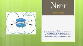
- 1. Nmrspectroscopy By : Dr. UMESH KUMAR SHARMA & ARATHY S A. DEPARTMENT OF PHARMACEUTICS MAR DIOSCORUS COLLEGE OF PHARMACY, THIRUVANANTHAPURAM, KERALA, INDIA 1
- 2. NMR SPECTROSCOPY CHEMICAL SHIFT FACTORS INFLUENCING CHEMICAL SHIFT SPIN –SPIN COUPLING COUPLING CONSTANT 2 Nmr spectroscopy
- 3. CHEMICAL SHIFT Chemical shift is the difference between absorption position of a sample proton & absorption position of a reference compound. Theoretically for any organic compound only one NMR should be recorded for all protons present. But this does not happen in practice since all hydrogen atoms are not in same environment. Magnetic field applied is not felt by all hydrogen atoms uniformly. This is due to the presence or nearness to double bond or triple bond, aromatic / hetrocyclic /alicyclic ring systems, electronegative atoms. 3 Nmr spectroscopy
- 4. Due to differences in magnetic field felt by H atoms, each proton has different processional frequency. Different range of frequency are required for excitation. Different peaks or signals for each kind of proton are obtained. The signal positions in NMR spectra are chemical shifts. Nucleus is surrounded by constantly revolving electrons, it produce spherically symmetrical electromagnetic charge cloud. When magnetic field is applied electron charge cloud circulate in a direction to produce induced field, which is opposed to applied field. Induced field shied the nuclei from external field. 4 Nmr spectroscopy
- 5. Induced field is directly proportional to applied field strength. B induced=σ B applied Constant σ is called shielding or screening constant. B effective = B applied-B induced Where B effective=Bo(1-σ), Bo is applied magnetic field Shielding causes small difference in absorption frequency. Chemical shift is measured by comparing positions of peaks of reference compound. Frequency of proton=ϒBo/2Ϡ. Reference standard like Tetra methyl Silane are used. Resonance frequencies of sample are quoted with respect to TMS. 5 Nmr spectroscopy
- 7. Shielding of electrons: High electron density around nucleus shields nucleus from external magnetic field. Signals are up-field in NMR spectrum. De-shielding of electrons: Lower electron density around nucleus de-shield nucleus from external magnetic field. Signals are downfield in NMR spectrum. 7 Nmr spectroscopy
- 8. NMR spectra shows applied field strength increasing from left to right. Left part is downfield & right part is up-field. Nuclei which absorb on up-field side are strongly shielded & downfield side are weakly shielded. Shielded nuclei do not sense magnetic field as large as de-shielded do. 8 Nmr spectroscopy
- 9. As a result energy difference between α & β spin states is much lower for shielded nuclei. They resonate at lower frequency. De-shielded nuclei have higher energy difference between α & β spin states. They resonate at higher frequency. 9 Nmr spectroscopy
- 10. Frequency at which a nucleus come into resonance with in a magnetic field, υ = ϒ𝐻𝑜 2π Resonance frequencies of nuclei in sample is measured & compared to a reference standard Position of peaks in NMR spectrum relative to reference peaks in terms of chemical shift 𝛿 = 𝐻𝑜 𝑟𝑒𝑓𝑒𝑟𝑒𝑛𝑐𝑒 −𝐻𝑜(𝑠𝑎𝑚𝑝𝑙𝑒)∗106 ppm Ho(reference) 10 Nmr spectroscopy
- 11. Tetra methyl Silane (TMS) as reference compound : TMS has 12 equivalent protons & gives a single intense signal. Chemically inert. Low boiling point. Volatile. Can be easily recovered from any sample. Soluble in most organic solvents. 11 Nmr spectroscopy
- 12. FACTORS AFFECTING CHEMICAL SHIFT ELECTRO NEGATIVITY Chemical shift arises from secondary magnetic field produced by circulation of electrons in molecule. Electrons bonding proton passes around the nucleus in a plane perpendicular to applied field. It creates a secondary field which opposes applied field. Nucleus experience resultant field smaller than applied field. This shielding effect lowers processional frequency. In TMS since Si is electropositive, electron pushes towards methyl group by positive inductive effect. Powerful shielding causes protons of TMS to resonate at lower frequency. As electronegativity increases, electron withdrawal from methyl group increases. 12 Nmr spectroscopy
- 13. Electronegative groups attached to C-H system decrease electron density around protons. Its de-shielding effect causes proton to experience higher magnetic field & higher resonance frequency. As electronegativity increases, de-shielding increases Absorption occurs downfield or at higher resonance frequency. Additional electronegative atoms cause increase in chemical shift. Effect decreases with distance. More electronegative atoms de-shield more & give larger shift values. Diamagnetism. Produces secondary field that opposes applied field. Paramagnetism. Produces secondary field in the direction of applied field. 13 Nmr spectroscopy
- 14. MAGNETIC ANISOTROPY: π electrons of aromatic compounds, alkenes, alkynes, carbonyls etc. interact with applied field which induces magnetic field. They cause magnetic anisotropy. Causes both shielding & de-shielding. Eg : benzene ALKENES : When alkene group is so oriented that plane of double bond is at 90 to the direction of applied field. Induced circulation of pie electrons generates a secondary field. It is diamagnetic around carbon atom but paramagnetic in region of alkene protons. 14 Nmr spectroscopy
- 15. ALKENES When alkene group is so oriented that plane of double bond is at 90 to direction of applied field. Induced circulation of pie electrons generates a secondary field. It is diamagnetic around carbon atom but paramagnetic in region of alkene protons. Pie electrons of double bond generates a magnetic field that oppose applied magnetic field in middle of molecule. It reinforces the applied field on outside where vinylic protons are located. Reinforcement de-shield vinylic protons & shift down fields. 15 Nmr spectroscopy
- 17. ALKYNES When terminal triple bond is aligned in direction of magnetic field, electrons circulate to create induced magnetic field. Acetyl proton lies along the axis of field & opposes external field. Acetyl protons are shielded. 17 Nmr spectroscopy
- 18. AROMATIC COMPOUNDS: Pie electrons of aromatic ring are delocalized cyclically over aromatic ring. It induce a secondary field, that is diamagnetic at centre & paramagnetic at periphery where protons present. They experience a higher magnetic field & so higher resonance frequency. 18 Nmr spectroscopy
- 19. HYDROGEN BONDING: In compounds capable of hydrogen bonding there is greater de-shielding of protons. Shieft down fields. When sample temperature increases hydrogen bond weakens & up-field shift. Increasing solute concentration increases de-shielding & down field shift occurs. Intra molecular hydrogen bonding is unchanged by dilution. NMR spectra of such systems is unaltered by varying concentration. Effect of secondary field affects chemical shift in one the ways like a. Positive shielding b. Negative shielding Two types of shielding phenomena modifies resonance peak in NMR spectrum by a. Local shielding b. Low range shielding 19 Nmr spectroscopy
- 20. SPIN-SPIN COUPLING: Interaction between spins of neighbouring nuclei in a molecule cause splitting of lines in NMR spectrum. It occurs by means of a slight un-pairing of bonded electrons. Number of lines observed in NMR signals for a group of protons is related to number of protons in neighbouring groups. Splitting is related to number of possible spin orientations that neighbouring group can adopt. Different spin states and resultant magnetic moment of neighbouring protons modifies actual magnetic field experienced by a proton. Mutual influence between protons is not transmitted through space ,but via electrons of intervening bonds. 20 Nmr spectroscopy
- 22. CAUSES OF SPLITTING: Splitting of signals can be determined by its environment of absorbing protons with respect to neighbouring protons. Due to different spin states & resultant magnetic moments of neighbouring protons actual magnetic field experienced by proton gets modified. Absorption proton experience each of the modified field. Its absorption signal split into different peaks. 22 Nmr spectroscopy
- 23. COUPLING CONSTANT(J) It is the distance between peaks in a given multiplet. It is a measure of magnitude of splitting effect. Value of J is independent of applied magnetic field. Depends on molecular structure. For geminal protons value of J varies from 0 - 20Hz. Depends on bond angle & overall structure of molecule. For vicinal protons J varies from 2 -18Hz depending on spatial position of protons & molecular structure. In freely rotating groups, protons with anti conformation have J value 5 - 12Hz. Protons with gauche conformation have J value 2 - 4Hz. For cis protons J value is 6 - 14Hz & trans protons 11 - 18Hz. Coupling constants are a measure of effectiveness of spin spin coupling. Useful in getting information from NMR spectra of complex molecular structures. 23 Nmr spectroscopy
- 24. PROTON NMR SPECTRA OF ETHANOL 24 Nmr spectroscopy
- 25. HNMR SPECTRA OF BENZENE 25 Nmr spectroscopy
- 26. INTERPRETATION OF HNMR SPECTRA The number of signals shows how many different kinds of protons are present. The location of the signals shows how shielded or de-shielded the proton is. The intensity of the signal shows the number of protons of equivalent protons. Signal splitting shows the number of protons on adjacent atoms. 26 Nmr spectroscopy
- 27. REFERENCE Organic spectroscopy by William Kemp Instrumental method of chemical analysis by Chatwal Principles of Instrumental analysis. Doglas A Skoog, James Holler.5th edition Instrumental methods of analysis by Willard Wikipedia.org 27 Nmr spectroscopy
- 28. For Feedback / Comments Write to umeshpanditjp@gmail.com 28